at dilution of 1:6000 incubated at room temperature for 1.5 hours.")


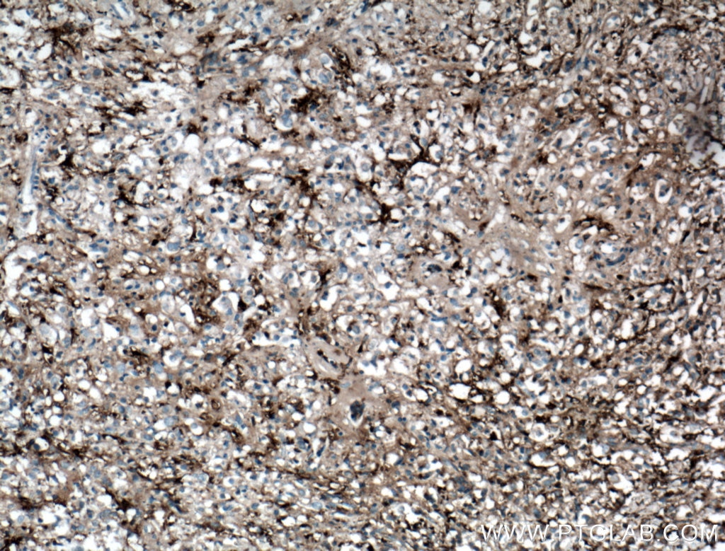
 at dilution of 1:500 (under 10x lens. Heat mediated antigen retrieval with Tris-EDTA buffer (pH 9.0).")
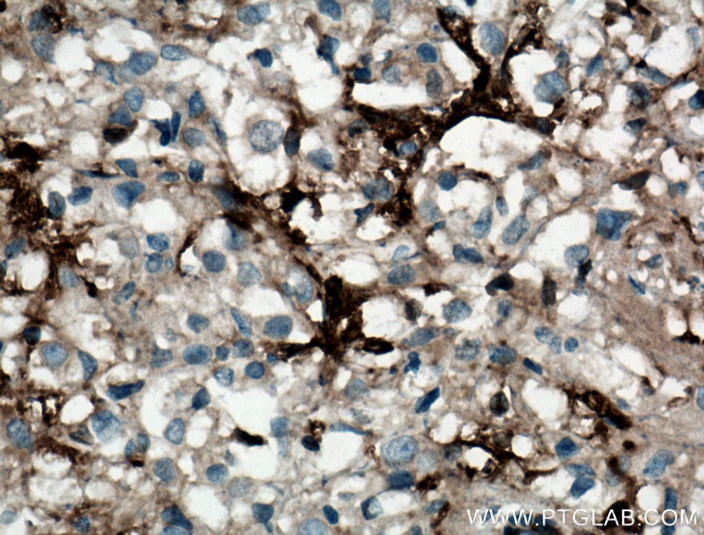
 at dilution of 1:500 (under 40x lens. Heat mediated antigen retrieval with Tris-EDTA buffer (pH 9.0).")
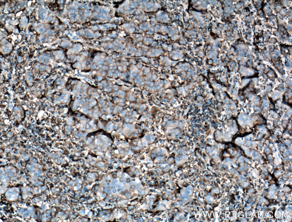
 at dilution of 1:500 (under 10x lens. Heat mediated antigen retrieval with Tris-EDTA buffer (pH 9.0).")
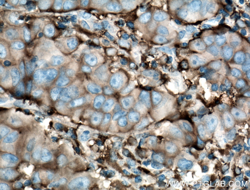
 at dilution of 1:500 (under 40x lens. Heat mediated antigen retrieval with Tris-EDTA buffer (pH 9.0).")
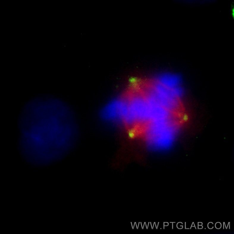
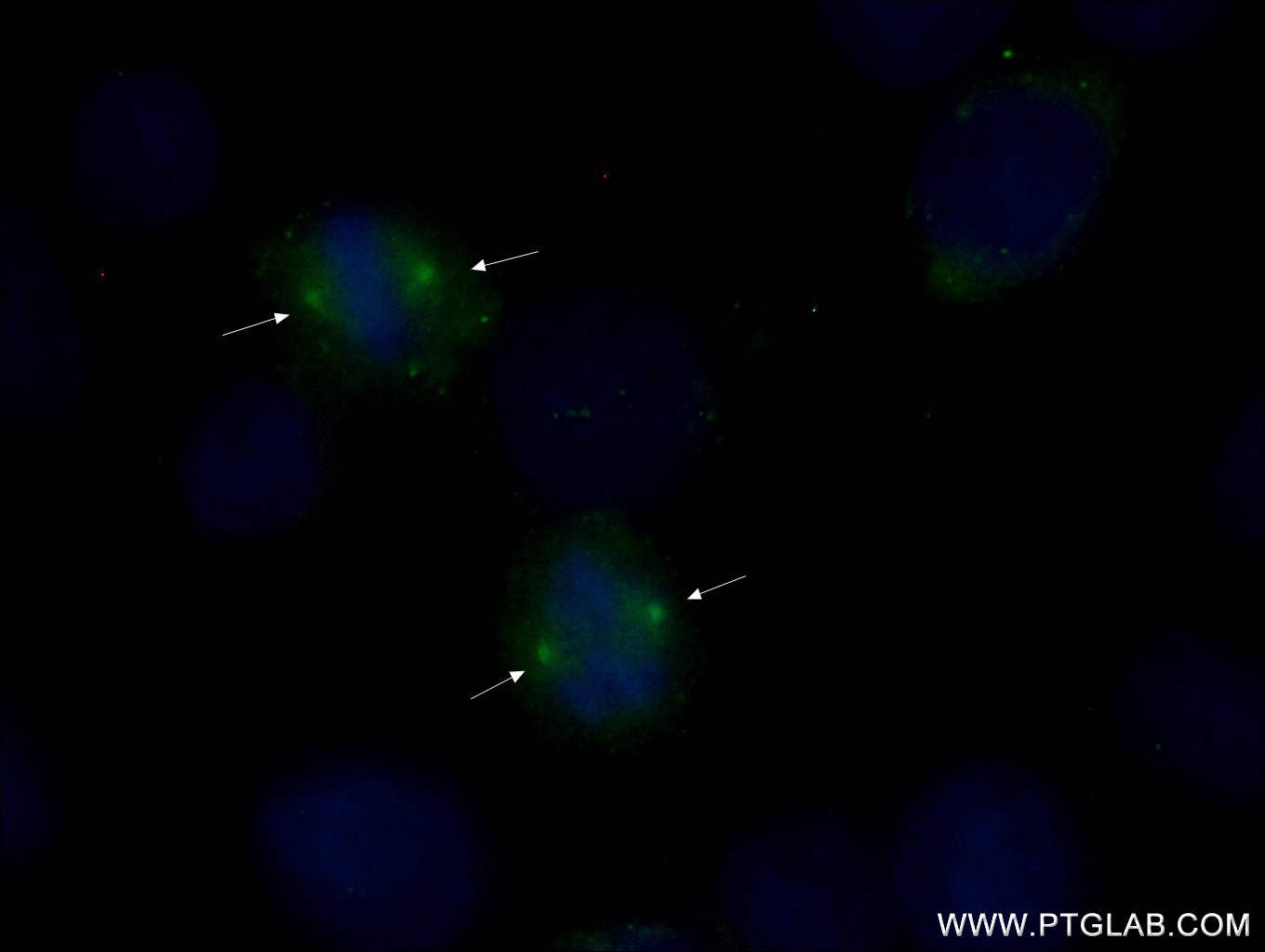
 fixed MDCK cells using 66320-1-Ig (gamma tubulin antibody) at dilution of 1:200 and CoraLite488-Conjugated AffiniPure Goat Anti-Mouse IgG(H+L).")
 fixed MDCK cells using 66320-1-Ig (gamma tubulin antibody) at dilution of 1:200 and CoraLite488-Conjugated AffiniPure Goat Anti-Mouse IgG(H+L).")
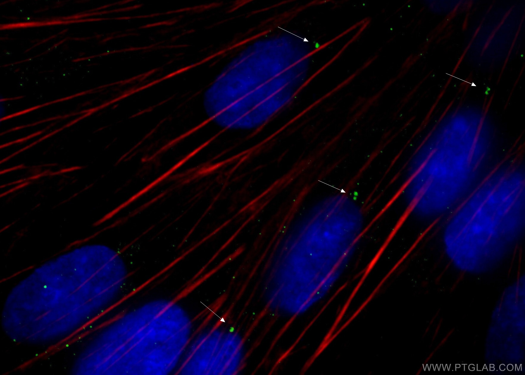
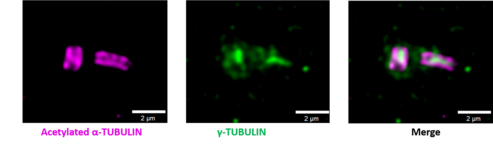
 fixed A549 cells using Gamma Tubulin antibody (66320-1-Ig, Clone: 3F9H8 ) at dilution of 1:1000 and CoraLite®488-Conjugated AffiniPure Goat Anti-Mouse IgG(H+L), Alpha Tubulin antibody (11224-1-AP, red).")
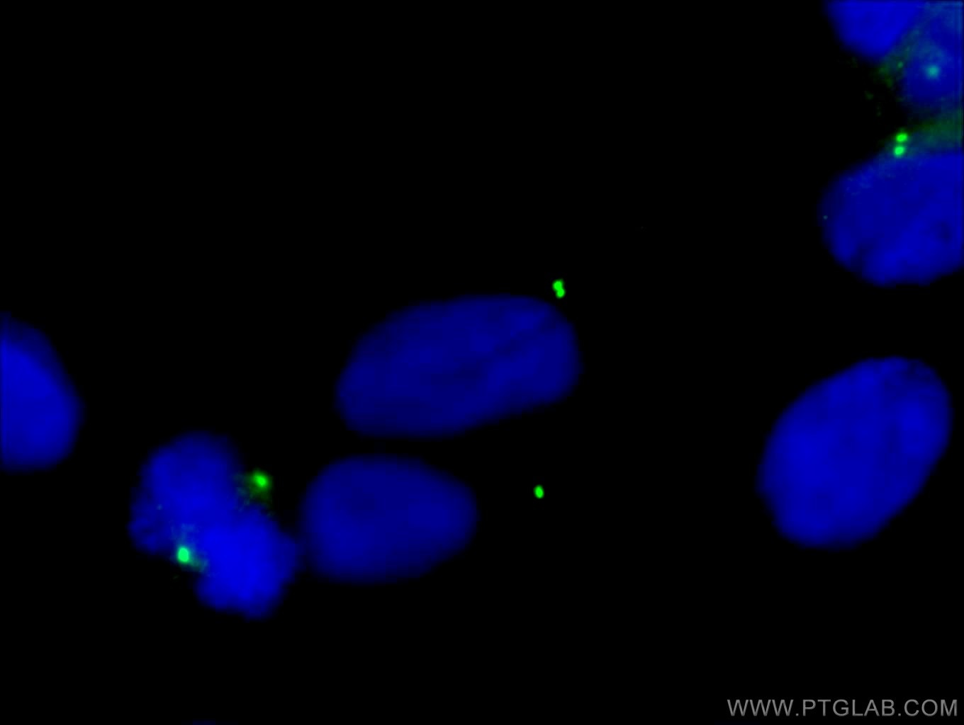
 fixed HeLa cells using 66320-1-Ig (gamma tubulin antibody) at dilution of 1:100 and Alexa Fluor 488-Conjugated AffiniPure Goat Anti-Mouse IgG(H+L).")
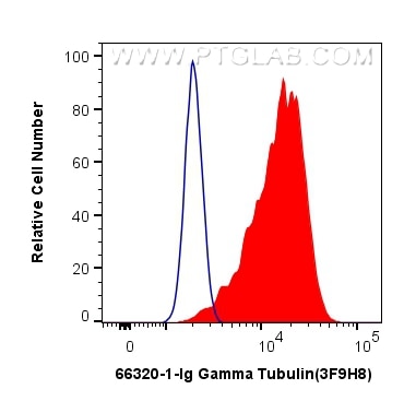
 and CoraLite®488-Conjugated AffiniPure Goat Anti-Mouse IgG(H+L) at dilution 1:1000 (red), or 0.4 ug Mouse IgG2a Isotype Control (C1.18.4) (65208-1-Ig, Clone: C1.18.4) (blue). Cells were fixed with 4% PFA and permeabilized with Flow Cytometry Perm Buffer.")
"Gamma Tubulin Antibodies" Comparison
View side-by-side comparison of Gamma Tubulin antibodies from other vendors to find the one that best suits your research needs.
Tested Applications
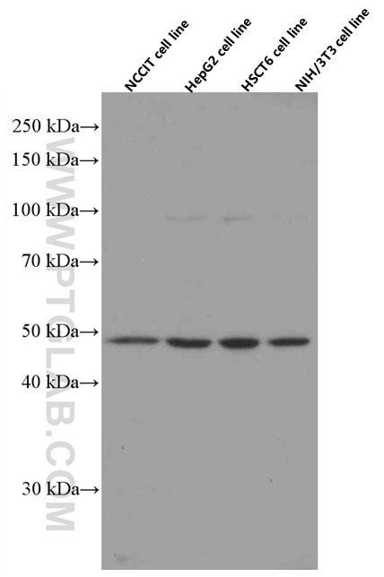
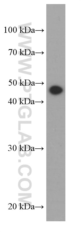
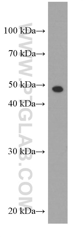
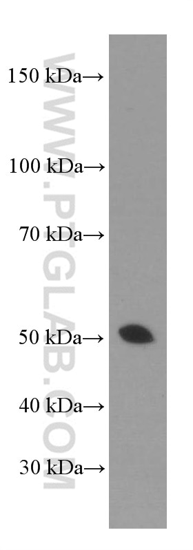
| Positive WB detected in | NCCIT cells, HepG2 cells, EC109 cells, K-562 cells, HSCT6 cells, NIH3T3 cells |
| Positive IHC detected in | human breast cancer tissue, human prostate cancer tissue Note: suggested antigen retrieval with TE buffer pH 9.0; (*) Alternatively, antigen retrieval may be performed with citrate buffer pH 6.0 |
| Positive IF/ICC detected in | MDCK cells, HeLa cells, A549 cells |
| Positive FC (Intra) detected in | HeLa cells |
Recommended dilution
| Application | Dilution |
|---|---|
| Western Blot (WB) | WB : 1:2000-1:12000 |
| Immunohistochemistry (IHC) | IHC : 1:200-1:1000 |
| Immunofluorescence (IF)/ICC | IF/ICC : 1:50-1:500 |
| Flow Cytometry (FC) (INTRA) | FC (INTRA) : 0.40 ug per 10^6 cells in a 100 µl suspension |
| It is recommended that this reagent should be titrated in each testing system to obtain optimal results. | |
| Sample-dependent, Check data in validation data gallery. | |
Published Applications
| WB | See 13 publications below |
| IF | See 23 publications below |
Product Information
66320-1-Ig targets Gamma Tubulin in WB, IHC, IF/ICC, FC (Intra), ELISA applications and shows reactivity with human, mouse, rat, canine samples.
| Tested Reactivity | human, mouse, rat, canine |
| Cited Reactivity | human, mouse |
| Host / Isotype | Mouse / IgG2a |
| Class | Monoclonal |
| Type | Antibody |
| Immunogen |
CatNo: Ag23913 Product name: Recombinant human tubulin-gamma protein Source: e coli.-derived, PET28a Tag: 6*His Domain: 150-451 aa of BC000619 Sequence: GSYLLERLNDRYPKKLVQTYSVFPNQDEMSDVVVQPYNSLLTLKRLTQNADCVVVLDNTALNRIATDRLHIQNPSFSQINQLVSTIMSASTTTLRYPGYMNNDLIGLIASLIPTPRLHFLMTGYTPLTTDQSVASVRKTTVLDVMRRLLQPKNVMVSTGRDRQTNHCYIAILNIIQGEVDPTQVHKSLQRIRERKLANFIPWGPASIQVALSRKSPYLPSAHRVSGLMMANHTSISSLFERTCRQYDKLRKREAFLEQFRKEDMFKDNFDEMDTSREIVQQLIDEYHAATRPDYISWGTQEQ Predict reactive species |
| Full Name | tubulin, gamma 1 |
| Calculated Molecular Weight | 51 kDa |
| Observed Molecular Weight | 48-55 kDa |
| GenBank Accession Number | BC000619 |
| Gene Symbol | Gamma Tubulin |
| Gene ID (NCBI) | 7283 |
| RRID | AB_2857350 |
| Conjugate | Unconjugated |
| Form | Liquid |
| Purification Method | Protein A purification |
| UNIPROT ID | P23258 |
| Storage Buffer | PBS with 0.02% sodium azide and 50% glycerol, pH 7.3. |
| Storage Conditions | Store at -20°C. Stable for one year after shipment. Aliquoting is unnecessary for -20oC storage. 20ul sizes contain 0.1% BSA. |
Background Information
Gamma tubulin is a member of tubulin superfamily and is a key component required for microtubule nucleation and stabilization. It is concentrated at the pericentriolar material of centrosomes in interphase cells, predominantly on spindle poles in mitotic cells, while found in midbodies during cytokinesis. Overexpression of gamma tubulin has been found in various cancers including breast cancer and gliomas. This antibody can well label the centrosome structure.
Protocols
| Product Specific Protocols | |
|---|---|
| FC protocol for Gamma Tubulin antibody 66320-1-Ig | Download protocol |
| IF protocol for Gamma Tubulin antibody 66320-1-Ig | Download protocol |
| IHC protocol for Gamma Tubulin antibody 66320-1-Ig | Download protocol |
| WB protocol for Gamma Tubulin antibody 66320-1-Ig | Download protocol |
| Standard Protocols | |
|---|---|
| Click here to view our Standard Protocols |
Publications
| Species | Application | Title |
|---|---|---|
EMBO Mol Med Mutations in GRK2 cause Jeune syndrome by impairing Hedgehog and canonical Wnt signaling. | ||
Cell Rep MYO10 regulates genome stability and cancer inflammation through mediating mitosis | ||
Elife Respiratory syncytial virus co-opts host mitochondrial function to favour infectious virus production. | ||
J Nanobiotechnology GRP75-driven, cell-cycle-dependent macropinocytosis of Tat/pDNA-Ca2+ nanoparticles underlies distinct gene therapy effect in ovarian cancer. |
Reviews
The reviews below have been submitted by verified Proteintech customers who received an incentive for providing their feedback.
FH Lola (Verified Customer) (10-23-2025) | This antibody works well. Suitable for immunofluorescence and expansion microscopy. We use cold MeOH fixation.
 |