Tested Applications
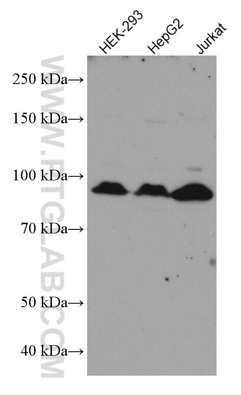
| Positive WB detected in | HEK-293 cells, HepG2 cells, Jurkat cells |
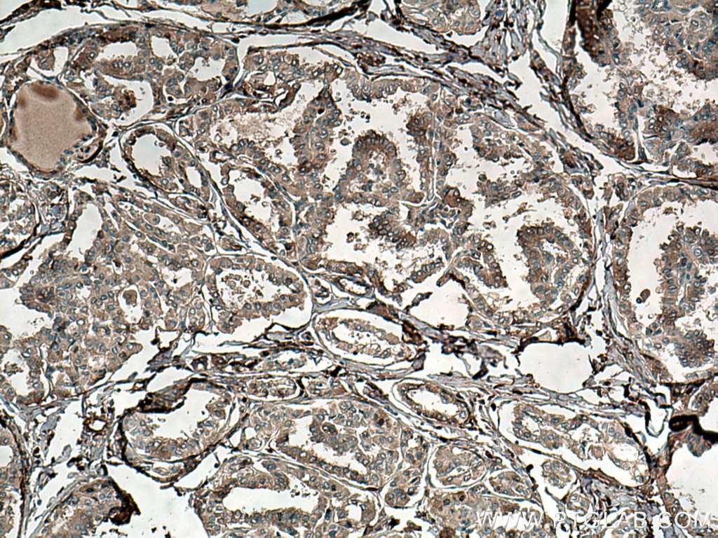
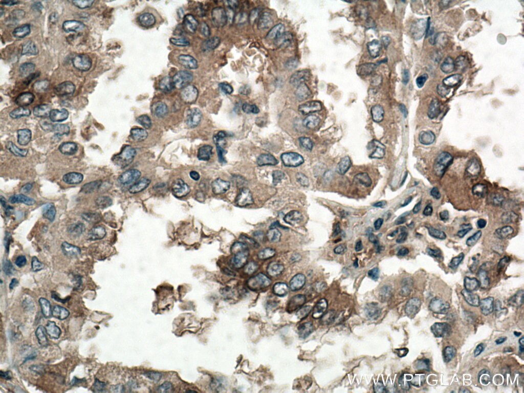
| Positive IHC detected in | human thyroid cancer tissue Note: suggested antigen retrieval with TE buffer pH 9.0; (*) Alternatively, antigen retrieval may be performed with citrate buffer pH 6.0 |
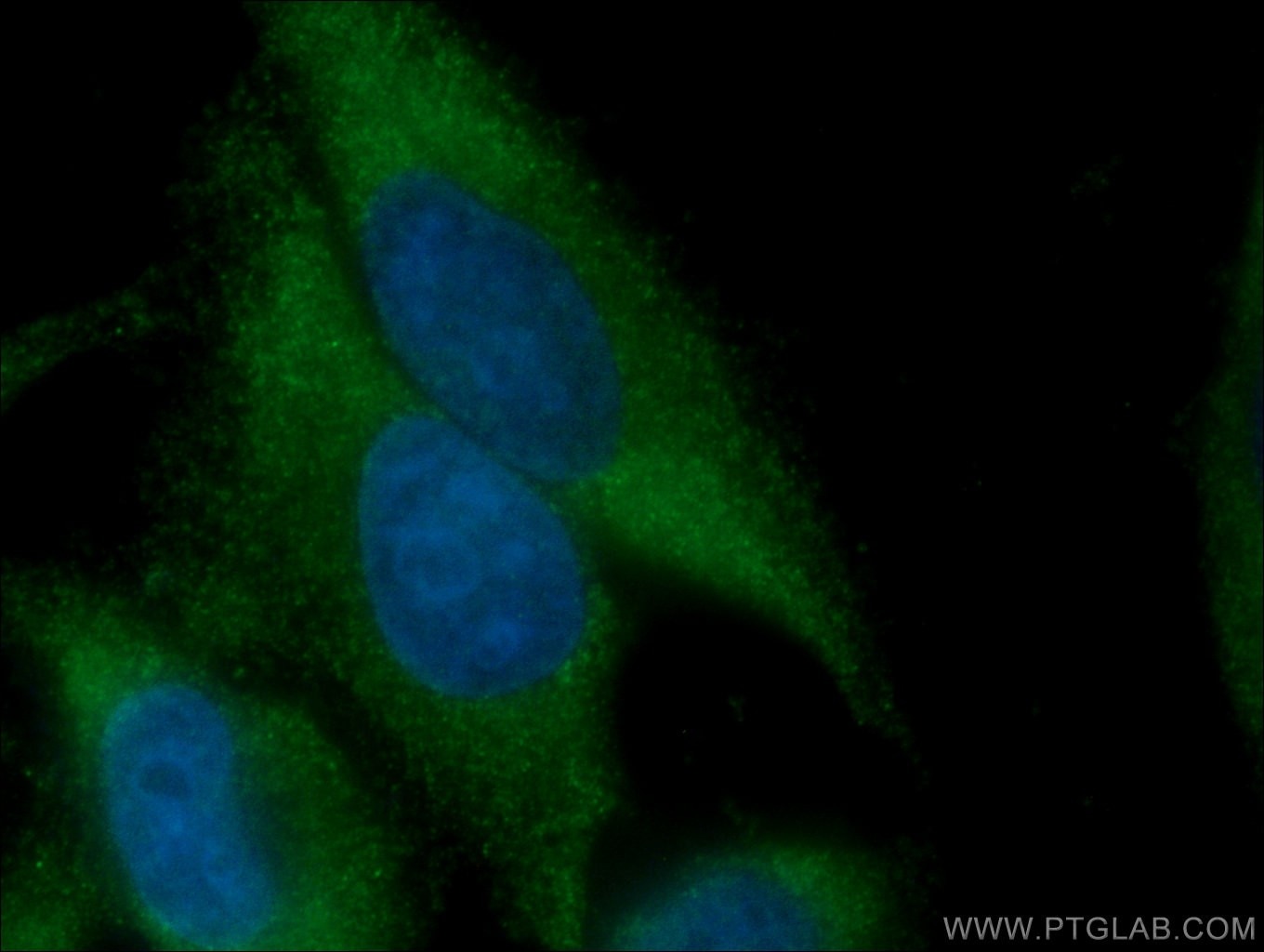
| Positive IF/ICC detected in | HepG2 cells |
Recommended dilution
| Application | Dilution |
|---|---|
| Western Blot (WB) | WB : 1:2000-1:10000 |
| Immunohistochemistry (IHC) | IHC : 1:300-1:1200 |
| Immunofluorescence (IF)/ICC | IF/ICC : 1:50-1:500 |
| It is recommended that this reagent should be titrated in each testing system to obtain optimal results. | |
| Sample-dependent, Check data in validation data gallery. | |
Published Applications
| WB | See 5 publications below |
| IF | See 4 publications below |
Product Information
67096-1-Ig targets ATG9A in WB, IHC, IF/ICC, ELISA applications and shows reactivity with Human samples.
| Tested Reactivity | Human |
| Cited Reactivity | human, mouse |
| Host / Isotype | Mouse / IgG1 |
| Class | Monoclonal |
| Type | Antibody |
| Immunogen |
CatNo: Ag24278 Product name: Recombinant human ATG9A protein Source: e coli.-derived, PET28a Tag: 6*His Domain: 427-528 aa of BC065534 Sequence: DQHMVFCPEQLLRVILAHIHYMPDHWQGVHFGRVAEPHCHTPHPHLLPAPTGPGDYRLLPKLHRGGRWCGRYLLLCSDGCSPAWSSPVAICWADRGLSVPAS Predict reactive species |
| Full Name | ATG9 autophagy related 9 homolog A (S. cerevisiae) |
| Calculated Molecular Weight | 837 aa, 94 kDa |
| Observed Molecular Weight | 94 kDa |
| GenBank Accession Number | BC065534 |
| Gene Symbol | ATG9A |
| Gene ID (NCBI) | 79065 |
| RRID | AB_2882401 |
| Conjugate | Unconjugated |
| Form | Liquid |
| Purification Method | Protein G purification |
| UNIPROT ID | Q7Z3C6 |
| Storage Buffer | PBS with 0.02% sodium azide and 50% glycerol, pH 7.3. |
| Storage Conditions | Store at -20°C. Stable for one year after shipment. Aliquoting is unnecessary for -20oC storage. 20ul sizes contain 0.1% BSA. |
Background Information
ATG9A is the only transmembrane ATG protein essential for autophagy. It plays a key role in the organization of the preautophagosomal structure/phagophore assembly site (PAS). It has been reported that ATG9A expression is increased in oral squamous cell carcinoma and breast cancers. The inhibition of ATG9A can lead to an inhibition of cancer cell proliferation and invasion.(PMID: 29437695, 29568063)
Protocols
| Product Specific Protocols | |
|---|---|
| IF protocol for ATG9A antibody 67096-1-Ig | Download protocol |
| IHC protocol for ATG9A antibody 67096-1-Ig | Download protocol |
| WB protocol for ATG9A antibody 67096-1-Ig | Download protocol |
| Standard Protocols | |
|---|---|
| Click here to view our Standard Protocols |
Publications
| Species | Application | Title |
|---|---|---|
NPJ Parkinsons Dis Regulators of proteostasis are translationally repressed in fibroblasts from patients with sporadic and LRRK2-G2019S Parkinson's disease | ||
Invest Ophthalmol Vis Sci HSF4 Transcriptionally Activates Autophagy by Regulating ATG9a During Lens Terminal Differentiation | ||
Autophagy WNT2B impairs endosomal trafficking via WASHC5 to inhibit autophagy: a novel non-secretory WNT pathway. | ||
Drug Resist Updat NDRG1 overcomes resistance to immunotherapy of pancreatic ductal adenocarcinoma through inhibiting ATG9A-dependent degradation of MHC-1 | ||