
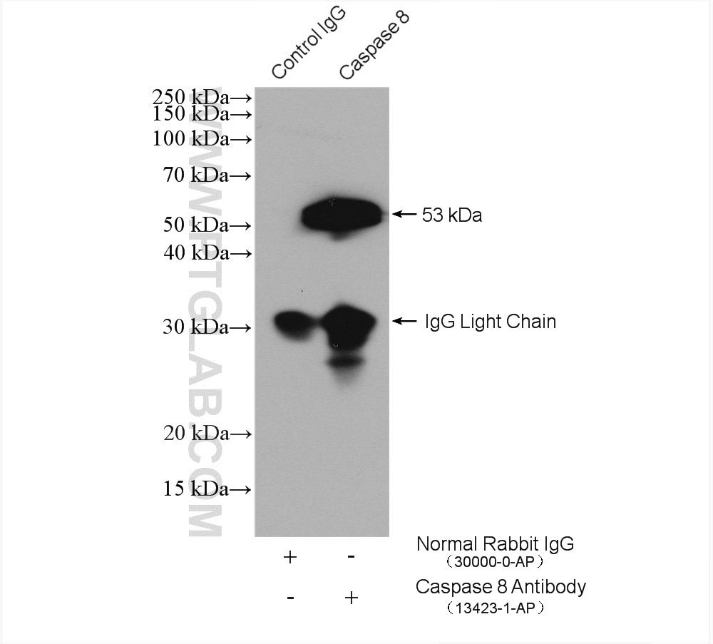
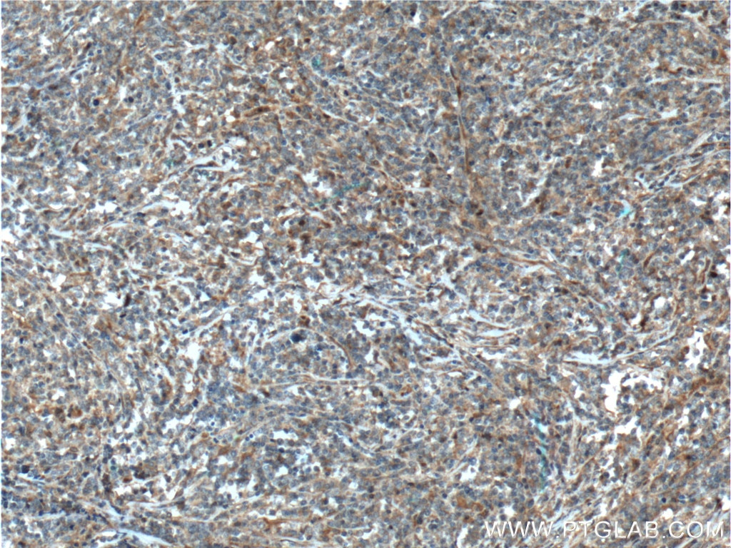
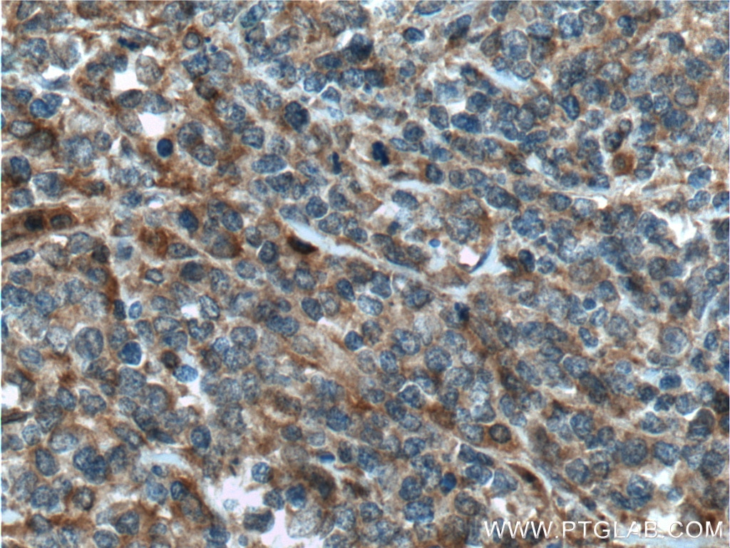
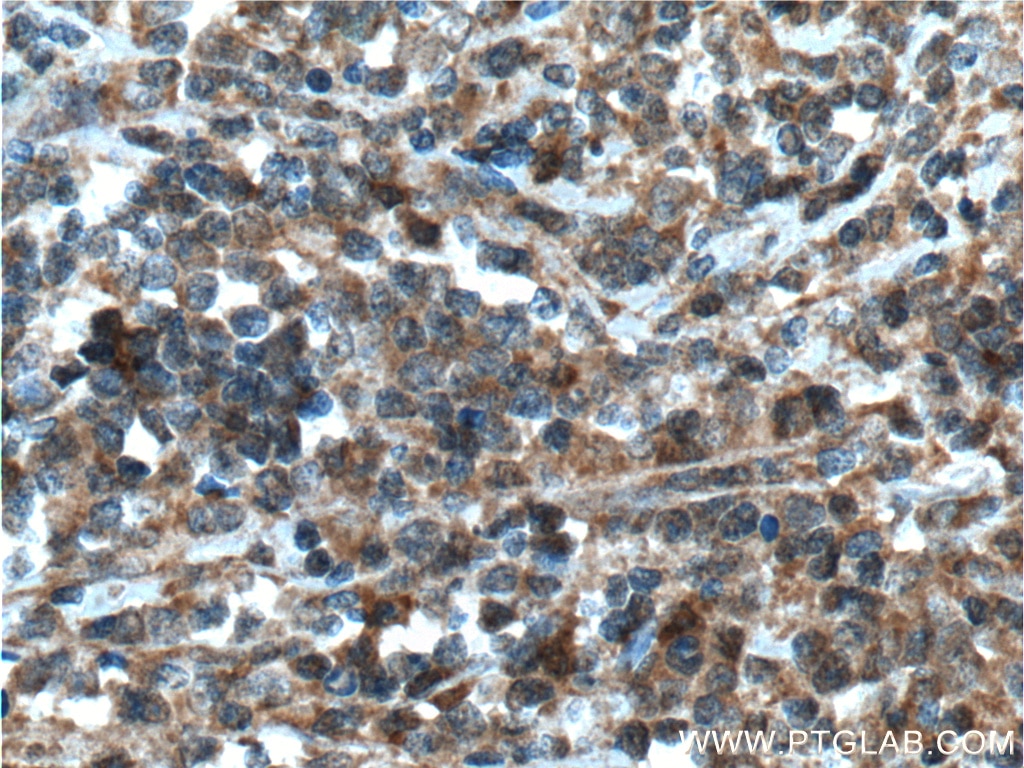

Tested Applications
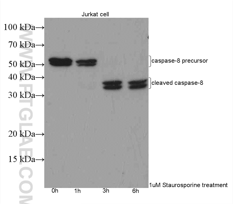
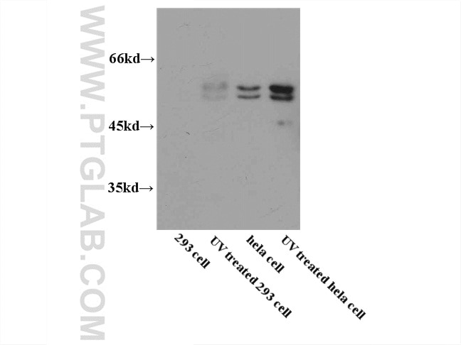
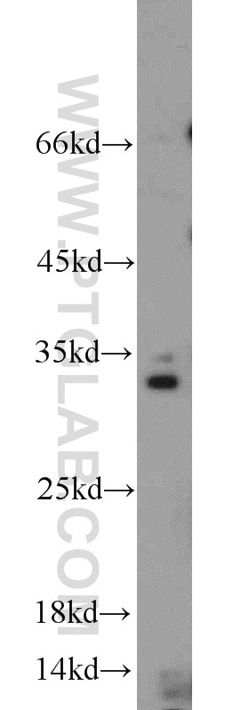
| Positive WB detected in | Staurosporine treated Jurkat cells, UV treated Hela, RAW264.7 |
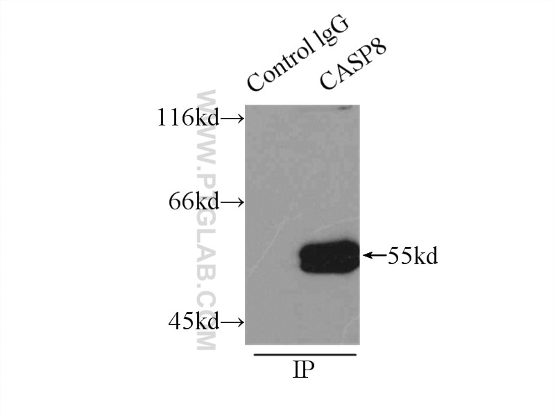
| Positive IP detected in | HeLa cells, Jurkat cells |
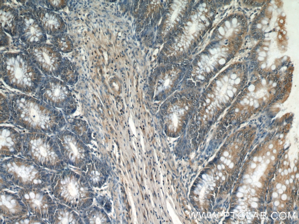
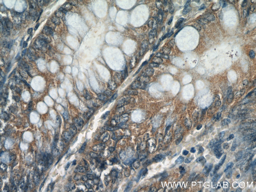
| Positive IHC detected in | human lymphoma tissue, human colon tissue Note: suggested antigen retrieval with TE buffer pH 9.0; (*) Alternatively, antigen retrieval may be performed with citrate buffer pH 6.0 |
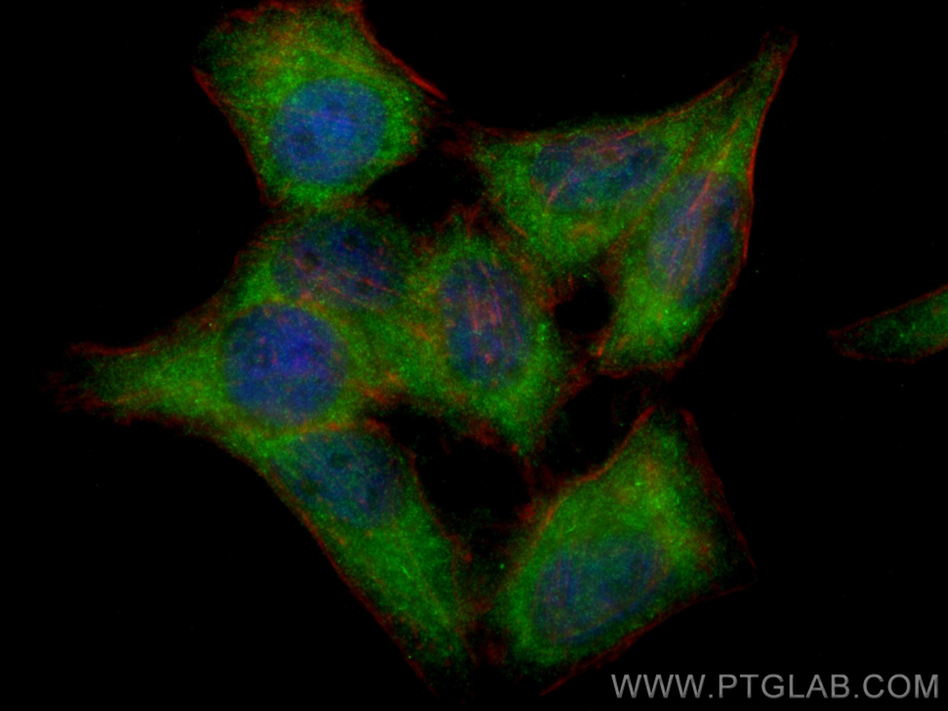
| Positive IF/ICC detected in | HepG2 cells |
Recommended dilution
| Application | Dilution |
|---|---|
| Western Blot (WB) | WB : 1:500-1:1000 |
| Immunoprecipitation (IP) | IP : 0.5-4.0 ug for 1.0-3.0 mg of total protein lysate |
| Immunohistochemistry (IHC) | IHC : 1:50-1:500 |
| Immunofluorescence (IF)/ICC | IF/ICC : 1:50-1:500 |
| It is recommended that this reagent should be titrated in each testing system to obtain optimal results. | |
| Sample-dependent, Check data in validation data gallery. | |
Product Information
13423-1-AP targets Caspase 8/P43/P18 in WB, IHC, IF/ICC, IP, CoIP, RIP, ELISA applications and shows reactivity with human, mouse samples.
| Tested Reactivity | human, mouse |
| Cited Reactivity | human, mouse, rat, pig, rabbit, chicken, zebrafish, hamster, sheep |
| Host / Isotype | Rabbit / IgG |
| Class | Polyclonal |
| Type | Antibody |
| Immunogen |
CatNo: Ag4214 Product name: Recombinant human Caspase 8 protein Source: e coli.-derived, PGEX-4T Tag: GST Domain: 1-270 aa of BC028223 Sequence: MDFSRNLYDIGEQLDSEDLASLKFLSLDYIPQRKQEPIKDALMLFQRLQEKRMLEESNLSFLKELLFRINRLDLLITYLNTRKEEMERELQTPGRAQISAYRVMLYQISEEVSRSELRSFKFLLQEEISKCKLDDDMNLLDIFIEMEKRVILGEGKLDILKRVCAQINKSLLKIINDYEEFSKERSSSLEGSPDEFSNGEELCGVMTISDSPREQDSESQTLDKVYQMKSKPRGYCLIINNHNFAKAREKVPKLHSIRDRNGTHLDAGTV Predict reactive species |
| Full Name | caspase 8, apoptosis-related cysteine peptidase |
| Calculated Molecular Weight | 538 aa, 62 kDa |
| Observed Molecular Weight | 53-57 kDa, 32-45 kDa |
| GenBank Accession Number | BC028223 |
| Gene Symbol | Caspase 8 |
| Gene ID (NCBI) | 841 |
| RRID | AB_2068463 |
| Conjugate | Unconjugated |
| Form | Liquid |
| Purification Method | Antigen affinity purification |
| UNIPROT ID | Q14790 |
| Storage Buffer | PBS with 0.02% sodium azide and 50% glycerol, pH 7.3. |
| Storage Conditions | Store at -20°C. Stable for one year after shipment. Aliquoting is unnecessary for -20oC storage. 20ul sizes contain 0.1% BSA. |
Background Information
CASP8, also named as MCH5, MACH, FLICE, and CAP4, belongs to the peptidase C14A family. It may participate in the GZMB apoptotic pathways and functions as an upstream regulator in a-bisabolol-induced apoptosis. CASP8 catalyzes an essential intermediate step in the ubiquitination and proteasome-mediated degradation of IRF3(PMID:21816816 ). It may control diabetic embryopathy-associated apoptosis via regulation of the Bid-stimulated mitochondrion/caspase-9 pathway (PMID:19194987). CASP8 is expressed as nine isoforms by alternative splicing with the molecular mass from 26 kDa to 62 kDa. This antibody can recognize the pro- and cleaved-caspase 8.
Protocols
| Product Specific Protocols | |
|---|---|
| IF protocol for Caspase 8/P43/P18 antibody 13423-1-AP | Download protocol |
| IHC protocol for Caspase 8/P43/P18 antibody 13423-1-AP | Download protocol |
| IP protocol for Caspase 8/P43/P18 antibody 13423-1-AP | Download protocol |
| WB protocol for Caspase 8/P43/P18 antibody 13423-1-AP | Download protocol |
| Standard Protocols | |
|---|---|
| Click here to view our Standard Protocols |
Publications
What published studies show
Anti-Caspase-8 (Cat# 13423-1-AP) has 481 total citations. Publications appear in journals including Nature Communications, Cell Death & Disease, Cell Death & Differentiation, Autophagy, Cancer Research, EMBO Journal, and over 70 others. Research fields span apoptosis, PANoptosis, necroptosis, pyroptosis, oncology, cardiovascular disease, neurodegeneration, inflammation, mitochondrial biology, drug resistance, and traditional Chinese medicine applications.
| Species | Application | Title |
|---|---|---|
Signal Transduct Target Ther 2-APQC, a small-molecule activator of Sirtuin-3 (SIRT3), alleviates myocardial hypertrophy and fibrosis by regulating mitochondrial homeostasis | ||
Adv Mater Selective Organ-Targeting Hafnium Oxide Nanoparticles with Multienzyme-Mimetic Activities Attenuate Radiation-Induced Tissue Damage | ||
Cancer Res Foretinib is effective in acute myeloid leukemia by inhibiting FLT3 and overcoming secondary mutations that drive resistance to quizartinib and gilteritinib | ||
Autophagy Histone lactylation stimulated upregulation of PSMD14 alleviates neuron PANoptosis through deubiquitinating PKM/PKM2 to activate PINK1-mediated mitophagy after traumatic brain injury | ||
Autophagy Simultaneous inhibition of the ubiquitin-proteasome system and autophagy enhances apoptosis induced by ER stress aggravators in human pancreatic cancer cells. | ||
Nat Commun UDP-glucuronate metabolism controls RIPK1-driven liver damage in nonalcoholic steatohepatitis |
Reviews
What customers say
Both reviewers rated the antibody 4 out of 5 stars for Western Blot applications. One user reported strong, clean bands in kidney lysates at a 1:500 dilution. The other found it effective for detecting ischemia-reperfusion injury-induced apoptosis in mouse heart tissue. Overall, customers are satisfied with its performance in WB, noting reliable signal quality across different tissue types.
The reviews below have been submitted by verified Proteintech customers who received an incentive for providing their feedback.
Xiaoping (Verified Customer) (12-15-2021) | Got really good bands in kidney lysates at 1:500 dilution
|
WEI (Verified Customer) (01-17-2020) | Good antibody for detecting IR injury-induced apoptosis in mouse heart.
|












