
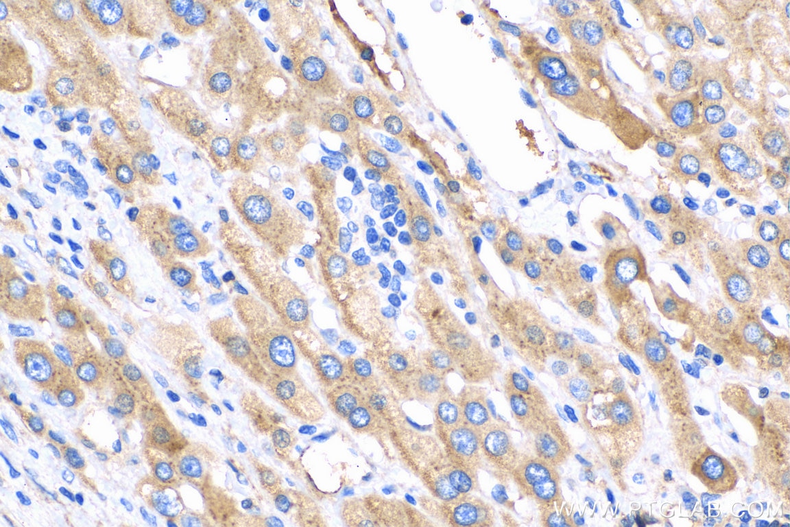
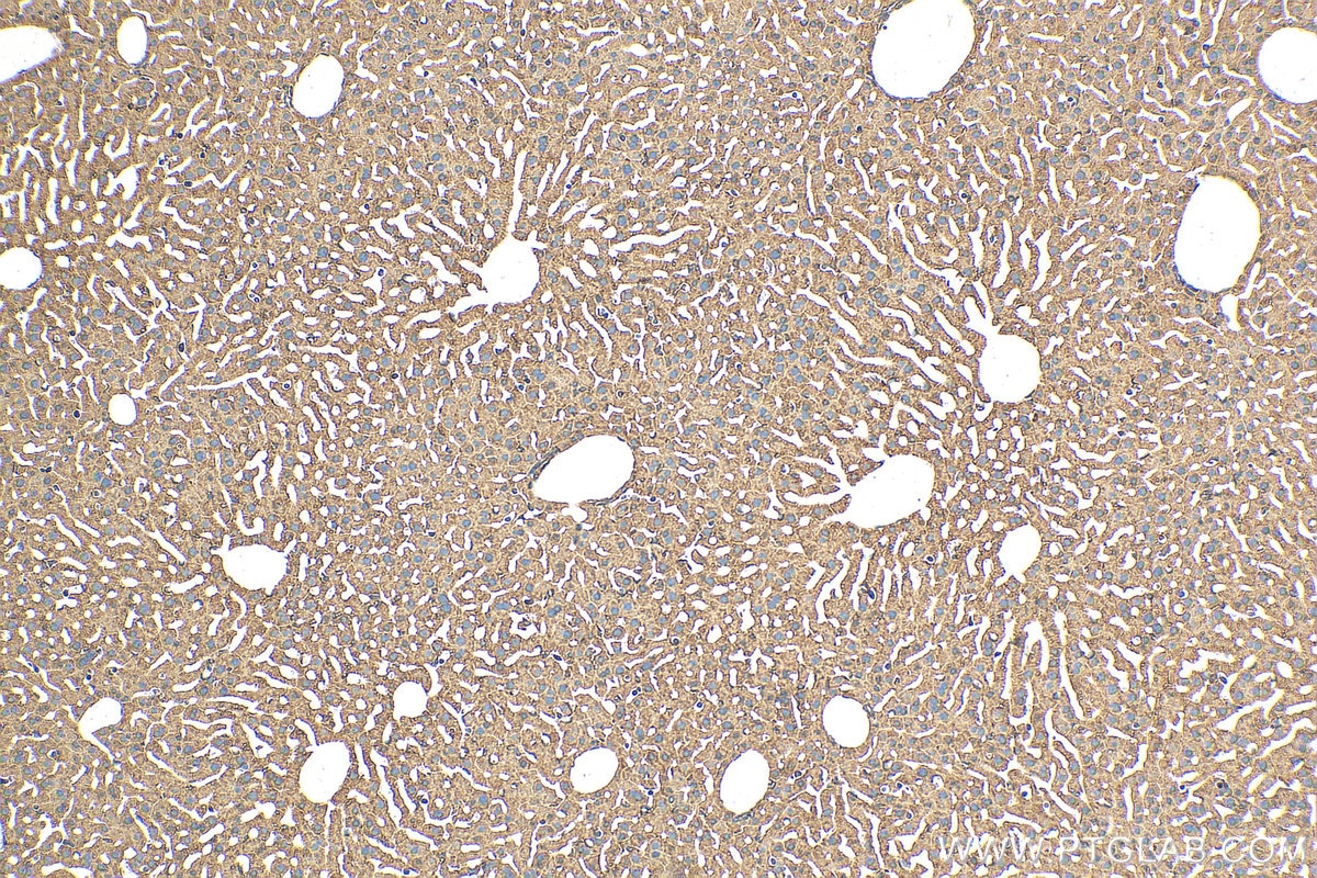
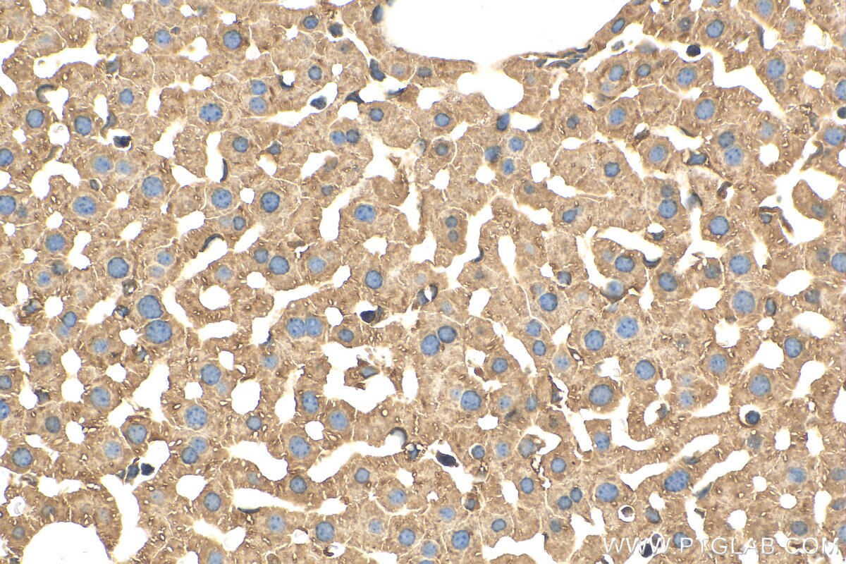

Tested Applications
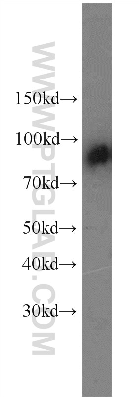
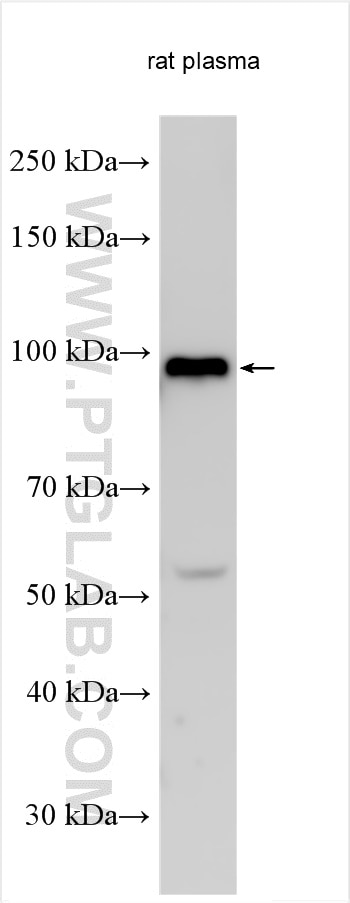
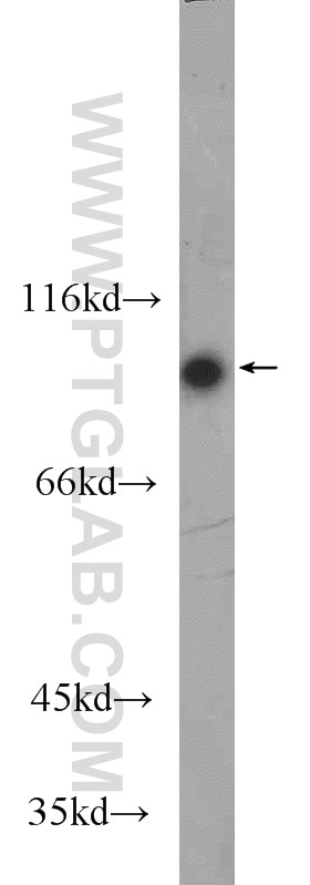
| Positive WB detected in | rat plasma, human plasma |
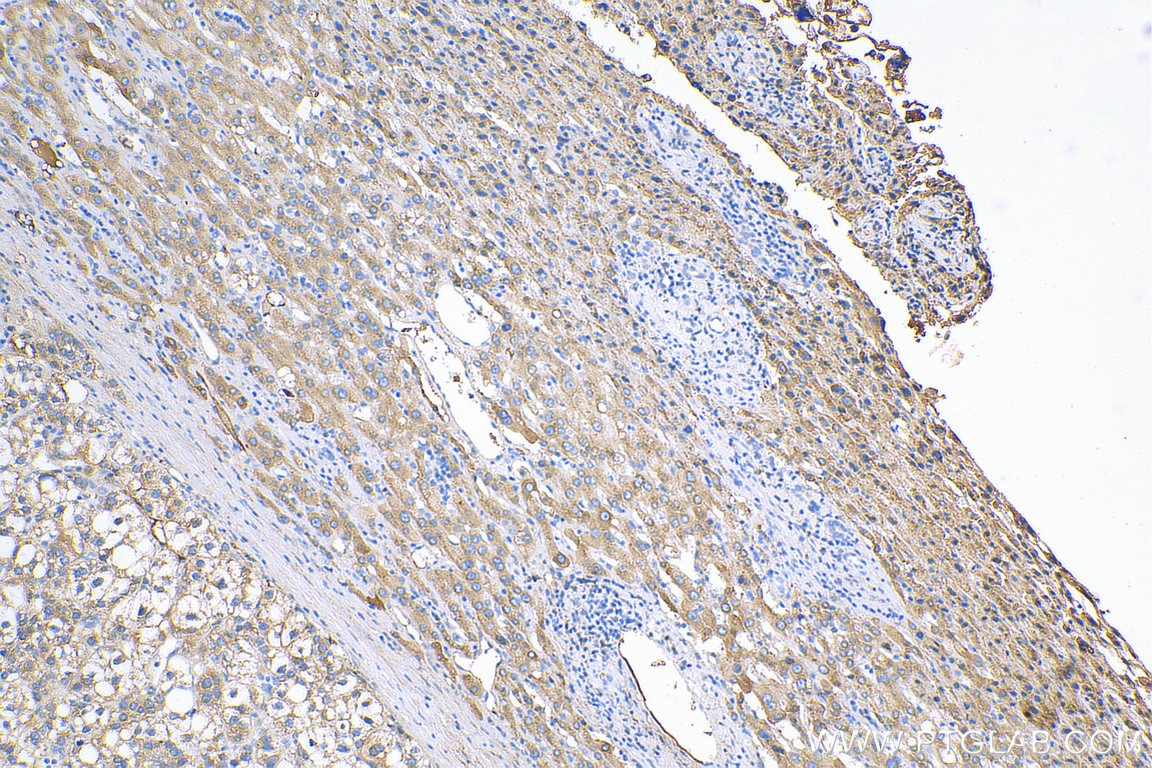
| Positive IHC detected in | human liver cancer tissue, mouse liver tissue Note: suggested antigen retrieval with TE buffer pH 9.0; (*) Alternatively, antigen retrieval may be performed with citrate buffer pH 6.0 |
Recommended dilution
| Application | Dilution |
|---|---|
| Western Blot (WB) | WB : 1:2000-1:16000 |
| Immunohistochemistry (IHC) | IHC : 1:50-1:500 |
| It is recommended that this reagent should be titrated in each testing system to obtain optimal results. | |
| Sample-dependent, Check data in validation data gallery. | |
Product Information
14554-1-AP targets C1S in WB, IHC, IF, ELISA applications and shows reactivity with human, mouse, rat samples.
| Tested Reactivity | human, mouse, rat |
| Cited Reactivity | human, mouse |
| Host / Isotype | Rabbit / IgG |
| Class | Polyclonal |
| Type | Antibody |
| Immunogen |
CatNo: Ag5977 Product name: Recombinant human C1S protein Source: e coli.-derived, PGEX-4T Tag: GST Domain: 166-688 aa of BC056903 Sequence: DDMKNCGVNCSGDVFTALIGEIASPNYPKPYPENSRCEYQIRLEKGFQVVVTLRREDFDVEAADSAGNCLDSLVFVAGDRQFGPYCGHGFPGPLNIETKSNALDIIFQTDLTGQKKGWKLRYHGDPMPCPKEDTPNSVWEPAKAKYVFRDVVQITCLDGFEVVEGRVGATSFYSTCQSNGKWSNSKLKCQPVDCGIPESIENGKVEDPESTLFGSVIRYTCEEPYYYMENGGGGEYHCAGNGSWVNEVLGPELPKCVPVCGVPREPFEEKQRIIGGSDADIKNFPWQVFFDNPWAGGALINEYWVLTAAHVVEGNREPTMYVGSTSVQTSRLAKSKMLTPEHVFIHPGWKLLEVPEGRTNFDNDIALVRLKDPVKMGPTVSPICLPGTSSDYNLMDGDLGLISGWGRTEKRDRAVRLKAARLPVAPLRKCKEVKVEKPTADAEAYVFTPNMICAGGEKGMDSCKGDSGGAFAVQDPNDKTKFYAAGLVSWGPQCGTYGLYTRVKNYVDWIMKTMQENSTPRED Predict reactive species |
| Full Name | complement component 1, s subcomponent |
| Calculated Molecular Weight | 77 kDa |
| Observed Molecular Weight | 88 kDa |
| GenBank Accession Number | BC056903 |
| Gene Symbol | C1S |
| Gene ID (NCBI) | 716 |
| RRID | AB_2243610 |
| Conjugate | Unconjugated |
| Form | Liquid |
| Purification Method | Antigen affinity purification |
| UNIPROT ID | P09871 |
| Storage Buffer | PBS with 0.02% sodium azide and 50% glycerol, pH 7.3. |
| Storage Conditions | Store at -20°C. Stable for one year after shipment. Aliquoting is unnecessary for -20oC storage. 20ul sizes contain 0.1% BSA. |
Background Information
The first component of complement is a calcium-dependent complex of the 3 subcomponents C1q, C1r, and C1s. The serine protease subunits C1r and C1s form a Ca(2+)-dependent C1s-C1r-C1r-C1s tetramer. C1r activates C1s so that it can, in turn, activate C2 and C4. C1s is a glycoprotein of approximately 88 kDa, which is activated by cleavage into an A chain and a B chain.
Protocols
| Product Specific Protocols | |
|---|---|
| IHC protocol for C1S antibody 14554-1-AP | Download protocol |
| WB protocol for C1S antibody 14554-1-AP | Download protocol |
| Standard Protocols | |
|---|---|
| Click here to view our Standard Protocols |
Publications
What published studies show
The C1S Polyclonal antibody (Cat# 14554-1-AP) has 7 citations published in journals including J Transl Med, Cell Reports, Cancer Cell International, Int J Mol Sci, Sci Rep, and Exp Eye Res. Research fields span renal cell carcinoma prognostic signatures, retinal complement biology, esophageal squamous cell carcinoma, uric acid crystal–mediated inflammation, and corneal stroma proteomics related to myopia progression. Applications cited include WB, IHC, and IF in human and mouse samples.
| Species | Application | Title |
|---|---|---|
J Transl Med Comprehensive investigation of malignant epithelial cell-related genes in clear cell renal cell carcinoma: development of a prognostic signature and exploration of tumor microenvironment interactions | ||
Cancer Cell Int Association between cancer-associated fibroblasts and prognosis of neoadjuvant chemoradiotherapy in esophageal squamous cell carcinoma: a bioinformatics analysis based on single-cell RNA sequencing | ||
Int J Mol Sci Sodium Iodate-Induced Degeneration Results in Local Complement Changes and Inflammatory Processes in Murine Retina | ||
Sci Rep C-reactive protein (CRP) recognizes uric acid crystals and recruits proteases C1 and MASP1. | ||
Reviews
What customers say
The single review for this antibody reports a 4/5 rating. The user applied it at a 1:100 dilution for immunohistochemistry and immunofluorescence on hiPSC-derived microglia. They noted that the staining signal was clear with minimal background on both the cells and surrounding tissue, indicating good specificity and low non-specific binding in their application.
The reviews below have been submitted by verified Proteintech customers who received an incentive for providing their feedback.
Asimenia (Verified Customer) (11-14-2024) | The signal of this staining was clear and with minimal background on the cells or the surrounding tissue.
 |








