
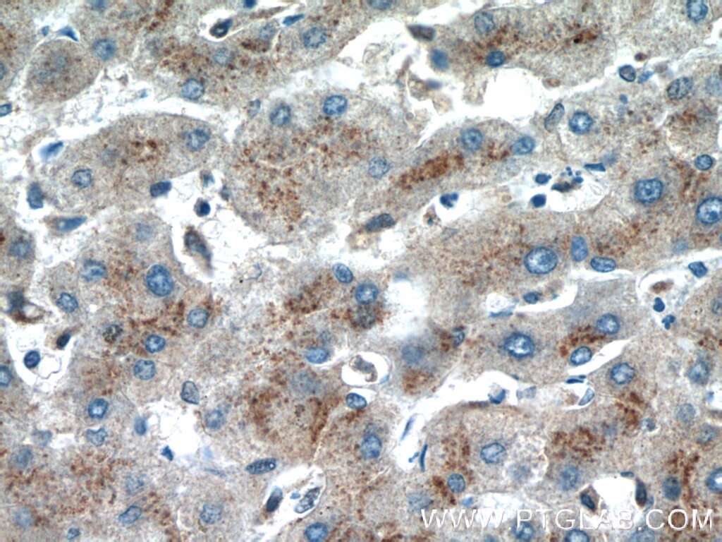

Tested Applications
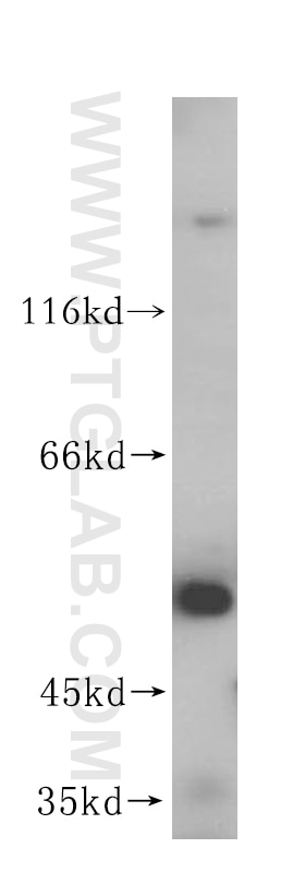
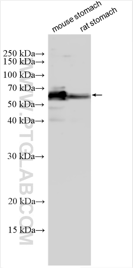
| Positive WB detected in | human heart tissue, mouse stomach tissue, rat stomach tissue |
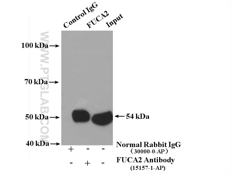
| Positive IP detected in | mouse heart tissue |
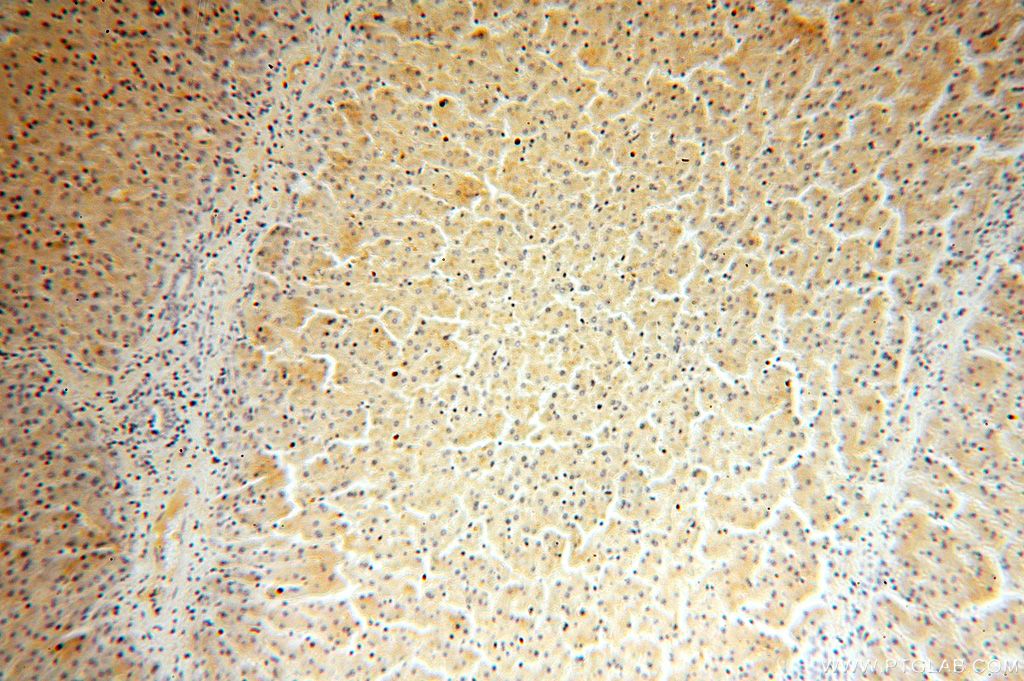
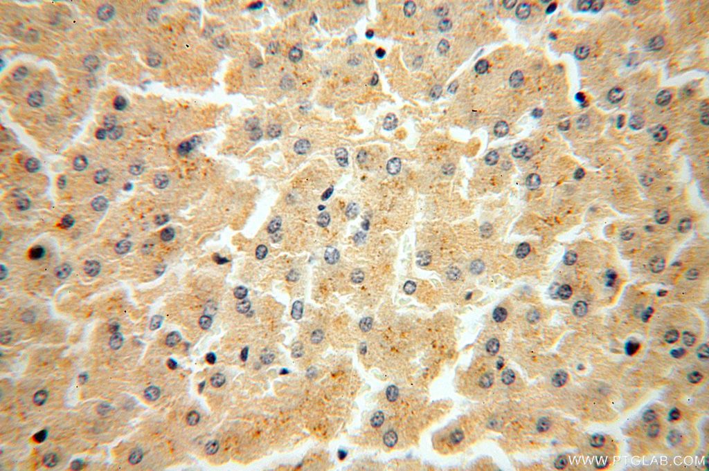
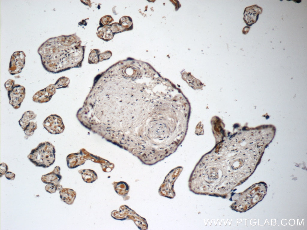
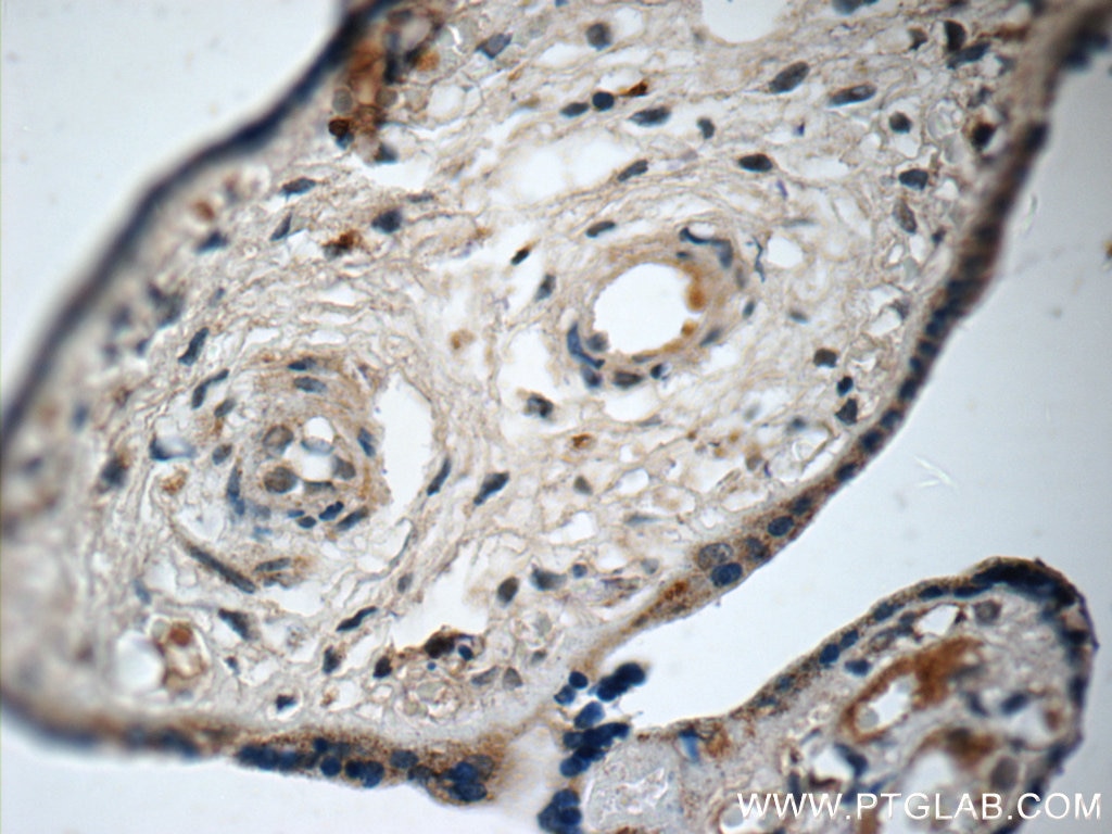
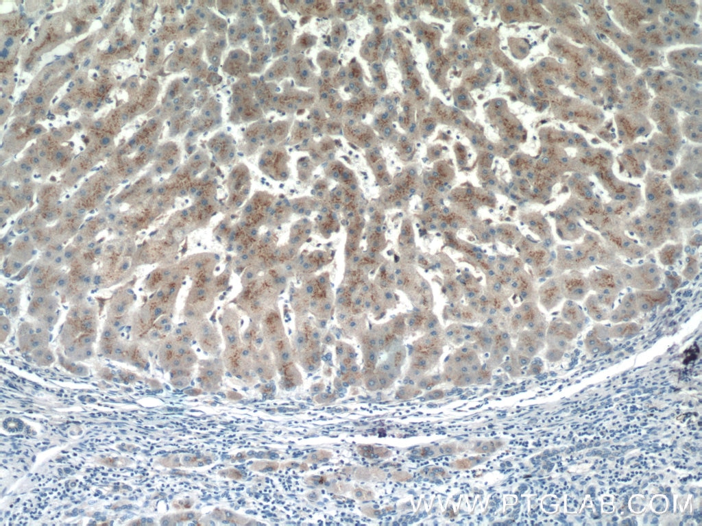
| Positive IHC detected in | human liver tissue, human hepatocirrhosis tissue, human placenta tissue Note: suggested antigen retrieval with TE buffer pH 9.0; (*) Alternatively, antigen retrieval may be performed with citrate buffer pH 6.0 |
Recommended dilution
| Application | Dilution |
|---|---|
| Western Blot (WB) | WB : 1:500-1:1000 |
| Immunoprecipitation (IP) | IP : 0.5-4.0 ug for 1.0-3.0 mg of total protein lysate |
| Immunohistochemistry (IHC) | IHC : 1:20-1:200 |
| It is recommended that this reagent should be titrated in each testing system to obtain optimal results. | |
| Sample-dependent, Check data in validation data gallery. | |
Product Information
15157-1-AP targets FUCA2 in WB, IHC, IF, IP, ELISA applications and shows reactivity with human, mouse, rat samples.
| Tested Reactivity | human, mouse, rat |
| Cited Reactivity | human, mouse |
| Host / Isotype | Rabbit / IgG |
| Class | Polyclonal |
| Type | Antibody |
| Immunogen |
CatNo: Ag7250 Product name: Recombinant human FUCA2 protein Source: e coli.-derived, PGEX-4T Tag: GST Domain: 124-467 aa of BC003060 Sequence: SGAKYIVLTSKHHEGFTLWGSEYSWNWNAIDEGPKRDIVKELEVAIRNRTDLRFGLYYSLFEWFHPLFLEDESSSFHKRQFPVSKTLPELYELVNNYQPEVLWSDGDGGAPDQYWNSTGFLAWLYNESPVRGTVVTNDRWGAGSICKHGGFYTCSDRYNPGHLLPHKWENCMTIDKLSWGYRREAGISDYLTIEELVKQLVETVSCGGNLLMNIGPTLDGTISVVFEERLRQVGSWLKVNGEAIYETYTWRSQNDTVTPDVWYTSKPKEKLVYAIFLKWPTSGQLFLGHPKAILGATEVKLLGHGQPLNWISLEQNGIMVELPQLTIHQMPCKWGWALALTNVI Predict reactive species |
| Full Name | fucosidase, alpha-L- 2, plasma |
| Calculated Molecular Weight | 54 kDa |
| Observed Molecular Weight | 54-60 kDa |
| GenBank Accession Number | BC003060 |
| Gene Symbol | FUCA2 |
| Gene ID (NCBI) | 2519 |
| RRID | AB_2104956 |
| Conjugate | Unconjugated |
| Form | Liquid |
| Purification Method | Antigen affinity purification |
| UNIPROT ID | Q9BTY2 |
| Storage Buffer | PBS with 0.02% sodium azide and 50% glycerol, pH 7.3. |
| Storage Conditions | Store at -20°C. Stable for one year after shipment. Aliquoting is unnecessary for -20oC storage. 20ul sizes contain 0.1% BSA. |
Background Information
Alpha-L-fucosidase 2 (FUCA2) is a gene that codes for a secreted non-lysosomal enzyme and belongs to the glycosyl hydrolase 29 (GH29) family. The enzyme is responsible for the removal of the alpha-1,6-fucose linked to N-acetylglucosamine residue of glycoproteins. Serum FUCA2 is a potential biomarker used for early detection of esophageal squamous cell carcinoma (ESCC) and hepatocellular carcinoma (HCC). FUCA2 has 2 isoforms with the molecular mass of 20 and 54 kDa (PMID: 34935631). Sometimes higher molecular weight around 60 kDa can also be observed, which may be a modified variant of FUCA2.
Protocols
| Product Specific Protocols | |
|---|---|
| IHC protocol for FUCA2 antibody 15157-1-AP | Download protocol |
| IP protocol for FUCA2 antibody 15157-1-AP | Download protocol |
| WB protocol for FUCA2 antibody 15157-1-AP | Download protocol |
| Standard Protocols | |
|---|---|
| Click here to view our Standard Protocols |
Publications
What published studies show
The FUCA2 Polyclonal antibody (Cat# 15157-1-AP) has 3 citations. They appear in Nature Communications, Cells, and BMC Cancer. Research fields span ER-associated degradation and GPI-anchored protein biogenesis, α-L-fucosidase activity in human and mouse tissues, and FUCA1/FUCA2 isoenzymes as prognostic markers in gliomas. Applications cited include WB, IF, and IHC, with reactivity in human and mouse.
| Species | Application | Title |
|---|---|---|
Nat Commun Proteomic screens of SEL1L-HRD1 ER-associated degradation substrates reveal its role in glycosylphosphatidylinositol-anchored protein biogenesis | ||
Cells Deciphering α-L-Fucosidase Activity Contribution in Human and Mouse: Tissue α-L-Fucosidase FUCA1 Meets Plasma α-L-Fucosidase FUCA2. | ||
BMC Cancer α-L-fucosidase isoenzymes (FUCA1/FUCA2) as prognostic markers in gliomas: a comprehensive study.
|
Reviews
What customers say
The single review for 15157-1-AP rates the antibody 4 out of 5. Used at a 1:2000 dilution with 30 µg of melanoma cell lysate for Western Blot, the antibody produced sharp, strong bands that developed in just 3 seconds. However, the reviewer noted considerable unspecific background bands, some of which were stronger than the target band. Overall, the antibody performs well for detection but may require optimization to reduce nonspecific binding.
The reviews below have been submitted by verified Proteintech customers who received an incentive for providing their feedback.
Gretchen (Verified Customer) (02-01-2019) | I did a 1:2000 dilution in 30ug of lysate protein. The bands were sharp and strong and it took 3 seconds to develop. However, I had a lot of unspecific bands and not only at 86, they were some stronger than others.
|










