

Tested Applications
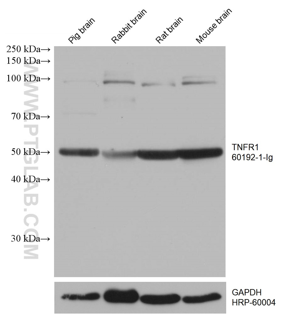
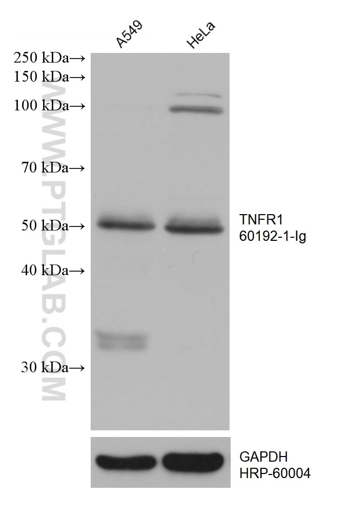
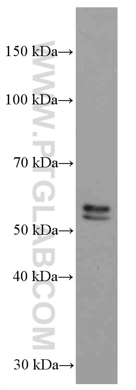
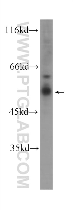
| Positive WB detected in | pig brain tissue, A549 cells, Daudi cells, HL-60 cells, HeLa cells, rabbit brain tissue, rat brain tissue, mouse brain tissue |
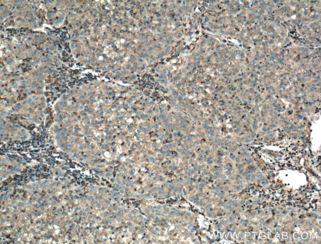
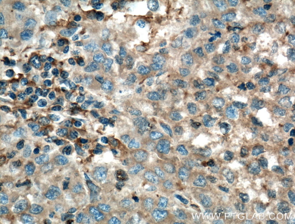
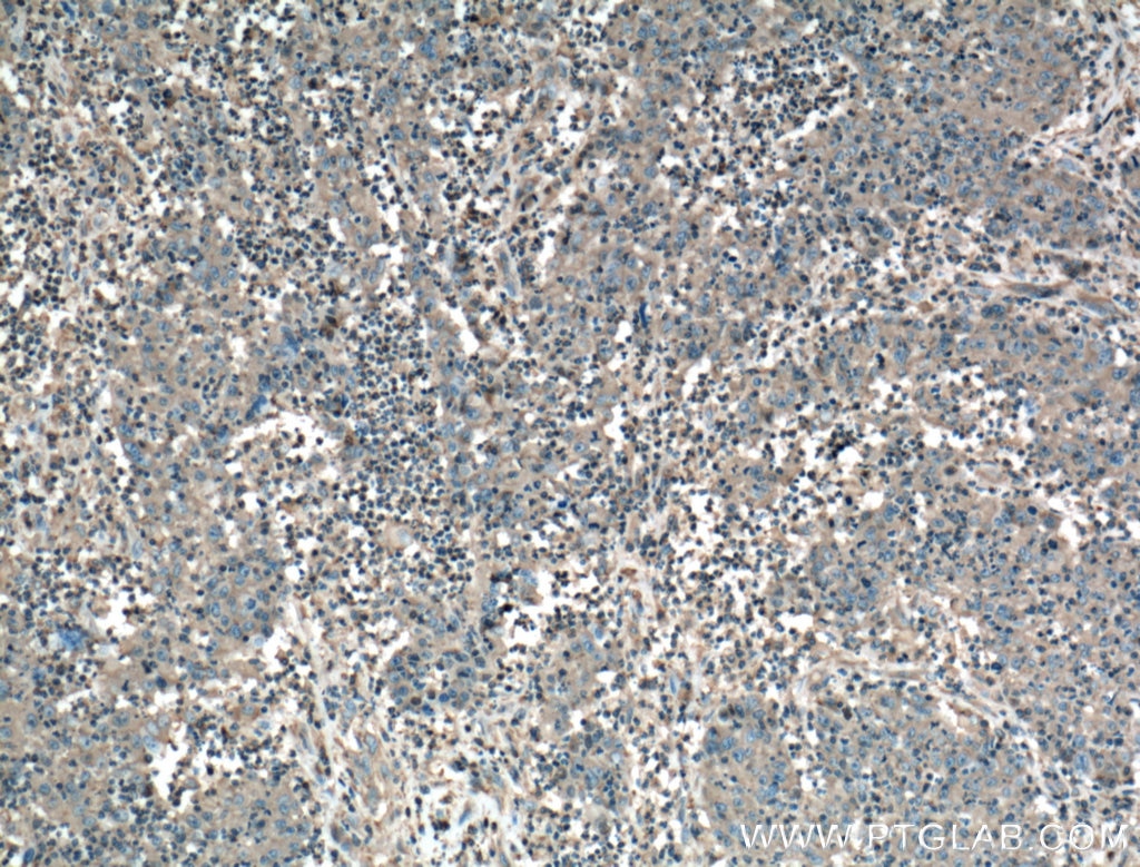
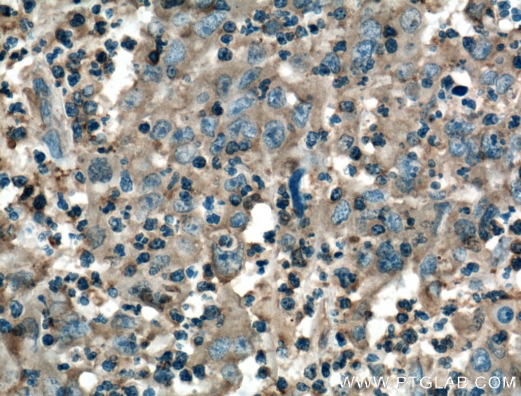
| Positive IHC detected in | human liver cancer tissue, human colon cancer tissue Note: suggested antigen retrieval with TE buffer pH 9.0; (*) Alternatively, antigen retrieval may be performed with citrate buffer pH 6.0 |
Recommended dilution
| Application | Dilution |
|---|---|
| Western Blot (WB) | WB : 1:5000-1:50000 |
| Immunohistochemistry (IHC) | IHC : 1:50-1:500 |
| It is recommended that this reagent should be titrated in each testing system to obtain optimal results. | |
| Sample-dependent, Check data in validation data gallery. | |
Product Information
60192-1-Ig targets TNFR1/CD120a in WB, IHC, IF, ELISA applications and shows reactivity with human, mouse, rat, pig samples.
| Tested Reactivity | human, mouse, rat, pig |
| Cited Reactivity | human, mouse, rat |
| Host / Isotype | Mouse / IgG1 |
| Class | Monoclonal |
| Type | Antibody |
| Immunogen |
CatNo: Ag16112 Product name: Recombinant human TNFR1 protein Source: e coli.-derived, PGEX-4T Tag: GST Domain: 235-455 aa of BC010140 Sequence: RYQRWKSKLYSIVCGKSTPEKEGELEGTTTKPLAPNPSFSPTPGFTPTLGFSPVPSSTFTSSSTYTPGDCPNFAAPRREVAPPYQGADPILATALASDPIPNPLQKWEDSAHKPQSLDTDDPATLYAVVENVPPLRWKEFVRRLGLSDHEIDRLELQNGRCLREAQYSMLATWRRRTPRREATLELLGRVLRDMDLLGCLEDIEEALCGPAALPPAPSLLR Predict reactive species |
| Full Name | tumor necrosis factor receptor superfamily, member 1A |
| Calculated Molecular Weight | 455 aa, 50 kDa |
| Observed Molecular Weight | 50-55 kDa |
| GenBank Accession Number | BC010140 |
| Gene Symbol | TNFR1 |
| Gene ID (NCBI) | 7132 |
| RRID | AB_10897160 |
| Conjugate | Unconjugated |
| Form | Liquid |
| Purification Method | Protein G purification |
| UNIPROT ID | P19438 |
| Storage Buffer | PBS with 0.02% sodium azide and 50% glycerol, pH 7.3. |
| Storage Conditions | Store at -20°C. Stable for one year after shipment. Aliquoting is unnecessary for -20oC storage. 20ul sizes contain 0.1% BSA. |
Background Information
Tumor necrosis factor (TNF) is a multifunctional cytokine that plays a key role in regulating inflammation, immune functions, host defense, and apoptosis (PMID: 16407280). TNF exists in soluble and membrane-bound forms. TNF signals through two distinct cell surface receptors, TNFR1 (TNFRSF1A, CD120a) and TNFR2 (TNFRSF1B, CD120b). Whereas TNFR1 is widely expressed, expression of TNFR2 is limited to cells of the immune system, endothelial cells, and nerve cells (PMID: 22053109). TNFR1, which contains a death domain (DD) within its intracytoplasmic region, is thought to be the key receptor for TNF signaling (PMID: 16407280). This receptor can activate NF-kappaB, mediate apoptosis, and function as a regulator of inflammation. Antiapoptotic protein BCL2-associated athanogene 4 (BAG4/SODD) and adaptor proteins TRADD and TRAF2 have been shown to interact with this receptor, and thus play regulatory roles in the signal transduction mediated by the receptor.
Protocols
| Product Specific Protocols | |
|---|---|
| IHC protocol for TNFR1/CD120a antibody 60192-1-Ig | Download protocol |
| WB protocol for TNFR1/CD120a antibody 60192-1-Ig | Download protocol |
| Standard Protocols | |
|---|---|
| Click here to view our Standard Protocols |
Publications
What published studies show
The TNFR1/CD120a Monoclonal antibody (Cat# 60192-1-Ig) has 24 citations across journals including Nature Communications, Molecular Therapy, Cell Reports, Frontiers in Immunology, International Immunopharmacology, Phytomedicine, Materials Today Bio, and Scientific Reports. Research fields encompass oncology (hepatocellular carcinoma, glioblastoma, lung cancer), immunology and inflammation (ulcerative colitis, autoimmune myocarditis, acute lung injury), neurology (astrocyte-neuron crosstalk, dry eye pain), metabolism and obesity, cardiovascular disease, and programmed cell death (ferroptosis, necroptosis, apoptosis).
| Species | Application | Title |
|---|---|---|
MedComm (2020) Silibinin, a potential fasting mimetic, inhibits hepatocellular carcinoma by triggering extrinsic apoptosis | ||
Mol Ther Tumor necrosis factor alpha delivers exogenous inflammation-related microRNAs to recipient cells with functional targeting capabilities. | ||
Phytomedicine Rhoifolin inhibits ferroptosis and alleviates pulmonary arterial hypertension via the TNF-α/TNF-R1/CASP8/CASP3 pathway. | ||
Mater Today Bio Nanoparticles coated with osteoblast-like cell membranes as curcumin delivery vehicles for targeted postmenopausal osteoporosis therapy. | ||
J Transl Med CRISPR screening of E3 ubiquitin ligases reveals Ring Finger Protein 185 as a novel tumor suppressor in glioblastoma repressed by promoter hypermethylation and miR-587. |
Reviews
What customers say
The single available review reports poor performance of this rabbit TNFR1 antibody (used at 1:1000) in U2OS cells for both Western blot and immunofluorescence. In IF, it stained microtubules only, and in WB it produced no clear bands. The reviewer rated it 1 out of 5. Overall, the feedback is negative, indicating the antibody did not work well for either application in this cell line.
The reviews below have been submitted by verified Proteintech customers who received an incentive for providing their feedback.
Lianna (Verified Customer) (06-12-2025) | Rabbit TNFR1 antibody (1:1000) did not work well for IF or WB in U2OS cells. In IF it solely stained microtubules and for WB it was poor as well with no clear bands.
|









