

Tested Applications
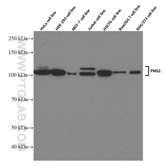
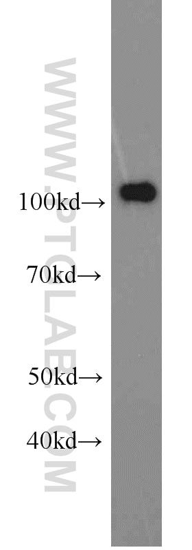
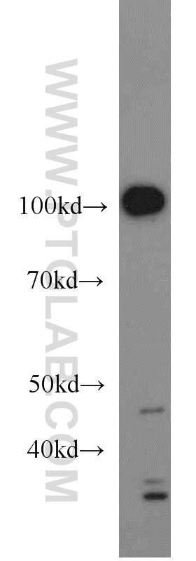
| Positive WB detected in | A431 cells, HeLa cells |
Recommended dilution
| Application | Dilution |
|---|---|
| Western Blot (WB) | WB : 1:500-1:2000 |
| It is recommended that this reagent should be titrated in each testing system to obtain optimal results. | |
| Sample-dependent, Check data in validation data gallery. | |
Product Information
66075-1-Ig targets PMS2 in WB, ELISA applications and shows reactivity with human, mouse, rat samples.
| Tested Reactivity | human, mouse, rat |
| Cited Reactivity | human |
| Host / Isotype | Mouse / IgG2a |
| Class | Monoclonal |
| Type | Antibody |
| Immunogen |
CatNo: Ag12661 Product name: Recombinant human PMS2 protein Source: e coli.-derived, PET28a Tag: 6*His Domain: 513-862 aa of BC093921 Sequence: SHCSSEYAASSPGDRGSQEHVDSQEKAPETDDSFSDVDCHSNQEDTGCKFRVLPQPTNLATPNTKRFKKEEILSSSDICQKLVNTQDMSASQVDVAVKINKKVVPLDFSISSLAKRIKQLHHEAQQSEGEQNYRKFRAKICPGENQAAEDELRKEISKTMFAEMEIIGQFNLGFIITKLNEDIFIVDQHATDEKYNFEMLQQHTVLQGQRLIAPQTLNLTAVNEAVLIENLEIFRKNGFDFVIDENAPVTERAKLISLPTSKNWTFGPQDVDELIFMLSDSPGVMCRPSRVKQMFASRACRKSVMIGTALNTSEMKKLITHMGEMDHPWNCPHGRPTMRHIANLGVISQN Predict reactive species |
| Full Name | PMS2 postmeiotic segregation increased 2 (S. cerevisiae) |
| Calculated Molecular Weight | 862 aa, 96 kDa |
| Observed Molecular Weight | 100 kDa |
| GenBank Accession Number | BC093921 |
| Gene Symbol | PMS2 |
| Gene ID (NCBI) | 5395 |
| RRID | AB_11182595 |
| Conjugate | Unconjugated |
| Form | Liquid |
| Purification Method | Protein A purification |
| UNIPROT ID | P54278 |
| Storage Buffer | PBS with 0.02% sodium azide and 50% glycerol, pH 7.3. |
| Storage Conditions | Store at -20°C. Stable for one year after shipment. Aliquoting is unnecessary for -20oC storage. 20ul sizes contain 0.1% BSA. |
Background Information
PMS2, also named as PMSL2, belongs to the DNA mismatch repair mutL/hexB family. It is a component of the post-replicative DNA mismatch repair system (MMR). It heterodimerizes with MLH1 to form MutL alpha. MulL alpha (MLH1-PMS2) interacts physically with the clamp loader subunits of DNA polymerase III, suggesting that it may play a role to recruit the DNA polymerase III to the site of the MMR. It also implicated in DNA damage signaling, a process which induces cell cycle arrest and can lead to apoptosis in case of major DNA damages. (PMID: 16873062, PMID: 18206974) Defects in PMS2 are the cause of hereditary non-polyposis colorectal cancer type 4 (HNPCC4). Defects in PMS2 are a cause of mismatch repair cancer syndrome (MMRCS).
Protocols
| Product Specific Protocols | |
|---|---|
| WB protocol for PMS2 antibody 66075-1-Ig | Download protocol |
| Standard Protocols | |
|---|---|
| Click here to view our Standard Protocols |
Publications
What published studies show
The PMS2 Monoclonal antibody (1G4E6), catalog number 66075-1-Ig, has 9 total citations. These appear in journals including Drug Resistance Updates, Cell Reports, EBioMedicine, Cells, Scientific Reports, Cell and Molecular Biology Letters, Molecular Carcinogenesis, Molecular Genetics & Genomic Medicine, and Veterinary and Comparative Oncology. Research fields span DNA mismatch repair, glioma, endometrial cancer, colon cancer, gastric cancer, ovarian cancer, Lynch syndrome, and feline oncology.
| Species | Application | Title |
|---|---|---|
Drug Resist Updat Intercellular adhesion molecule-1 suppresses TMZ chemosensitivity in acquired TMZ-resistant gliomas by increasing assembly of ABCB1 on the membrane | ||
EBioMedicine DNA methylation mediates the immunosuppressive tumour microenvironment in metastatic endometrial clear cell carcinoma. | ||
Cell Rep Phosphorylation of SIRT7 by ATM causes DNA mismatch repair downregulation and adaptive mutability during chemotherapy | ||
Cells TDGF1 Mediates the Oncogenic Effects of the OLMALINC/miR-3614-5p ceRNA Axis in Colon Cancer Through Nodal/Smad2 and Glypican-1/MAPK-AKT Signaling. | ||
Sci Rep Mismatch repair proteins play a role in ATR activation upon temozolomide treatment in MGMT-methylated glioblastoma. |
Reviews
What customers say
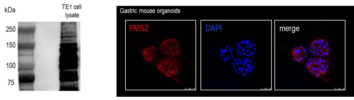
The single review for this product reports a low rating (1/5). The user tested the antibody in Western Blot (1:1000 dilution) and Immunofluorescence (1:100 dilution) on gastric mouse organoids and an oesophageal cancer cell line, and found that the bands in both WB and IF staining were aspecific. Overall, the feedback indicates poor specificity for the tested applications and sample types.
The reviews below have been submitted by verified Proteintech customers who received an incentive for providing their feedback.
Daniela (Verified Customer) (03-17-2022) | Bands in WB and IF staining were both aspecific.
 |




