

Tested Applications
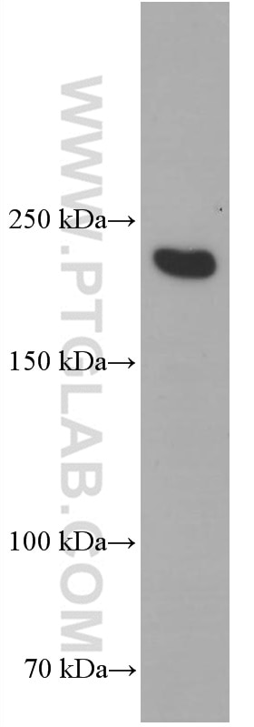
| Positive WB detected in | human skeletal muscle tissue |
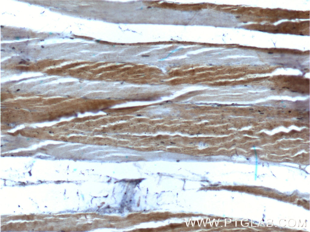
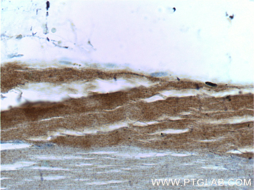
| Positive IHC detected in | human skeletal muscle tissue Note: suggested antigen retrieval with TE buffer pH 9.0; (*) Alternatively, antigen retrieval may be performed with citrate buffer pH 6.0 |
Recommended dilution
| Application | Dilution |
|---|---|
| Western Blot (WB) | WB : 1:5000-1:100000 |
| Immunohistochemistry (IHC) | IHC : 1:50-1:500 |
| It is recommended that this reagent should be titrated in each testing system to obtain optimal results. | |
| Sample-dependent, Check data in validation data gallery. | |
Product Information
66212-1-Ig targets MYH2-specific in WB, IHC, ELISA applications and shows reactivity with human samples.
| Tested Reactivity | human |
| Cited Reactivity | human, mouse, rat |
| Host / Isotype | Mouse / IgG2b |
| Class | Monoclonal |
| Type | Antibody |
| Immunogen |
Peptide Predict reactive species |
| Full Name | myosin, heavy chain 2, skeletal muscle, adult |
| Calculated Molecular Weight | 223 kDa |
| Observed Molecular Weight | 200 kDa |
| GenBank Accession Number | NM_017534 |
| Gene Symbol | MYH2 |
| Gene ID (NCBI) | 4620 |
| RRID | AB_2881603 |
| Conjugate | Unconjugated |
| Form | Liquid |
| Purification Method | Protein A purification |
| UNIPROT ID | Q9UKX2 |
| Storage Buffer | PBS with 0.02% sodium azide and 50% glycerol, pH 7.3. |
| Storage Conditions | Store at -20°C. Stable for one year after shipment. Aliquoting is unnecessary for -20oC storage. 20ul sizes contain 0.1% BSA. |
Background Information
MYH2 (Myosin II) is a member of the class II or conventional myosin heavy chains. Myosin II was first isolated from muscle but is also found in non muscle cells, and it is especially enriched in highly motile cell types such as amoebae. It forms bipolar filaments that interact with actin filaments to produce contraction. And it is the motor protein that generates force to drive muscle contraction. Functions in skeletal muscle contraction. The antibody is specific to MYH2, will not bind other myosins. It is a skeletal muscle specific antibody.
Protocols
| Product Specific Protocols | |
|---|---|
| IHC protocol for MYH2-specific antibody 66212-1-Ig | Download protocol |
| WB protocol for MYH2-specific antibody 66212-1-Ig | Download protocol |
| Standard Protocols | |
|---|---|
| Click here to view our Standard Protocols |
Publications
What published studies show
The MYH2-specific Monoclonal antibody (1F1B6), catalog number 66212-1-Ig, has 8 citations. These appear in journals including Scientific Reports, Journal of Biological Chemistry, Journal of Agricultural and Food Chemistry, Heliyon, Histochemistry and Cell Biology, Journal of Muscle Research and Cell Motility, and Bioscience, Biotechnology, and Biochemistry. The research fields span skeletal muscle biology, myogenic differentiation, muscle fiber remodeling, mitochondrial quality control, aging-related muscle changes, and oxidative metabolism.
| Species | Application | Title |
|---|---|---|
J Agric Food Chem Spermidine-Activated Satellite Cells Are Associated with Hypoacetylation in ACVR2B and Smad3 Binding to Myogenic Genes in Mice. | ||
Sci Rep Trimetazidine and exercise provide comparable improvements to high fat diet-induced muscle dysfunction through enhancement of mitochondrial quality control. | ||
Sci Rep Age-related thinning of orbicularis oculi muscle inside upper eyelid and its possible association with sunken upper eyelids | ||
J Biol Chem O-GlcNAcylation modification of MyoD regulates skeletal muscle fiber differentiation by antagonizing the UPF1 pathway | ||
Heliyon Light-phase time-restricted feeding disrupts the muscle clock and insulin sensitivity yet potentially induces muscle fiber remodeling in mice | ||
Histochem Cell Biol Fiber type-specific expression of LACTB leverages a function in oxidative metabolism. |




