

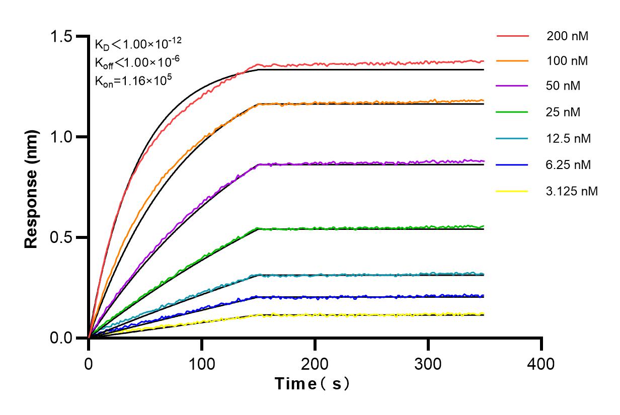

Tested Applications
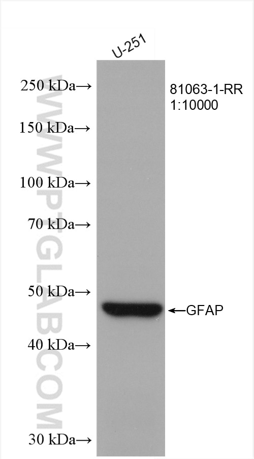
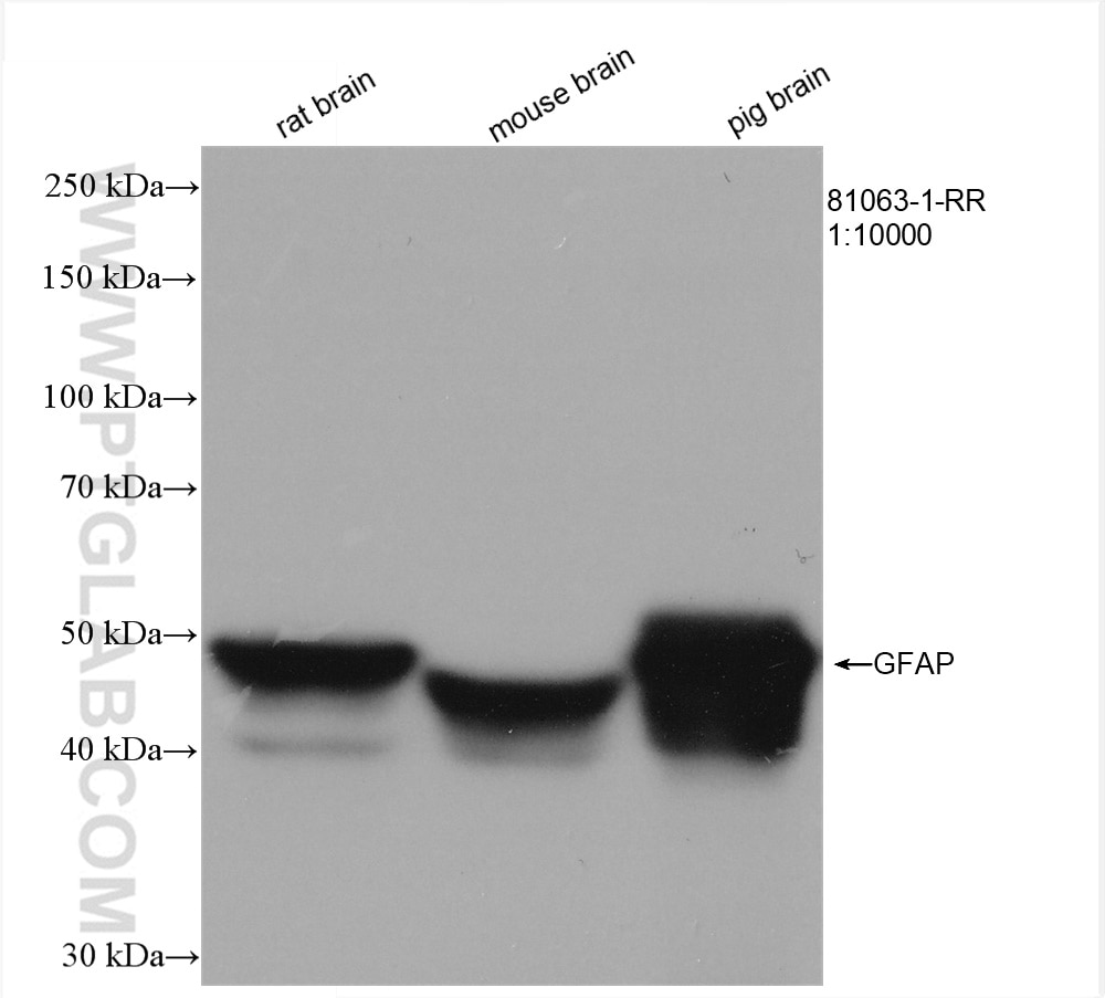
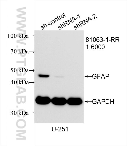
| Positive WB detected in | U-251 cells, rat brain tissue, mouse brain tissue, pig brain tissue |
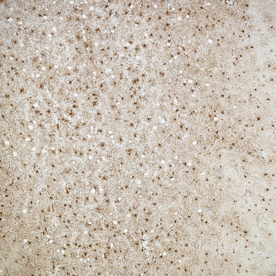
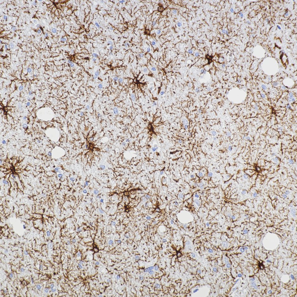
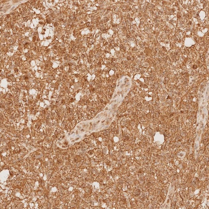
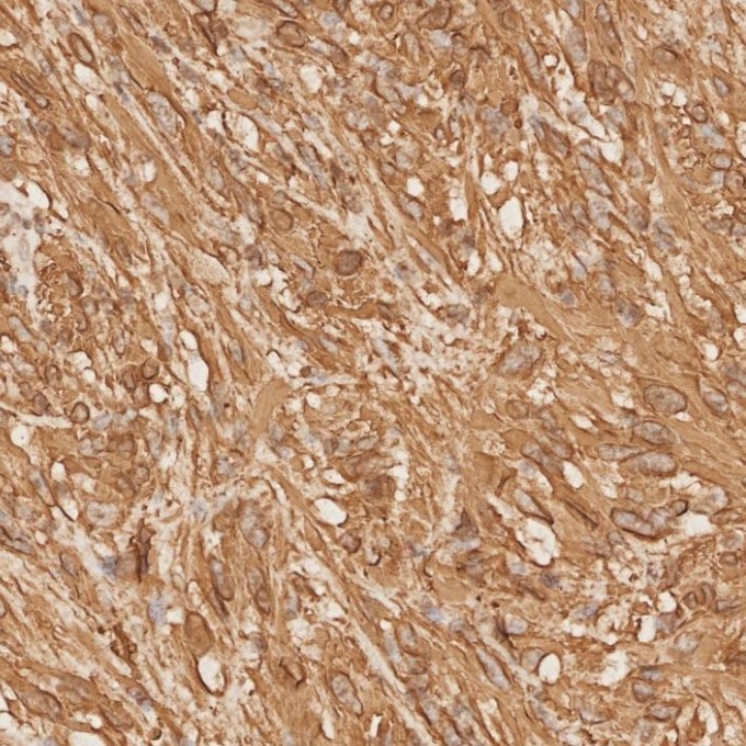
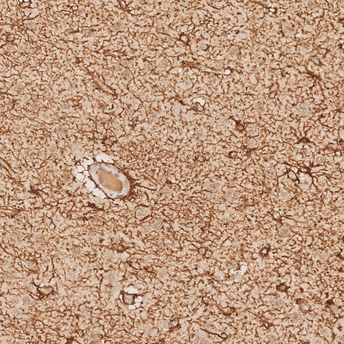
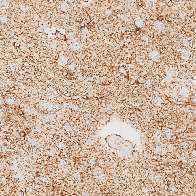
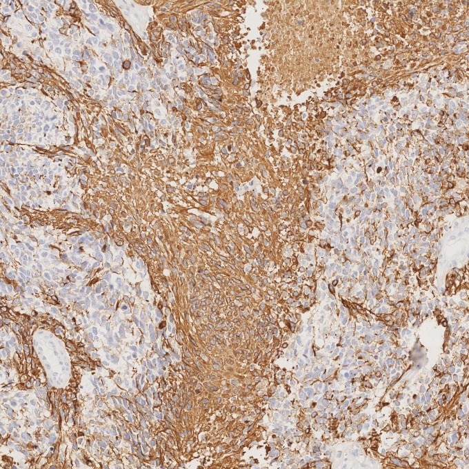
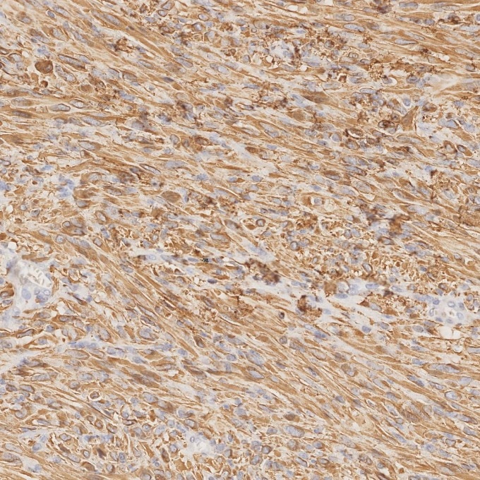
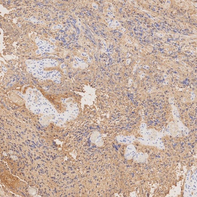
| Positive IHC-Autostainer detected in | human brain tissue, human gliomas tissue |
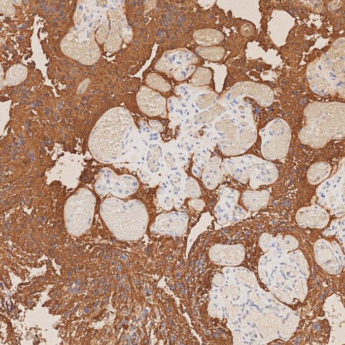
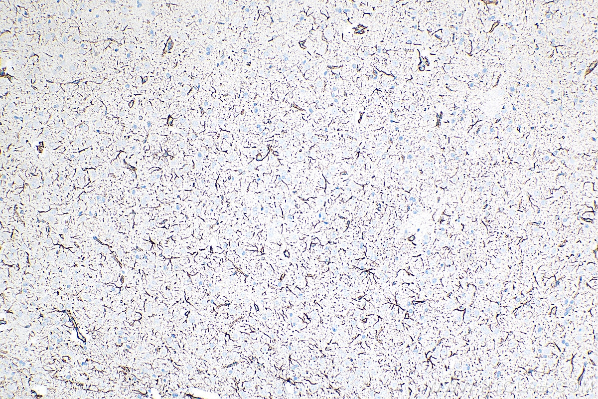
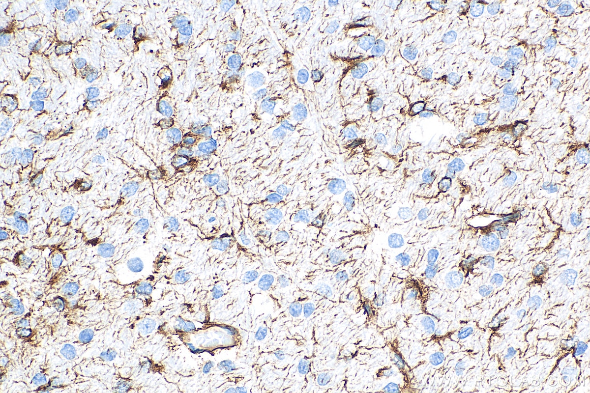
| Positive IHC detected in | rat brain tissue, human gliomas tissue Note: suggested antigen retrieval with TE buffer pH 9.0; (*) Alternatively, antigen retrieval may be performed with citrate buffer pH 6.0 |
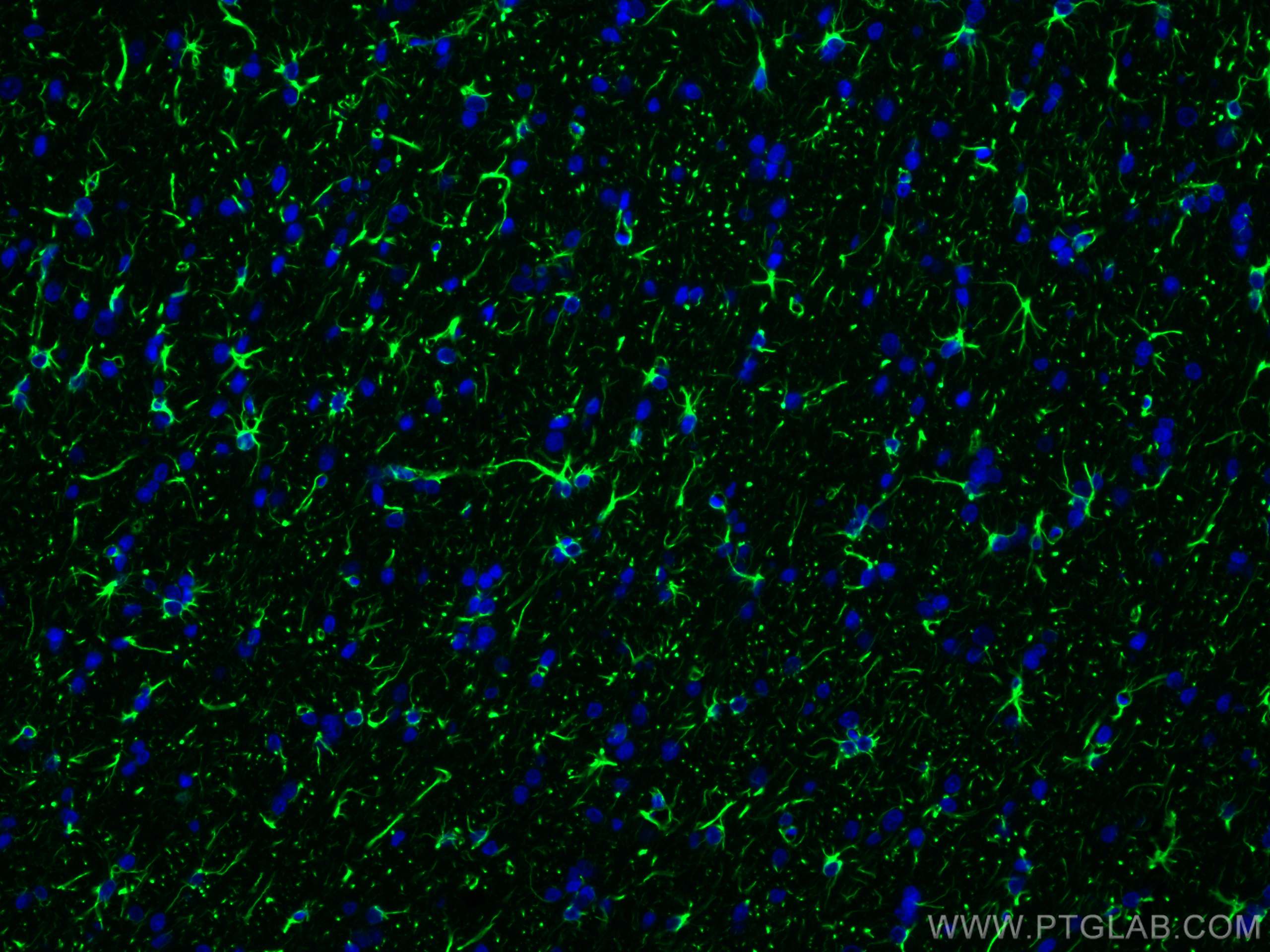
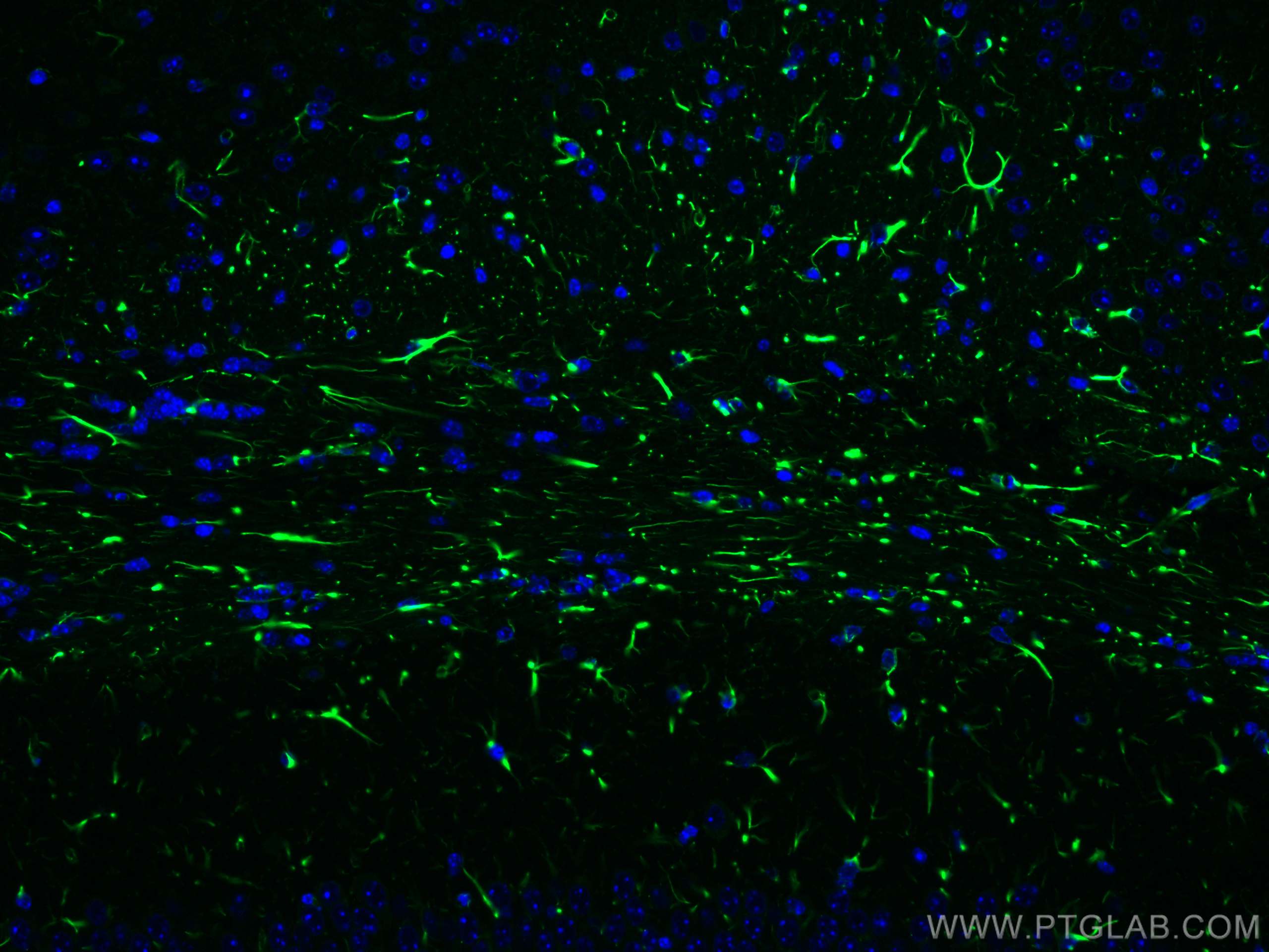
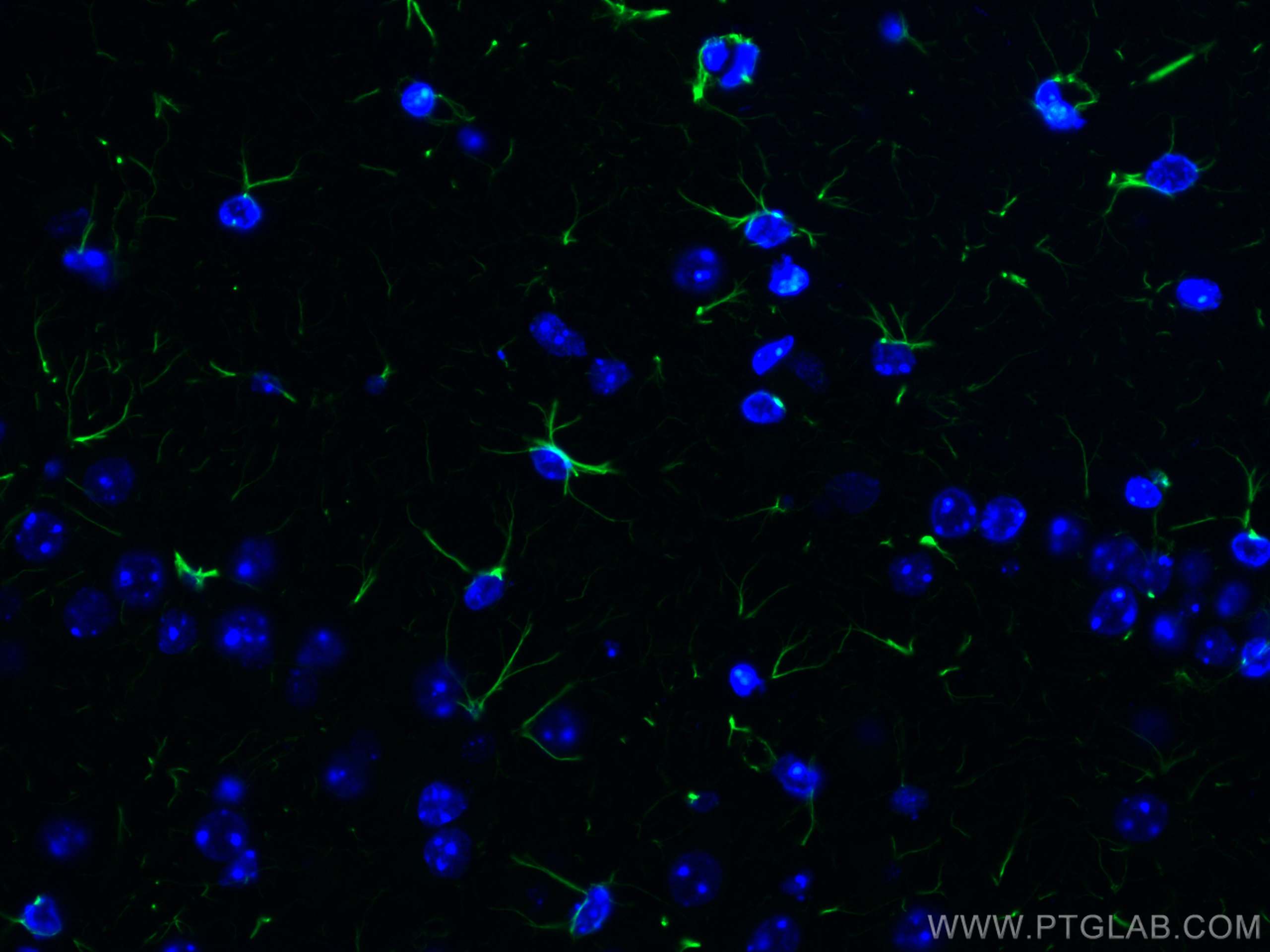
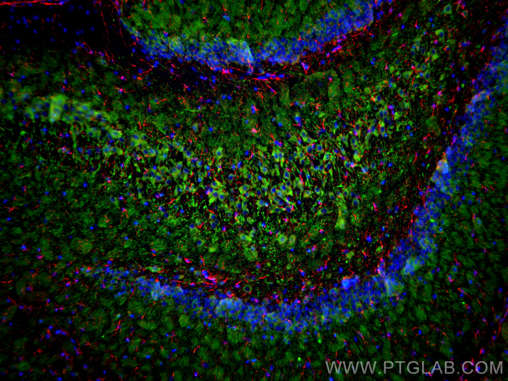
| Positive IF-P detected in | rat brain tissue, mouse brain tissue |
| Positive IF-Fro detected in | mouse brain tissue, rat brain tissue |
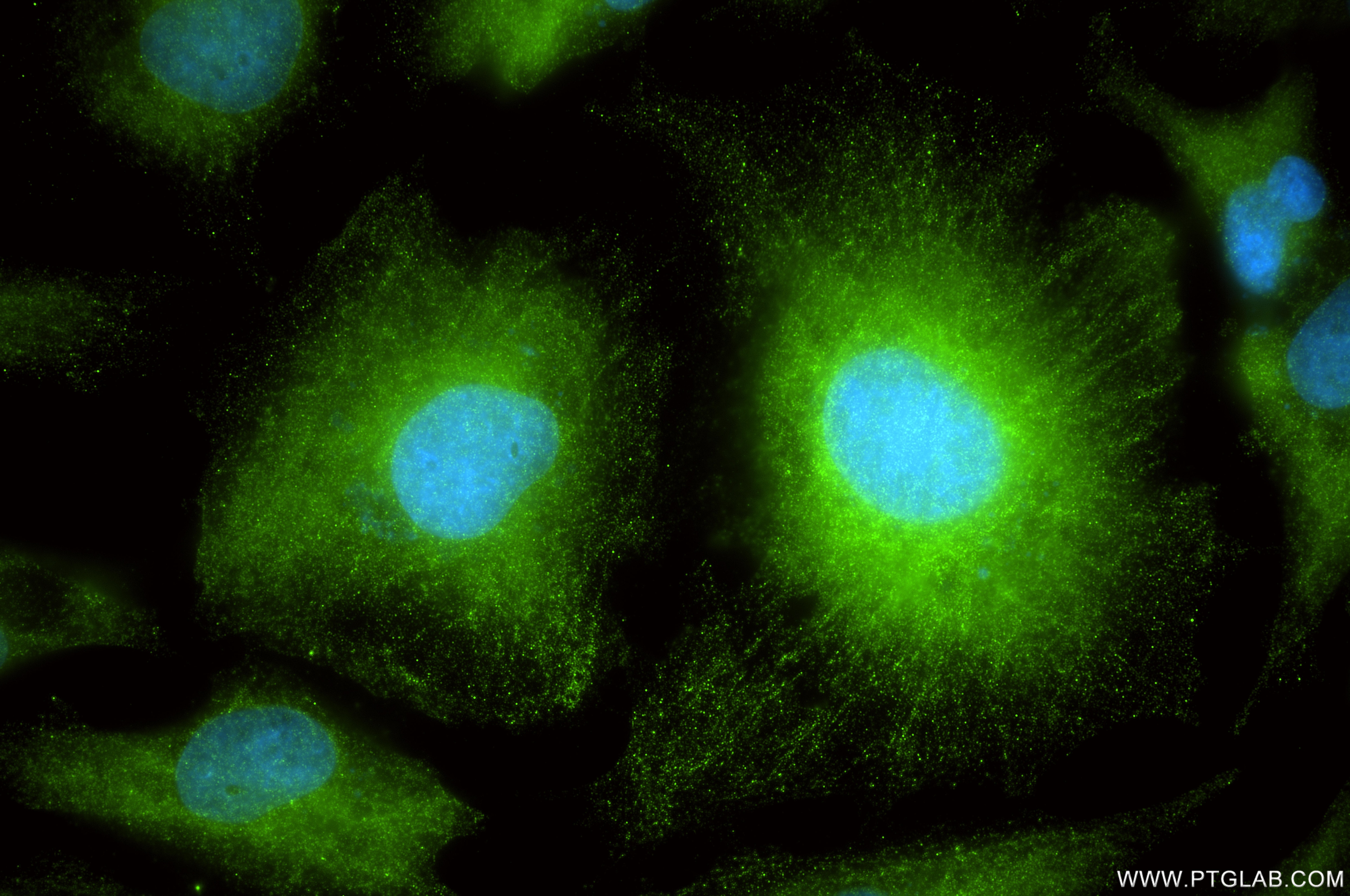
| Positive IF/ICC detected in | U-251 cells |
Recommended dilution
| Application | Dilution |
|---|---|
| Western Blot (WB) | WB : 1:5000-1:50000 |
| Immunohistochemistry (IHC)-AUTOSTAINER | IHC-AUTOSTAINER : 1:5000-1:20000 |
| Immunohistochemistry (IHC) | IHC : 1:4950-1:19800 |
| Immunofluorescence (IF)-P | IF-P : 1:50-1:500 |
| Immunofluorescence (IF)-FRO | IF-FRO : 1:50-1:500 |
| Immunofluorescence (IF)/ICC | IF/ICC : 1:200-1:800 |
| It is recommended that this reagent should be titrated in each testing system to obtain optimal results. | |
| Sample-dependent, Check data in validation data gallery. | |
Product Information
81063-1-RR targets GFAP in WB, IHC, IHC-Autostainer, IF/ICC, IF-P, IF-Fro, ELISA applications and shows reactivity with human, mouse, rat, pig samples.
| Tested Reactivity | human, mouse, rat, pig |
| Cited Reactivity | mouse |
| Host / Isotype | Rabbit / IgG |
| Class | Recombinant |
| Type | Antibody |
| Immunogen |
CatNo: Ag10423 Product name: Recombinant human GFAP protein Source: e coli.-derived, PGEX-4T Tag: GST Domain: 83-432 aa of BC013596 Sequence: YIEKVRFLEQQNKALAAELNQLRAKEPTKLADVYQAELRELRLRLDQLTANSARLEVERDNLAQDLATVRQKLQDETNLRLEAENNLAAYRQEADEATLARLDLERKIESLEEEIRFLRKIHEEEVRELQEQLARQQVHVELDVAKPDLTAALKEIRTQYEAMASSNMHEAEEWYRSKFADLTDAAARNAELLRQAKHEANDYRRQLQSLTCDLESLRGTNESLERQMREQEERHVREAASYQEALARLEEEGQSLKDEMARHLQEYQDLLNVKLALDIEIATYRKLLEGEENRITIPVQTFSNLQIRETSLDTKSVSEGHLKRNIVVKTVEMRDGEVIKESKQEHKDVM Predict reactive species |
| Full Name | glial fibrillary acidic protein |
| Calculated Molecular Weight | 432 aa, 50 kDa |
| Observed Molecular Weight | 45-50 kDa |
| GenBank Accession Number | BC013596 |
| Gene Symbol | GFAP |
| Gene ID (NCBI) | 2670 |
| RRID | AB_2923699 |
| Conjugate | Unconjugated |
| Form | Liquid |
| Purification Method | Protein A purification |
| UNIPROT ID | P14136 |
| Storage Buffer | PBS with 0.02% sodium azide and 50% glycerol, pH 7.3. |
| Storage Conditions | Store at -20°C. Stable for one year after shipment. Aliquoting is unnecessary for -20oC storage. 20ul sizes contain 0.1% BSA. |
Background Information
GFAP (Glial fibrillary acidic protein ), an intermediate-filament (IF) protein , is specifically expressed in cells of astroglial lineage and is widely used to mark astroglia in the brain. It is also used as a marker for intracranial and intraspinal tumors arising from astrocytes.
Protocols
| Product Specific Protocols | |
|---|---|
| IF protocol for GFAP antibody 81063-1-RR | Download protocol |
| IHC protocol for GFAP antibody 81063-1-RR | Download protocol |
| WB protocol for GFAP antibody 81063-1-RR | Download protocol |
| Standard Protocols | |
|---|---|
| Click here to view our Standard Protocols |
Publications
What published studies show
The GFAP Recombinant monoclonal antibody (4C6), catalog number 81063-1-RR, has 2 citations. These appear in the European Journal of Pharmacology and Cell Signaling. The cited research covers neuropharmacology (cerebral ischemia-reperfusion injury and NRF2/NF-κB/NLRP3 signaling) and neurodegeneration (Parkinson's disease, ferroptosis, and the SREBP1/SCD1 axis in dopaminergic neurons). The cited application is Western blot in mouse.
| Species | Application | Title |
|---|---|---|
Eur J Pharmacol Edaravone-dexborneol improves neurological function after cerebral ischemia-reperfusion injury by activating NRF2/p62 and suppressing NF-κB/NLRP3 signaling. | ||
Cell Signal FOXO1 inhibition attenuates ferroptosis-like changes and apoptosis in dopaminergic neurons with the involvement of the SREBP1/SCD1 axis in Parkinson's disease. |
Reviews
What customers say
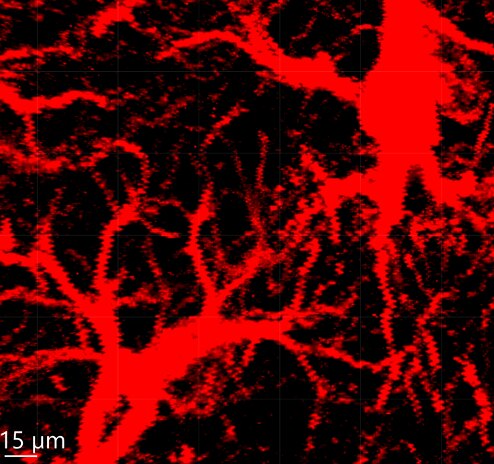
The single review for 81063-1-RR (GFAP) earned a 5-star rating for immunofluorescence. The user visualized astrocytes in fixed mouse brain tissue using 40 μm sections blocked with 3% normal goat serum. The primary antibody was applied at a 1:100 dilution and incubated overnight at 4°C, followed by an Alexa 633-labeled anti-rabbit fluorescent secondary antibody. Overall, the reviewer reported successful astrocyte staining with this protocol.
The reviews below have been submitted by verified Proteintech customers who received an incentive for providing their feedback.
Kateryna (Verified Customer) (01-23-2025) | We use the GFAP antibody to visualize astrocytes via immunostaining in mouse brain tissue. The mouse brain tissue is fixed, brain sections are 40 μm thick, and blocked with 3% normal goat serum. The incubation with the primary antibody (GFAP) is performed at 4°C overnight, followed by the corresponding Alexa 633-labeled anti-rabbit fluorescent secondary antibody.
 |























