

Tested Applications
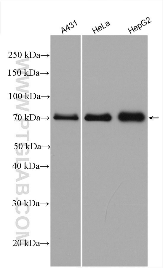
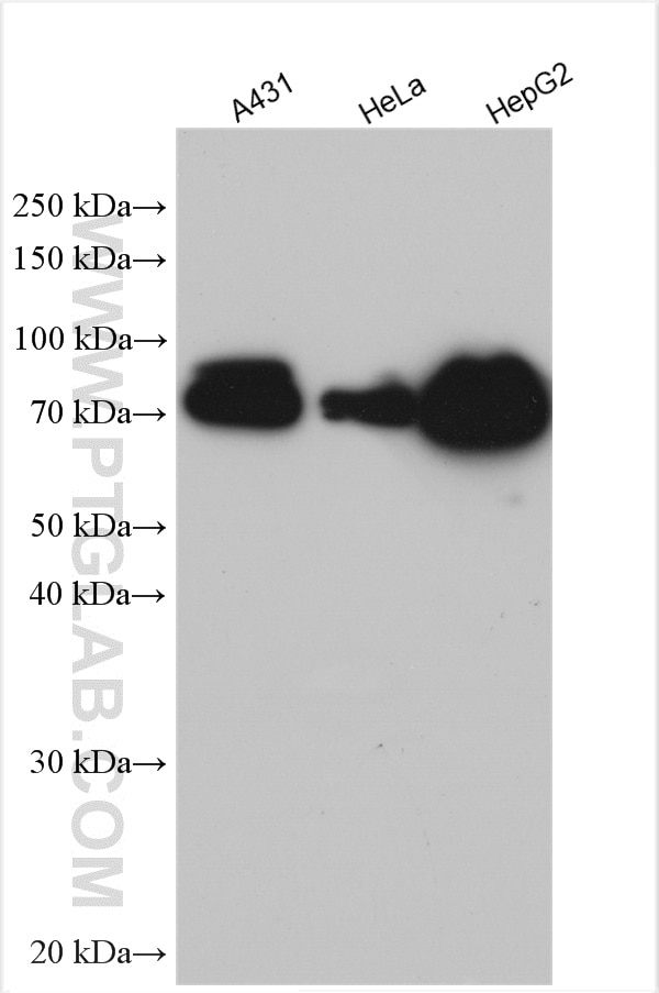
| Positive WB detected in | A431 cells, HeLa cells, HuH-7 cells, HepG2 cells |
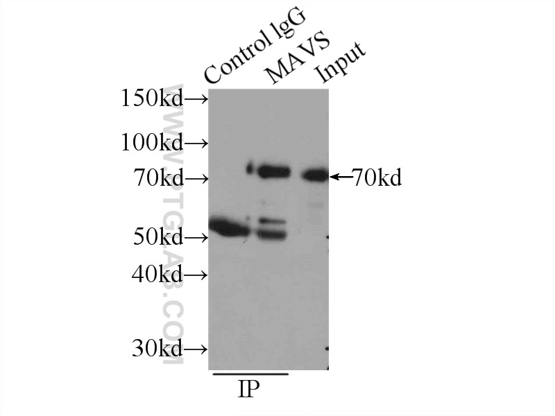
| Positive IP detected in | HEK-293 cells |
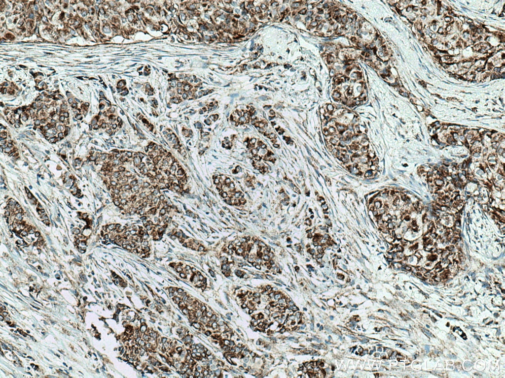
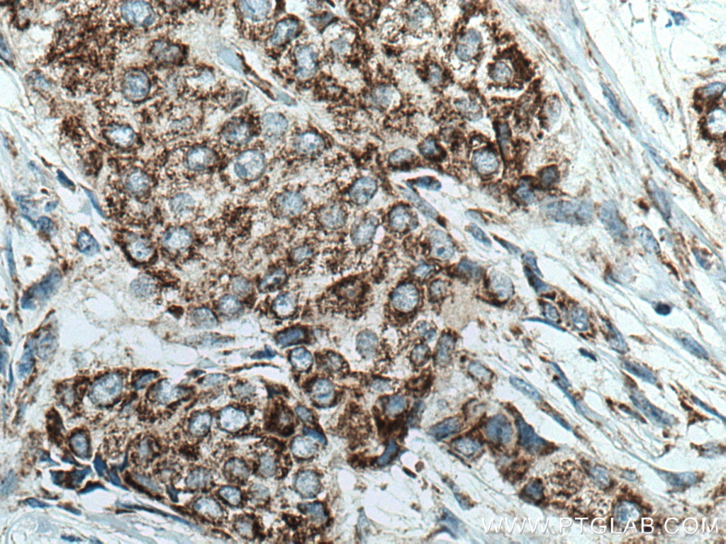
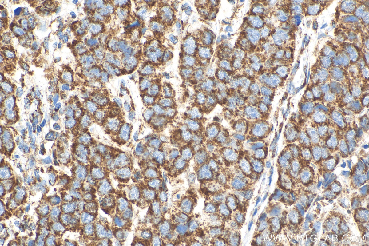
| Positive IHC detected in | human breast cancer tissue Note: suggested antigen retrieval with TE buffer pH 9.0; (*) Alternatively, antigen retrieval may be performed with citrate buffer pH 6.0 |
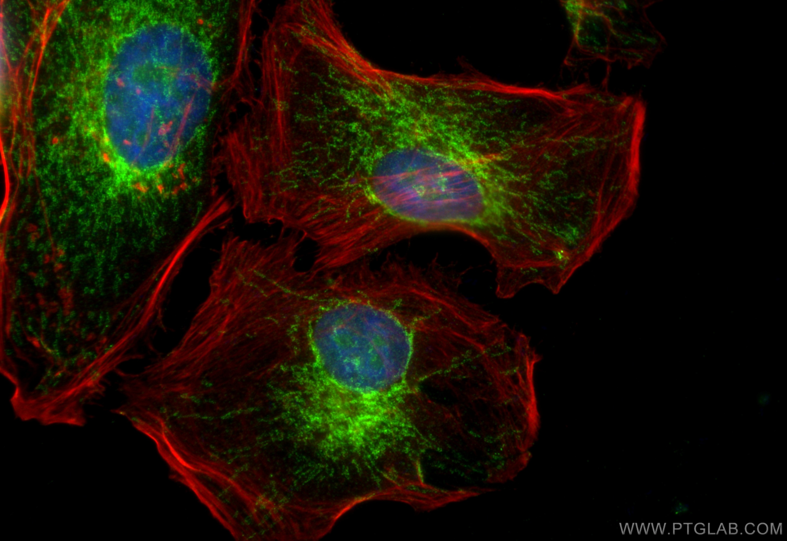
| Positive IF/ICC detected in | HeLa cells |
Recommended dilution
| Application | Dilution |
|---|---|
| Western Blot (WB) | WB : 1:2000-1:16000 |
| Immunoprecipitation (IP) | IP : 0.5-4.0 ug for 1.0-3.0 mg of total protein lysate |
| Immunohistochemistry (IHC) | IHC : 1:250-1:1000 |
| Immunofluorescence (IF)/ICC | IF/ICC : 1:50-1:500 |
| It is recommended that this reagent should be titrated in each testing system to obtain optimal results. | |
| Sample-dependent, Check data in validation data gallery. | |
Product Information
14341-1-AP targets MAVS; VISA in WB, IHC, IF/ICC, IP, CoIP, ELISA applications and shows reactivity with human samples.
| Tested Reactivity | human |
| Cited Reactivity | human, rat, pig, monkey, chicken, duck |
| Host / Isotype | Rabbit / IgG |
| Class | Polyclonal |
| Type | Antibody |
| Immunogen |
CatNo: Ag5655 Product name: Recombinant human MAVS; VISA protein Source: e coli.-derived, PGEX-4T Tag: GST Domain: 1-347 aa of BC044952 Sequence: MPFAEDKTYKYICRNFSNFCNVDVVEILPYLPCLTARDQDRLRATCTLSGNRDTLWHLFNTLQRRPGWVEYFIAALRGCELVDLADEVASVYESYQPRTSDRPPDPLEPPSLPAERPGPPTPAAAHSIPYNSCREKEPSYPMPVQETQAPESPGENSEQALQTLSPRAIPRNPDGGPLESSSDLAALSPLTSSGHQEKDTELGSTHTAGATSSLTPSRGPVSPSVSFQPLARSTPRASRLPGPTGSVVSTGTSFSSSSPGLASAGAAEGKQGAESDQAEPIICSSGAEAPANSLPSKVPTTLMPVNTVALKVPANPASVSTVPSKLPTSSKPPGAVPSNALTNPAPS Predict reactive species |
| Full Name | mitochondrial antiviral signaling protein |
| Calculated Molecular Weight | 57 kDa |
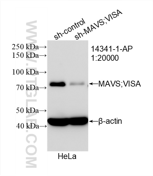
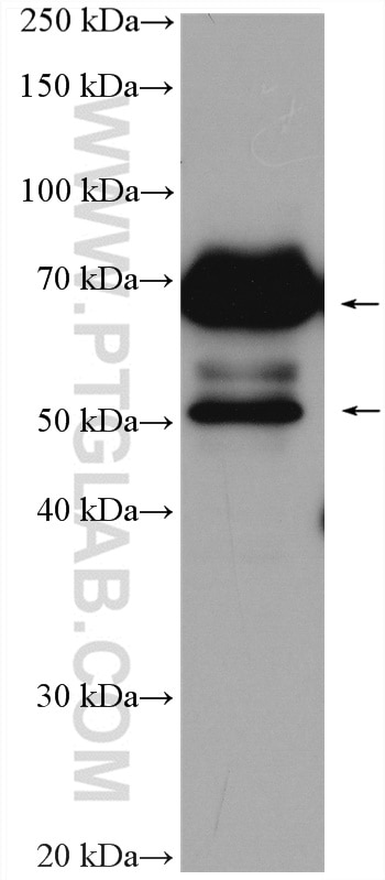
| Observed Molecular Weight | 50-55 kDa, 70-75 kDa |
| GenBank Accession Number | BC044952 |
| Gene Symbol | MAVS |
| Gene ID (NCBI) | 57506 |
| RRID | AB_10548408 |
| Conjugate | Unconjugated |
| Form | Liquid |
| Purification Method | Antigen affinity purification |
| UNIPROT ID | Q7Z434 |
| Storage Buffer | PBS with 0.02% sodium azide and 50% glycerol, pH 7.3. |
| Storage Conditions | Store at -20°C. Stable for one year after shipment. Aliquoting is unnecessary for -20oC storage. 20ul sizes contain 0.1% BSA. |
Background Information
Mitochondrial antiviral-signaling protein (MAVS) is also known as virus-induced-signaling adapter (VISA) or IFN-β promoter stimulator protein 1 (IPS-1), it is widely involved and required for innate immune defense against viruses. MAVS, present in T cells, monocytes, epithelial cells and hepatocytes, contains CARD and transmembrane domains which are essential for antiviral functions. MAVS is able to interact with various cellular proteins including DDX58/RIG-I, IFIH1/MDA5, TRAF2, TRAF6, TMEM173/MITA, IFIT3 and etc. It can undergoe phosphorylation on multiple sites and ubiquitination, which may together cause the molecular weight migrate to about 70 kDa despite the predicated 57 kDa.
Protocols
| Product Specific Protocols | |
|---|---|
| IF protocol for MAVS; VISA antibody 14341-1-AP | Download protocol |
| IHC protocol for MAVS; VISA antibody 14341-1-AP | Download protocol |
| IP protocol for MAVS; VISA antibody 14341-1-AP | Download protocol |
| WB protocol for MAVS; VISA antibody 14341-1-AP | Download protocol |
| Standard Protocols | |
|---|---|
| Click here to view our Standard Protocols |
Publications
What published studies show
MAVS; VISA Polyclonal Antibody (Cat# 14341-1-AP) has 114 citations across journals including Nature Immunology, Immunity, Nature Communications, Science Immunology, Autophagy, Cell Reports, Journal of Virology, PLoS Pathogens, EMBO Journal, and over 50 others. Research fields encompass antiviral innate immunity, RIG-I/MAVS signaling, type I interferon pathways, autophagy/mitophagy, viral infections (influenza, SARS-CoV-2, HSV, PRRSV, PEDV), cancer immunology, and neuroinflammation.
| Species | Application | Title |
|---|---|---|
Signal Transduct Target Ther TRAF3 activates STING-mediated suppression of EV-A71 and target of viral evasion | ||
Immunity Decreased Expression of the Host Long-Noncoding RNA-GM Facilitates Viral Escape by Inhibiting the Kinase activity TBK1 via S-glutathionylation. | ||
Nat Immunol ALOX15 orchestrates mitochondrial antiviral immunity and serves as a host target for anti-influenza therapy | ||
Cancer Res Targeting Catechol-O-Methyltransferase Induces Mitochondrial Dysfunction and Enhances the Efficacy of Radiotherapy in Glioma | ||
Reviews
What customers say
One reviewer rated this anti-MAVS antibody 2 out of 5 stars. Using it on HeLa cells at 1:2000 dilution for Western Blot, they reported unsatisfactory results. They expected to see two bands corresponding to short-MAVS (~55 kDa) and full-length MAVS (~72 kDa), but only observed a smear and a single band at 72 kDa. The anticipated 55 kDa band was consistently absent despite following the manufacturer's protocol under various conditions. The reviewer confirmed their lysates were intact, as other target proteins in the same samples showed clear bands.
The reviews below have been submitted by verified Proteintech customers who received an incentive for providing their feedback.
Damien (Verified Customer) (09-03-2019) | No convince by this anti-MAVS antibody.In fact, in human cells MAVS protein is expressed in two differents forms : short-MAVS (55kDa) and full-length MAVS (72 kDa). So I have to observe two bands by WB. However, after using different conditions with this antibody according to the manufacturer's protocol I always observe a smear of protein, and one band at 72 kDa. The expected on around 55 kDa never appear. Note that my cells lysates were not degraded since the others proteins that I want to reveal were great.
|










