



Tested Applications
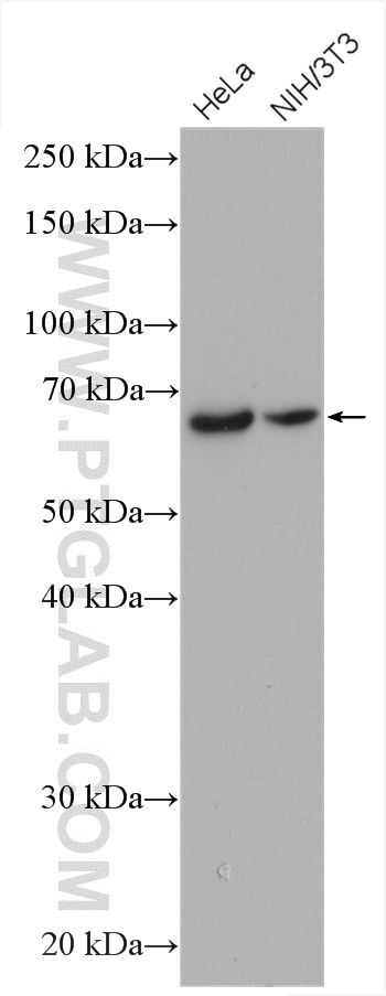
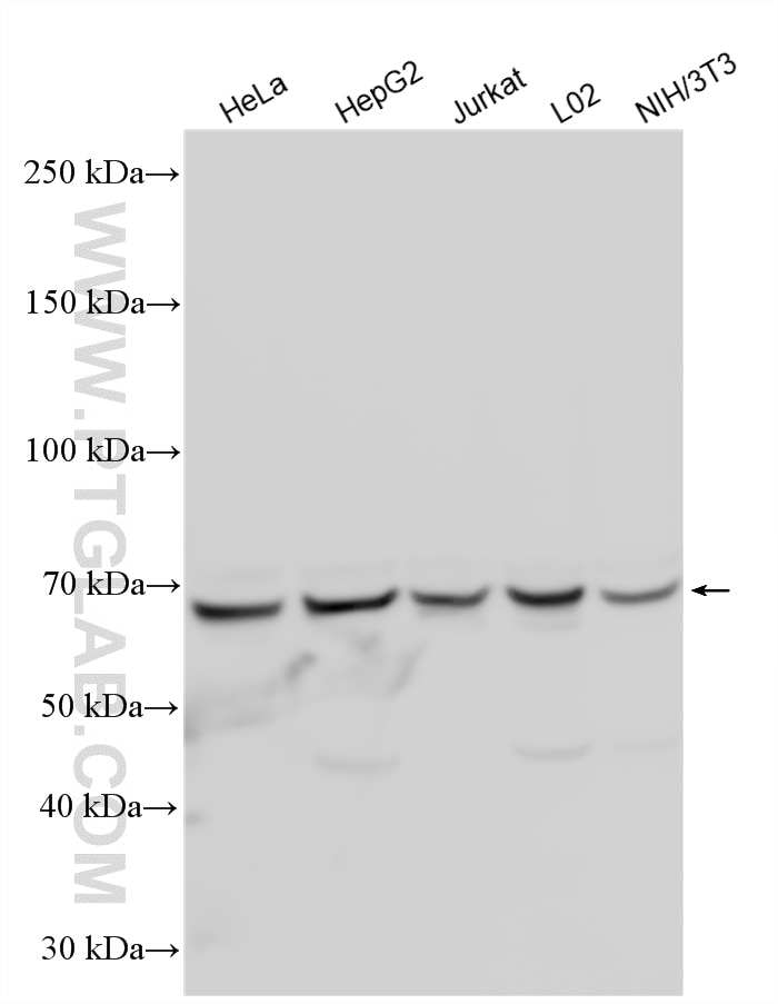
| Positive WB detected in | HeLa cells, NIH/3T3 cells, HepG2 cells, Jurkat cells, L02 cells |
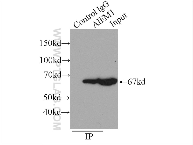
| Positive IP detected in | HeLa cells |
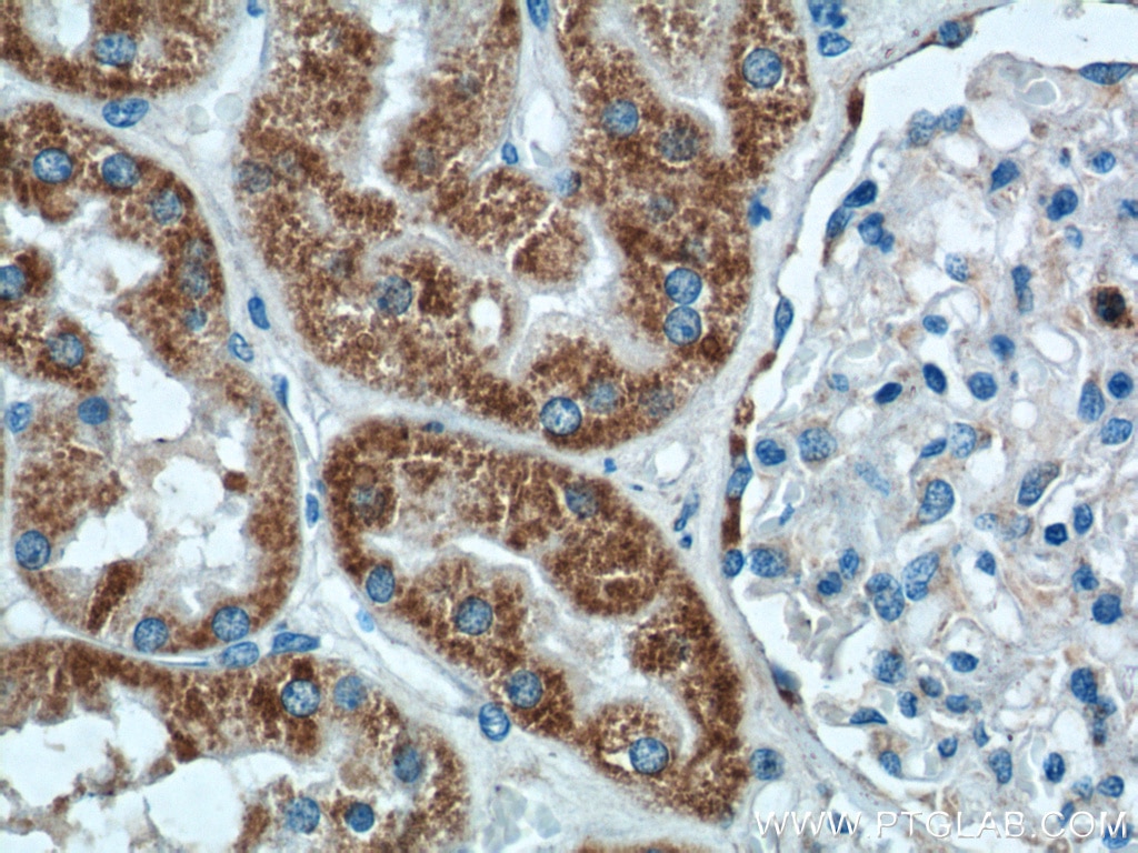
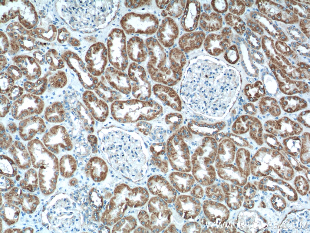
| Positive IHC detected in | human kidney tissue, mouse kidney tissue, mouse stomach tissue, rat kidney tissue Note: suggested antigen retrieval with TE buffer pH 9.0; (*) Alternatively, antigen retrieval may be performed with citrate buffer pH 6.0 |
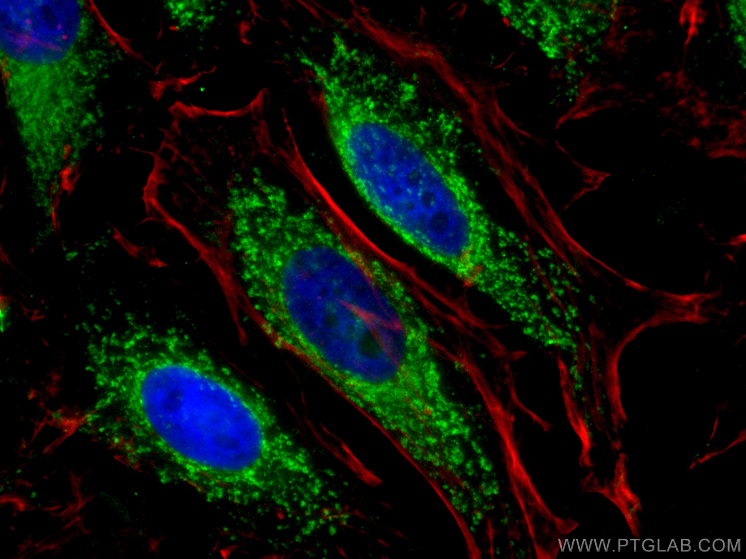
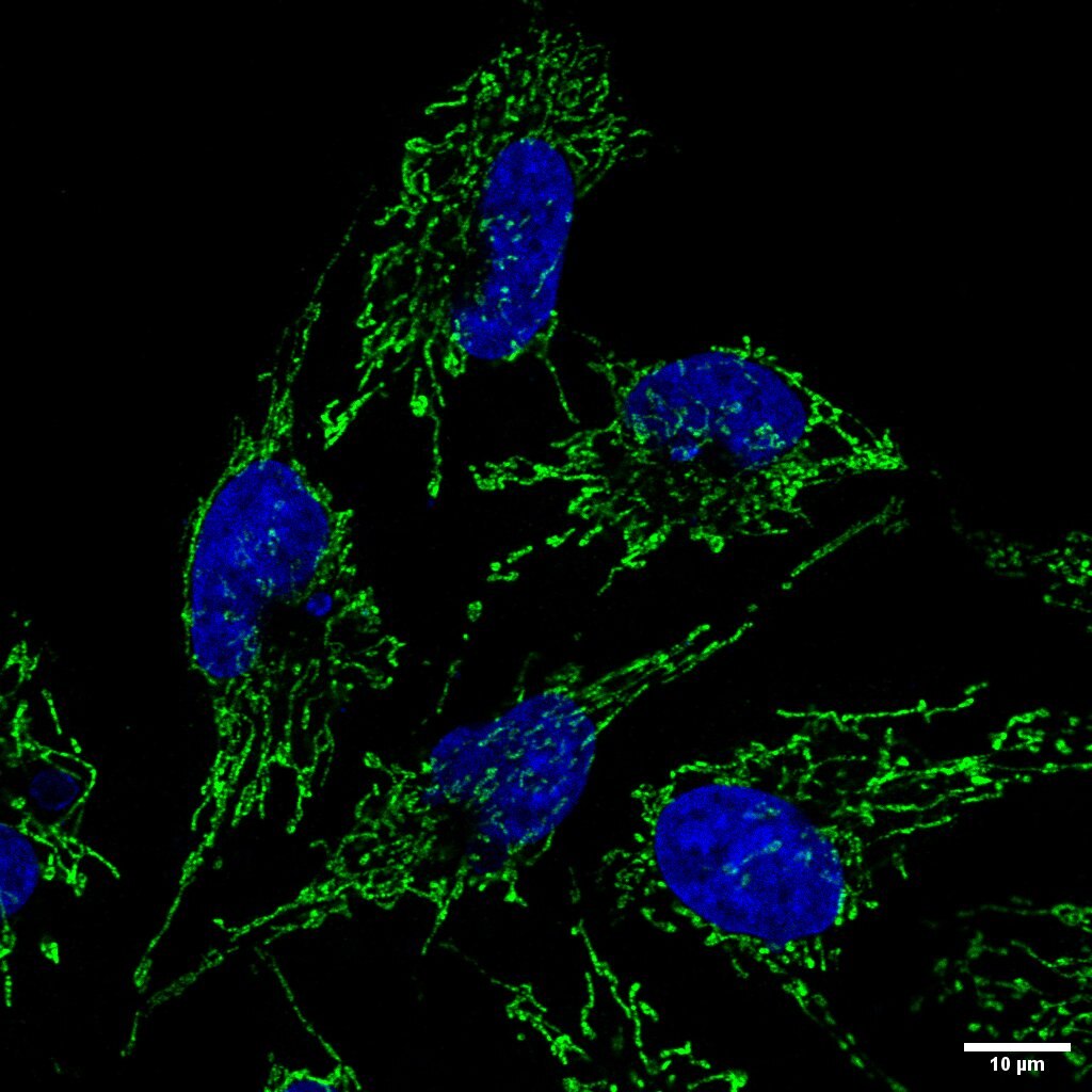
| Positive IF/ICC detected in | HeLa cells |
Recommended dilution
| Application | Dilution |
|---|---|
| Western Blot (WB) | WB : 1:5000-1:50000 |
| Immunoprecipitation (IP) | IP : 0.5-4.0 ug for 1.0-3.0 mg of total protein lysate |
| Immunohistochemistry (IHC) | IHC : 1:100-1:400 |
| Immunofluorescence (IF)/ICC | IF/ICC : 1:50-1:500 |
| It is recommended that this reagent should be titrated in each testing system to obtain optimal results. | |
| Sample-dependent, Check data in validation data gallery. | |
Product Information
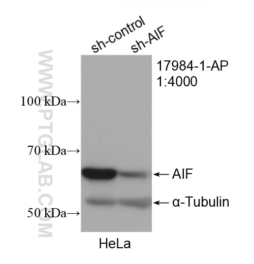
17984-1-AP targets AIF in WB, IHC, IF/ICC, IP, ELISA applications and shows reactivity with human, mouse, rat samples.
| Tested Reactivity | human, mouse, rat |
| Cited Reactivity | human, mouse, rat, pig, canine, sheep |
| Host / Isotype | Rabbit / IgG |
| Class | Polyclonal |
| Type | Antibody |
| Immunogen |
CatNo: Ag12400 Product name: Recombinant human AIF protein Source: e coli.-derived, PET28a Tag: 6*His Domain: 259-609 aa of BC111065 Sequence: TPRSLSAIDRAGAEVKSRTTLFRKIGDFRSLEKISREVKSITIIGGGFLGSELACALGRKARALGTEVIQLFPEKGNMGKILPEYLSNWTMEKVRREGVKVMPNAIVQSVGVSSGKLLIKLKDGRKVETDHIVAAVGLEPNVELAKTGGLEIDSDFGGFRVNAELQARSNIWVAGDAACFYDIKLGRRRVEHHDHAVVSGRLAGENMTGAAKPYWHQSMFWSDLGPDVGYEAIGLVDSSLPTVGVFAKATAQDNPKSATEQSGTGIRSESETESEASEITIPPSTPAVPQAPVQGEDYGKGVIFYLRDKVVVGIVLWNIFNRMPIARKIIKDGEQHEDLNEVAKLFNIHED Predict reactive species |
| Full Name | apoptosis-inducing factor, mitochondrion-associated, 1 |
| Calculated Molecular Weight | 609 aa, 66 kDa |
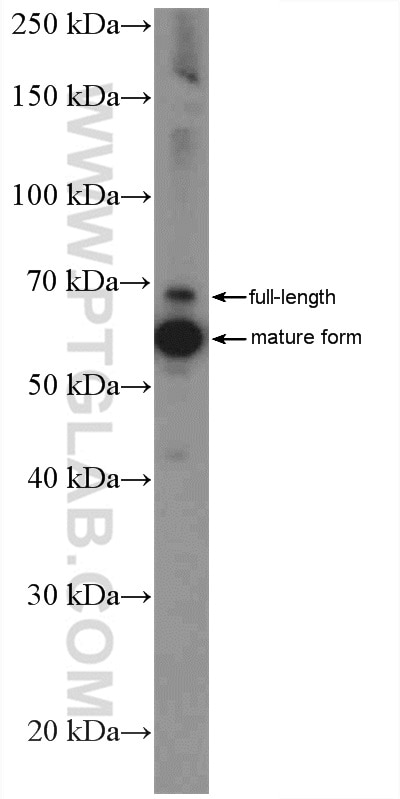
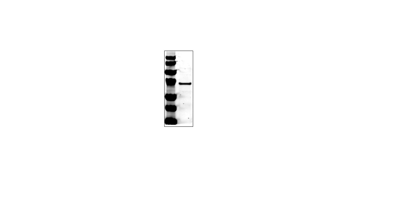
| Observed Molecular Weight | 67 kDa, 57 kDa |
| GenBank Accession Number | BC111065 |
| Gene Symbol | AIF |
| Gene ID (NCBI) | 9131 |
| RRID | AB_2224539 |
| Conjugate | Unconjugated |
| Form | Liquid |
| Purification Method | Antigen affinity purification |
| UNIPROT ID | O95831 |
| Storage Buffer | PBS with 0.02% sodium azide and 50% glycerol, pH 7.3. |
| Storage Conditions | Store at -20°C. Stable for one year after shipment. Aliquoting is unnecessary for -20oC storage. 20ul sizes contain 0.1% BSA. |
Background Information
Apoptosis-inducing factor (AIF) is one of the mitochondrial proteins to be released into the cytosol during apoptosis, and it is discovered as the first protein that regulates caspase-independent apoptosis(PMID:20494118). AIF is encoded as a 67 kDa protein that contains a mitochondrial localization signal (MLS) in the N-terminus.It is cleaved from the 62 kDa to the 57 kDa form following ischemic injury and translocated from the mitochondria to the nucleus in a calpain-dependent manner(PMID: 25101006).
Protocols
| Product Specific Protocols | |
|---|---|
| IF protocol for AIF antibody 17984-1-AP | Download protocol |
| IHC protocol for AIF antibody 17984-1-AP | Download protocol |
| IP protocol for AIF antibody 17984-1-AP | Download protocol |
| WB protocol for AIF antibody 17984-1-AP | Download protocol |
| Standard Protocols | |
|---|---|
| Click here to view our Standard Protocols |
Publications
What published studies show
The anti-AIFM1 antibody (Cat# 17984-1-AP) has accumulated 112 citations across journals including Autophagy, Redox Biology, Advanced Science, EMBO Journal, Cell Reports, Free Radical Biology and Medicine, Journal of Investigative Dermatology, Frontiers in Immunology, and PLoS Pathogens. Research fields span apoptosis, necroptosis, parthanatos, ferroptosis, mitochondrial dysfunction, oxidative stress, cerebral ischemia, cancer biology, neurodegeneration, inflammation, and pharmacology.
| Species | Application | Title |
|---|---|---|
Autophagy METTL3-dependent m6A modification of SNAP29 induces "autophagy-mitochondrial crisis" in the ischemic microenvironment after soft tissue transplantation | ||
Redox Biol Excessive SOX8 reprograms energy and iron metabolism to prime hepatocellular carcinoma for ferroptosis | ||
Redox Biol Isoliquiritigenin alleviates cerebral ischemia-reperfusion injury by reducing oxidative stress and ameliorating mitochondrial dysfunction via activating the Nrf2 pathway | ||
J Nanobiotechnology Multifunctional Fe-based coordination polymer nano-bomb modified with β-lapachone and CaO2 for targeted tumor dual chemodynamic therapy with enhanced ferroptosis and H2O2 self-supply | ||
Adv Sci (Weinh) Elevated TRIM25 Impairs Poly (ADP-ribose) Metabolism via PARG Degradation and Mediates Compression-Induced Intervertebral Disc Degeneration. |
Reviews
What customers say
Users consistently rate this antibody 4–5 stars across Western blot and immunofluorescence applications. Three reviewers used it for WB, describing it as reliable, excellent, and performing as expected. One reviewer provided a detailed IF protocol on HeLa cells at 1:100 dilution, reporting good results with standard permeabilization and blocking steps. Overall, customers express high satisfaction with the antibody's performance and consistency.
The reviews below have been submitted by verified Proteintech customers who received an incentive for providing their feedback.
Ziwei (Verified Customer) (01-04-2026) | works as it should be.
|
Ziwei (Verified Customer) (12-14-2025) | The antibody performs reliably in Western blot analysis.
|
Tom (Verified Customer) (11-13-2020) | HeLa cells permeablisied (0.1% Triton X-100 in PBS) then blocked (1% BSA in PBS). Cells incubated with primary antibody (1:100) for 1 hour, 3xPBS prior to 1 hour incubation with goat anti-rabbit 488 Alexa Fluor secondary antibody.
 |
Praveen (Verified Customer) (05-17-2019) | Excellent Antibody
 |











