

Tested Applications
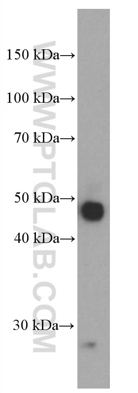
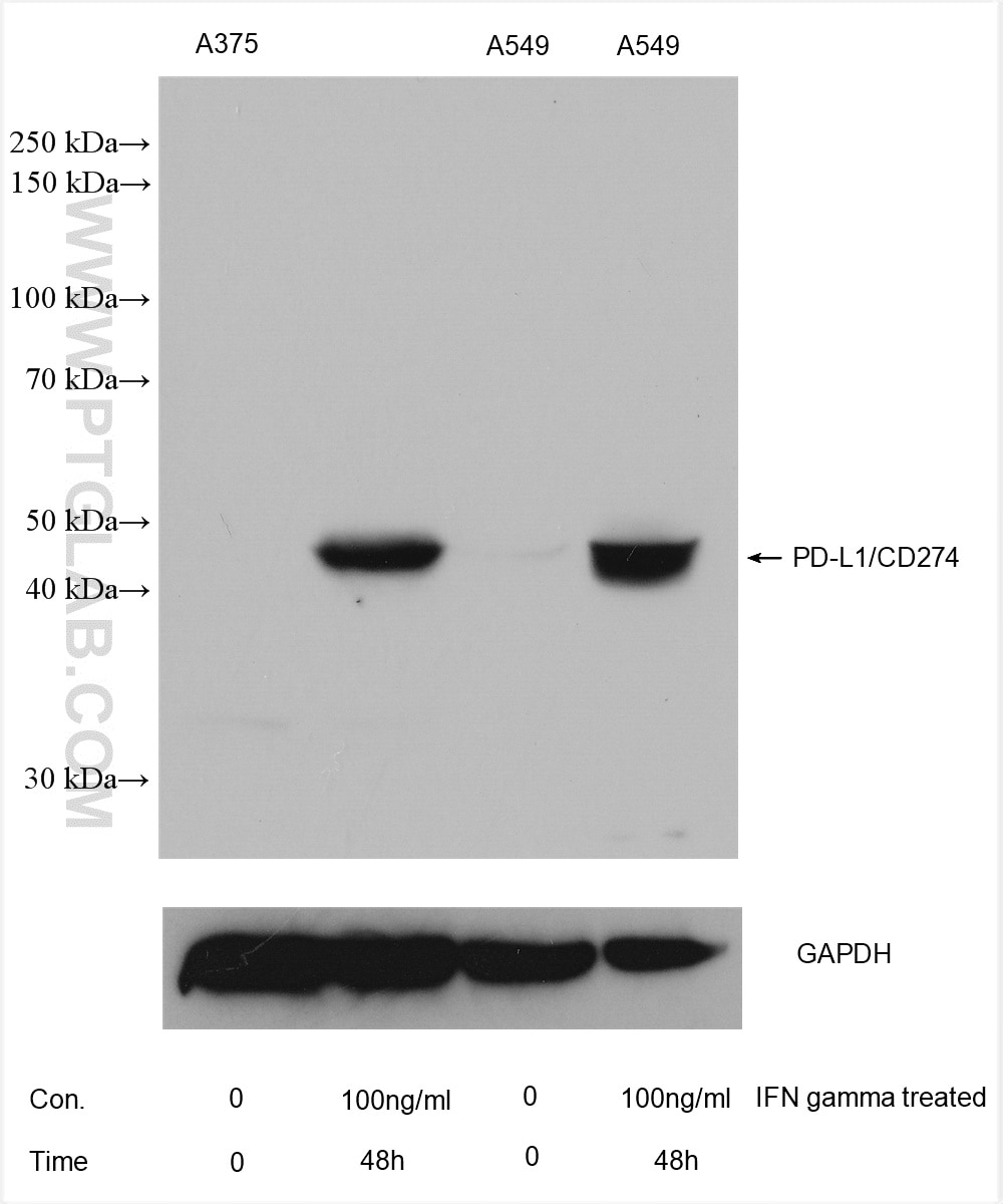
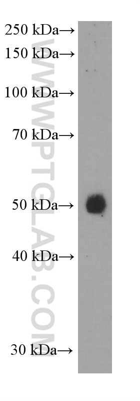
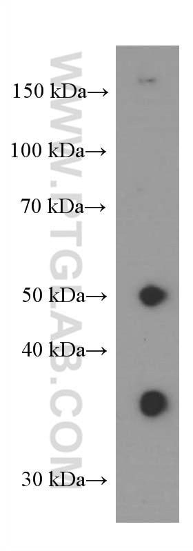
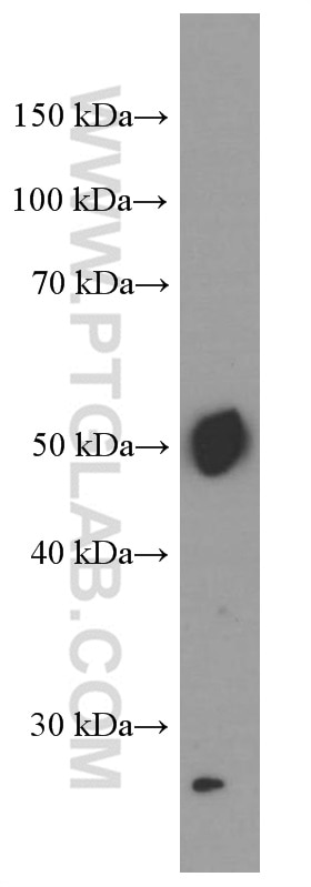
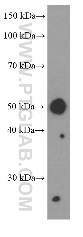
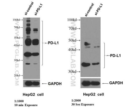
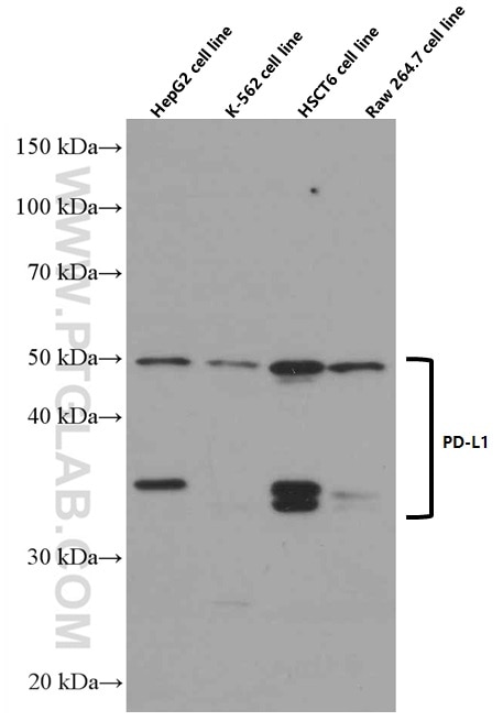
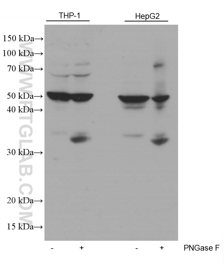
| Positive WB detected in | A375 cells, human placenta tissue, pig lung tissue, human skeletal muscle tissue, HepG2 cells, THP-1 cells, RAW 264.7 cells, A549 cells, K-562 cells, HSC-T6 cells |
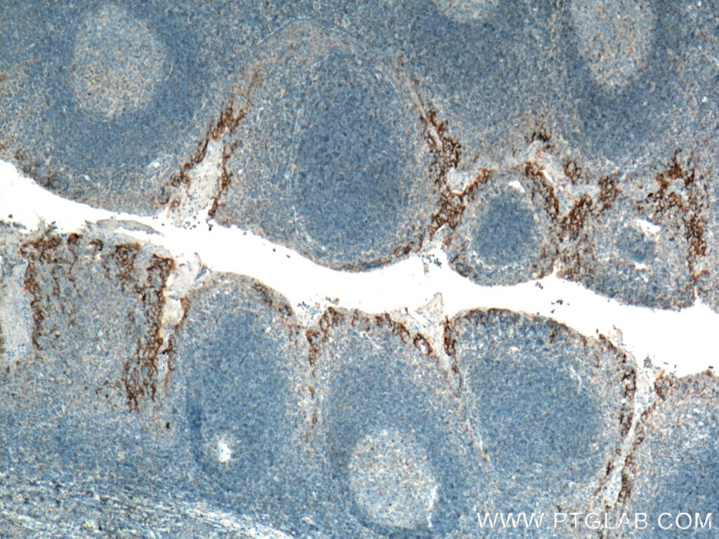
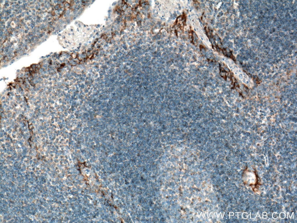
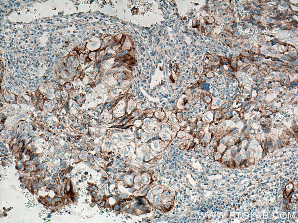
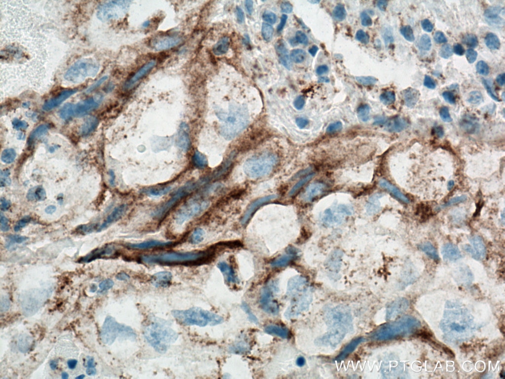
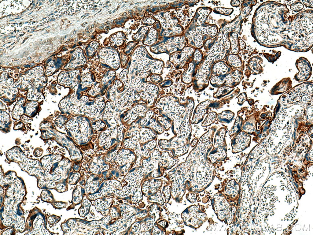
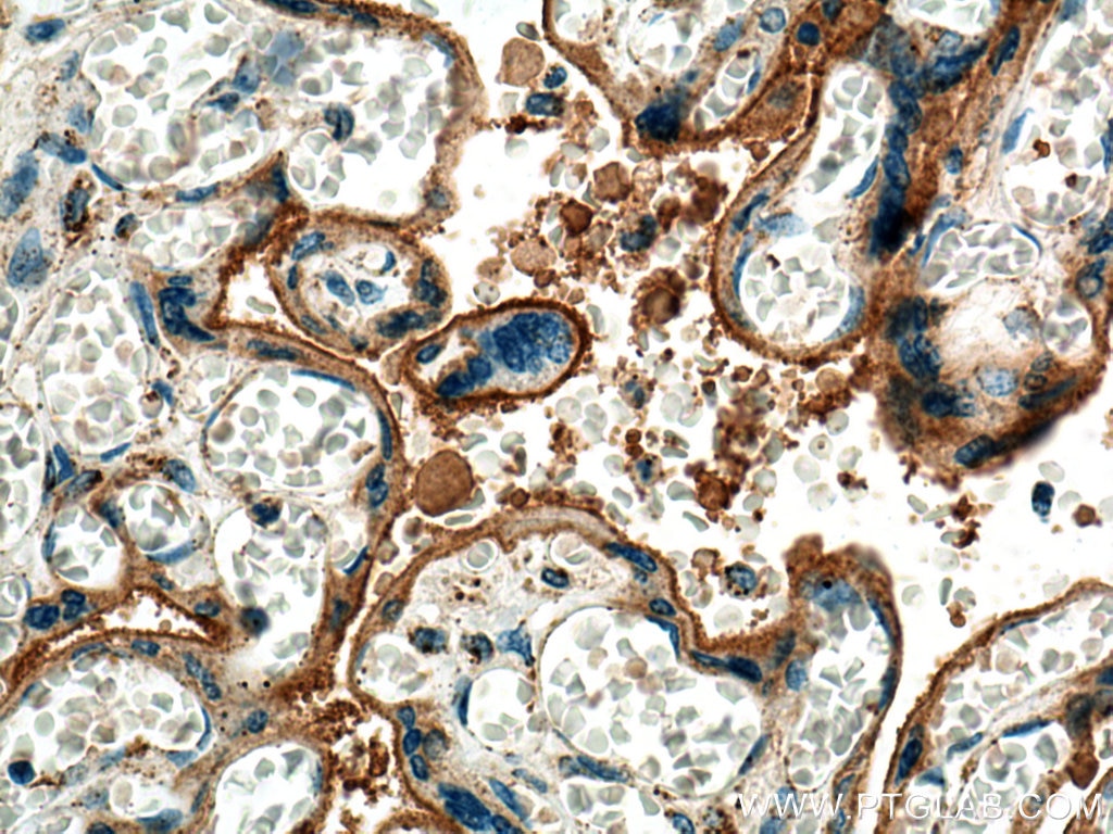
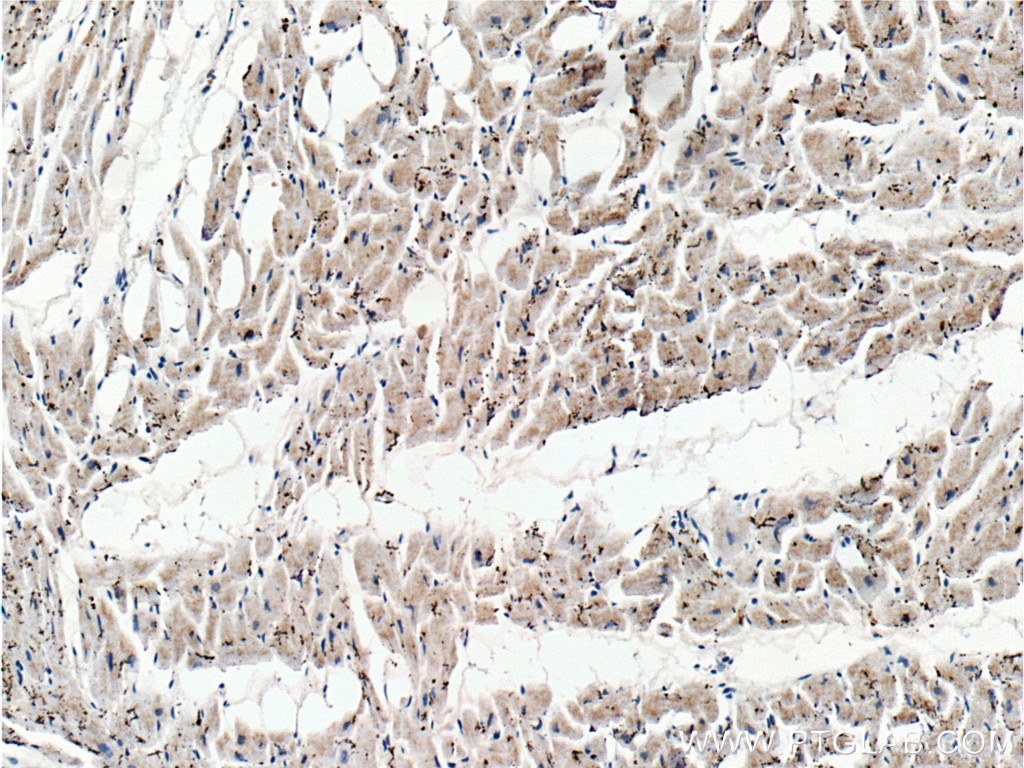
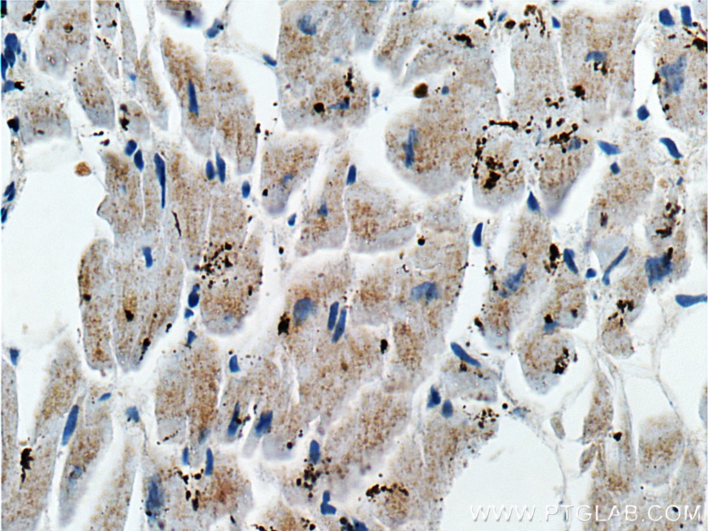
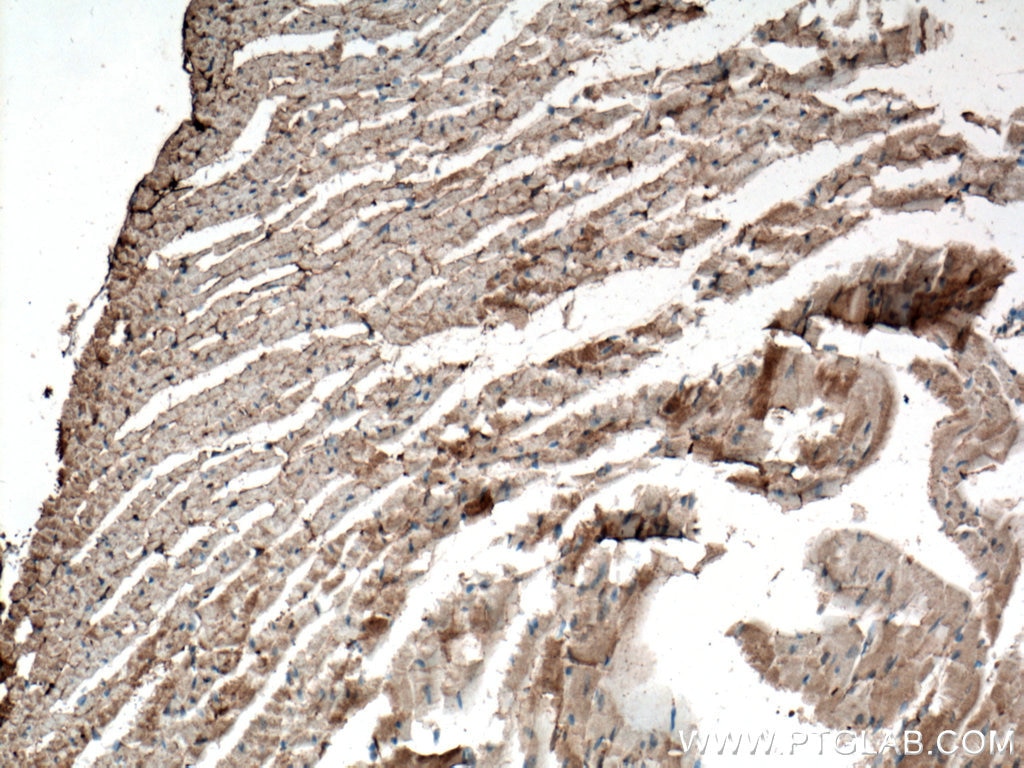
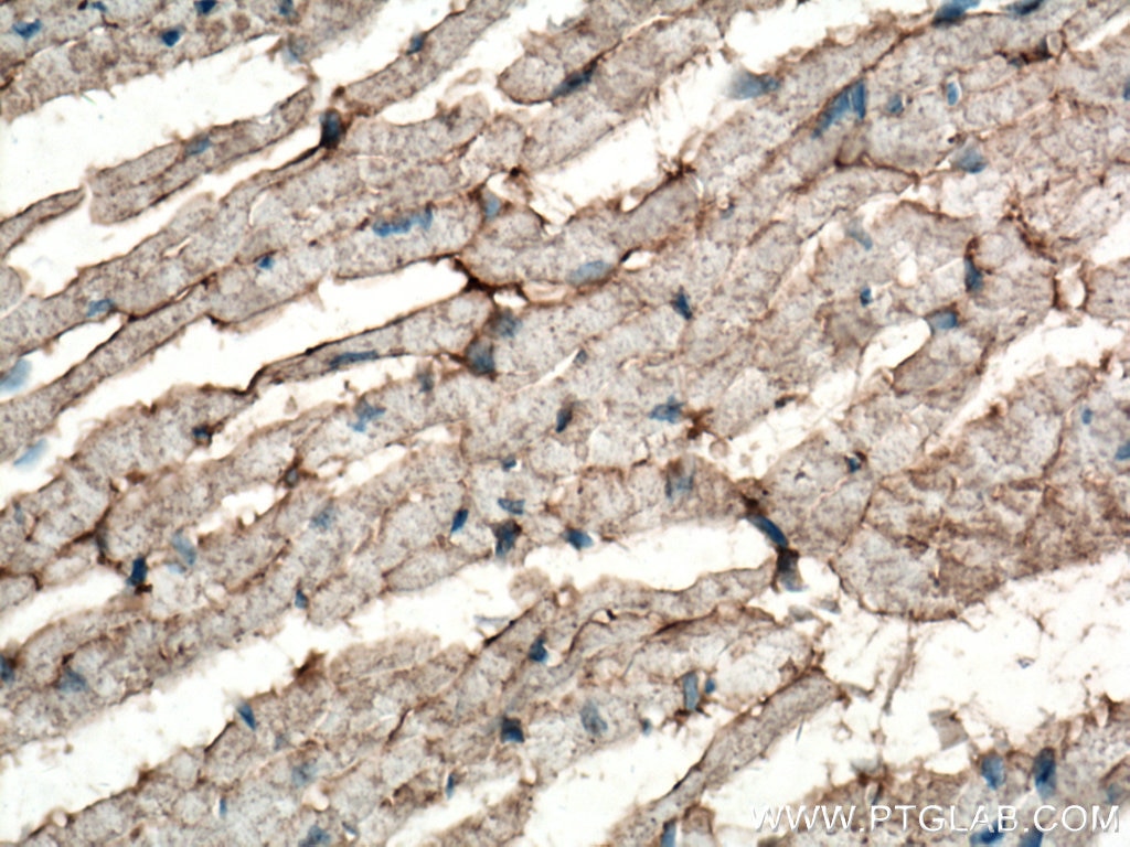
| Positive IHC detected in | human tonsillitis tissue, human heart tissue, human lung cancer tissue, human placenta tissue, mouse heart tissue Note: suggested antigen retrieval with TE buffer pH 9.0; (*) Alternatively, antigen retrieval may be performed with citrate buffer pH 6.0 |
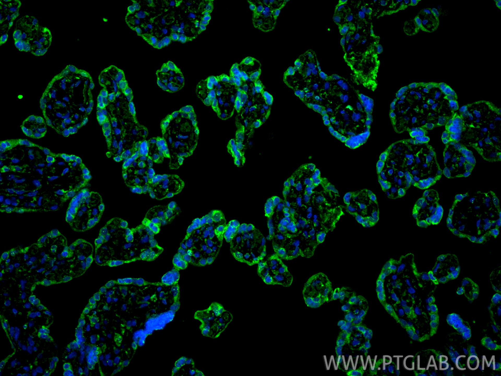
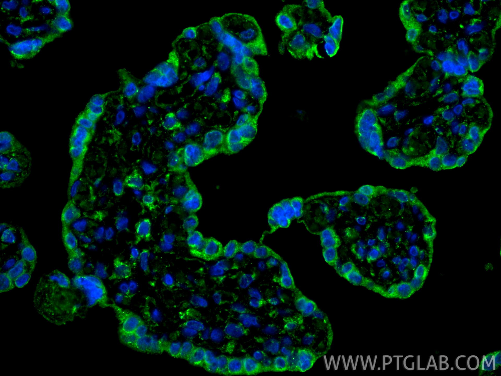
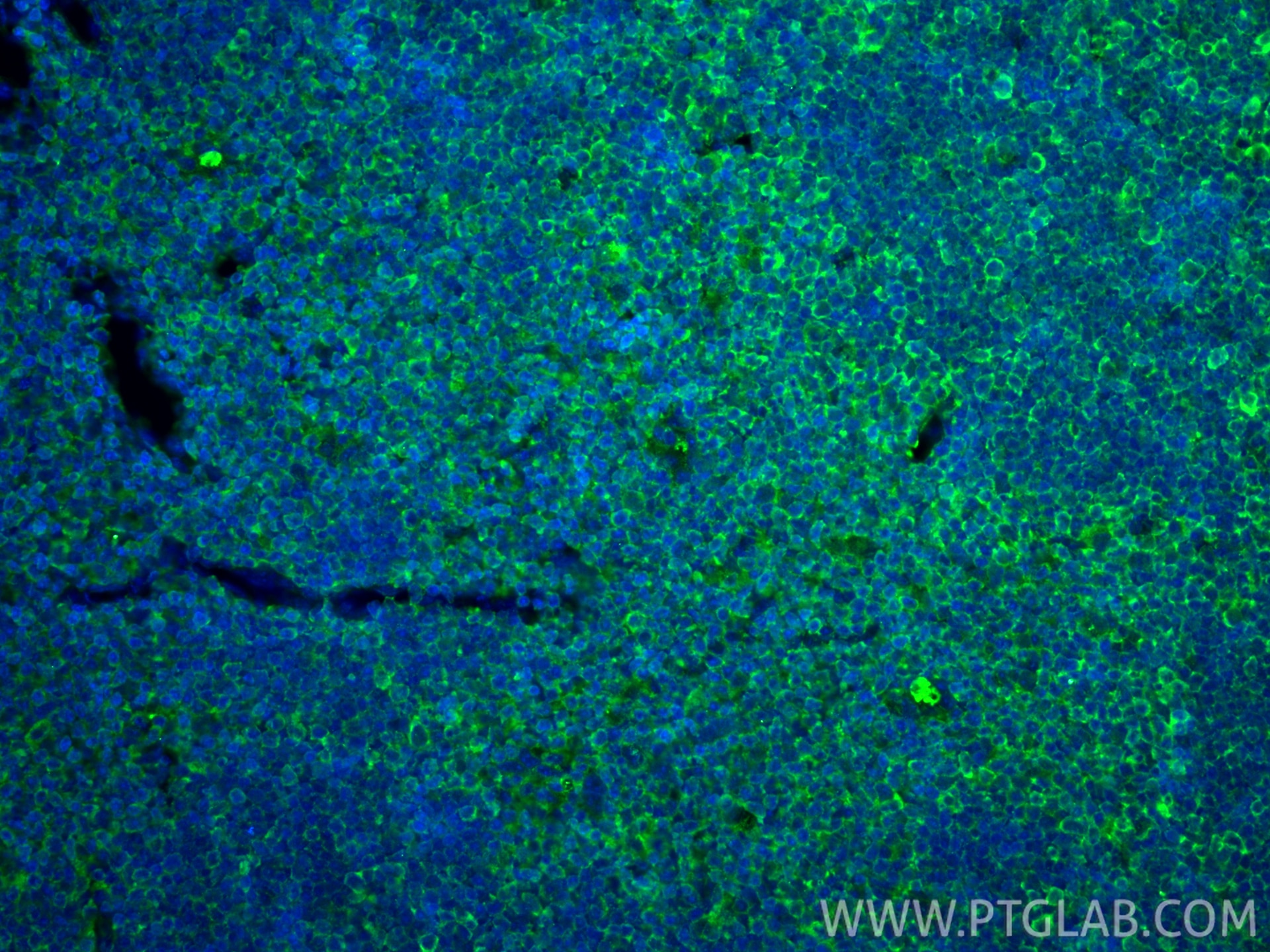
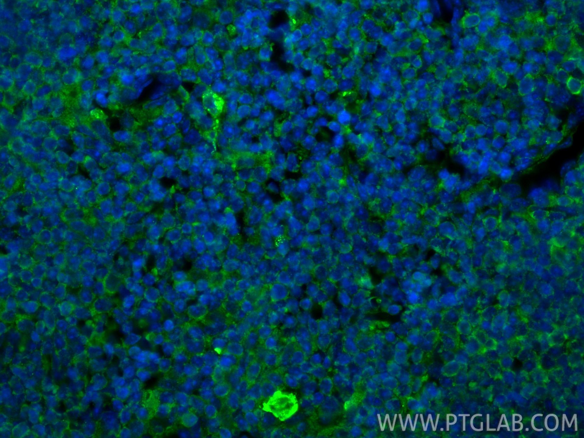
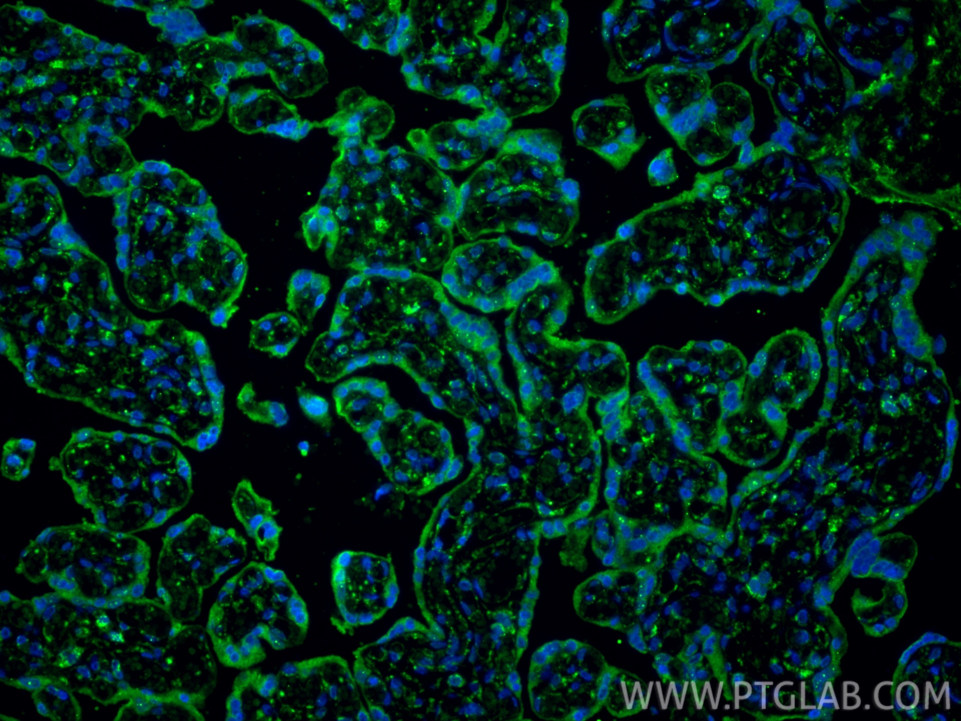
| Positive IF-P detected in | human placenta tissue, mouse thymus tissue |
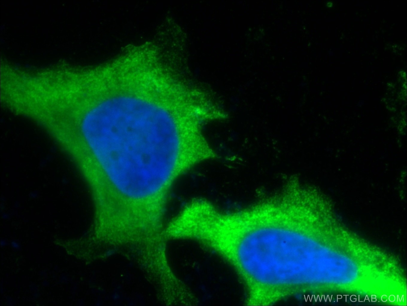
| Positive IF/ICC detected in | HeLa cells |
Recommended dilution
| Application | Dilution |
|---|---|
| Western Blot (WB) | WB : 1:2000-1:10000 |
| Immunohistochemistry (IHC) | IHC : 1:5000-1:20000 |
| Immunofluorescence (IF)-P | IF-P : 1:400-1:1600 |
| Immunofluorescence (IF)/ICC | IF/ICC : 1:50-1:500 |
| It is recommended that this reagent should be titrated in each testing system to obtain optimal results. | |
| Sample-dependent, Check data in validation data gallery. | |
Product Information
66248-1-Ig targets PD-L1/CD274 in WB, IHC, IF/ICC, IF-P, IP, CoIP, ChIP, ELISA, Blocking assay, Cell treatment applications and shows reactivity with human, mouse, rat, pig samples.
| Tested Reactivity | human, mouse, rat, pig |
| Cited Reactivity | human, mouse, rat, pig |
| Host / Isotype | Mouse / IgG1 |
| Class | Monoclonal |
| Type | Antibody |
| Immunogen |
CatNo: Ag12443 Product name: Recombinant human PD-L1/CD274 protein Source: e coli.-derived, PET28a Tag: 6*His Domain: 25-241 aa of BC074984 Sequence: KDLYVVEYGSNMTIECKFPVEKQLDLAALIVYWEMEDKNIIQFVHGEEDLKVQHSSYRQRARLLKDQLSLGNAALQITDVKLQDAGVYRCMISYGGADYKRITVKVNAPYNKINQRILVVDPVTSEHELTCQAEGYPKAEVIWTSSDHQVLSGKTTTTNSKREEKLFNVTSTLRINTTTNEIFYCTFRRLDPEENHTAELVIPELPLAHPPNERTHL Predict reactive species |
| Full Name | CD274 molecule |
| Calculated Molecular Weight | 290 aa, 33 kDa |
| Observed Molecular Weight | 45-50 kDa, 33 kDa |
| GenBank Accession Number | BC074984 |
| Gene Symbol | PD-L1 |
| Gene ID (NCBI) | 29126 |
| RRID | AB_2756526 |
| Conjugate | Unconjugated |
| Form | Liquid |
| Purification Method | Protein G purification |
| UNIPROT ID | Q9NZQ7 |
| Storage Buffer | PBS with 0.02% sodium azide and 50% glycerol, pH 7.3. |
| Storage Conditions | Store at -20°C. Stable for one year after shipment. Aliquoting is unnecessary for -20oC storage. 20ul sizes contain 0.1% BSA. |
Background Information
PD-L1, also known as CD274 or B7H1, stands for programmed cell death ligand 1. It is a type I transmembrane protein that is thought to repress immune responses by binding to its receptor (PD1), thus inhibiting T-cell activation, proliferation, and cytokine production. It contains V-like and C-like immunoglobulin domains. PD-L1 expression is regulated by various cytokines, such as TNF-α or LPS (ISSN: 1848-7718). Increased expression of this protein in certain types of cancers, e.g., renal cell carcinoma or colon cancer, correlates with poor prognosis.
What is the molecular weight of PD-L1?
Depending on the isoform, the calculated molecular weight of the protein varies between 20 and 33 kDa (176-290 aa).
What are the isoforms of PD-L1?
According to NCBI, three different isoforms have been identified. There are significant differences in the untranslated and protein coding regions.
What is the subcellular localization and tissue specificity of PD-L1?
It is predicted to localize in the plasma membrane of various cell types, with a particularly high expression in placental trophoblast and subsets of immune cells. High levels of PD-L1 protein have also been detected in lung and colon tissues.
What is the function of PD-L1 in immune responses?
PD-L1 is critical for the induction and maintenance of immune self-tolerance during infection or inflammation in normal tissues. The interaction of PD-L1 and its receptors is responsible for preventing auto-immune phenotypes and balancing the overall immune response in situations such as pregnancy or tissue allografts. The interaction between PD-L1 and PD-1 or B7.1 starts an inhibitory signaling cascade, which results in the decreased proliferation of antigen-specific T-cells and increased survival of regulatory T-cells (PMID: 15240681).
How can PD-L1's implication in cancer be used as a drug target?
In certain tumors, high expression of PD-L1 serves as a stop-sign to inhibit the recognition of cancer cells by T-cells (PMID: 23087408). The interaction between PD-L1 and its receptors (PD1 and B7.1) is a mechanism for the tumor to evade the host immune response (PMID: 29357948). Several mAbs have been developed to target that interaction and thus prevent the inactivation of cytotoxic T-cells by the tumor (PMIDs: 23890059, 18173375).
Protocols
| Product Specific Protocols | |
|---|---|
| IF protocol for PD-L1/CD274 antibody 66248-1-Ig | Download protocol |
| IHC protocol for PD-L1/CD274 antibody 66248-1-Ig | Download protocol |
| WB protocol for PD-L1/CD274 antibody 66248-1-Ig | Download protocol |
| Standard Protocols | |
|---|---|
| Click here to view our Standard Protocols |
Publications
What published studies show
Anti-PD-L1 antibody (Cat# 66248-1-Ig) has accumulated 639 total citations published in high-impact journals including Nature Communications, Cell Metabolism, Molecular Cancer, Cancer Cell, Advanced Science, Journal of Experimental & Clinical Cancer Research, Theranostics, and ACS Nano. The cited research primarily covers cancer immunotherapy, tumor immune evasion mechanisms, PD-L1 post-translational modification and regulation, nanomedicine-based immunotherapy strategies, and immune checkpoint biology, demonstrating extensive use in both fundamental and translational oncology research.
| Species | Application | Title |
|---|---|---|
Signal Transduct Target Ther Extracellular vesicle-derived miRNA-182-5p educates macrophages towards an immunosuppressive phenotype in pancreatic cancer. | ||
Cancer Cell ADORA1 Inhibition Promotes Tumor Immune Evasion by Regulating the ATF3-PD-L1 Axis. | ||
Mol Cancer Crosstalk between 5-methylcytosine and N6-methyladenosine machinery defines disease progression, therapeutic response and pharmacogenomic landscape in hepatocellular carcinoma | ||
Mol Cancer The miR-23a/27a/24 - 2 cluster drives immune evasion and resistance to PD-1/PD-L1 blockade in non-small cell lung cancer | ||
Mol Cancer Intercellular TIMP-1-CD63 signaling directs the evolution of immune escape and metastasis in KRAS-mutated pancreatic cancer cells |
Reviews
What customers say
Users report this anti-PD-L1 antibody performs well across Western Blot and Immunofluorescence applications. For WB, it detects overexpressed PD-L1 effectively at 1:1000–1:4000, though some users noted additional non-specific bands when detecting endogenous levels. For IF, results were positive on PC3 cells (1:200), FFPE prostate tissue (1:50), and HCC1937 breast cancer cells (1:300). Overall, reviewers rate the product favorably, with an average of 4 out of 5 stars, praising its specificity for overexpression studies while noting occasional background in endogenous detection.
The reviews below have been submitted by verified Proteintech customers who received an incentive for providing their feedback.
JIN-FENG (Verified Customer) (10-08-2025) | It works well in the PD-L1 overexpression cells. In terms of endogenous PD-L1 detection. There are a couple of non-specific bands around the target site.
 |
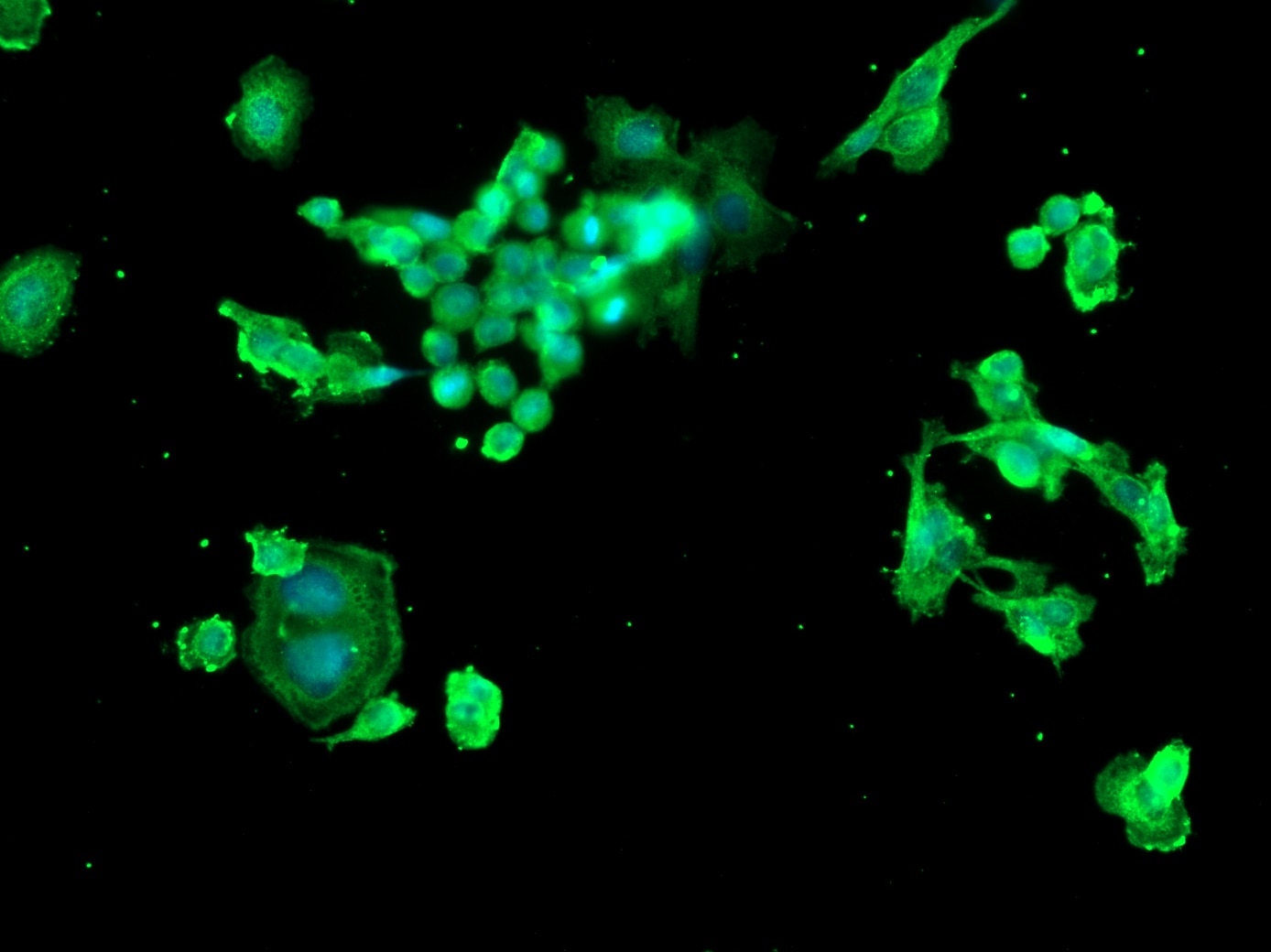
Margarita (Verified Customer) (11-20-2024) | Secondary antibody: AF 488
 |
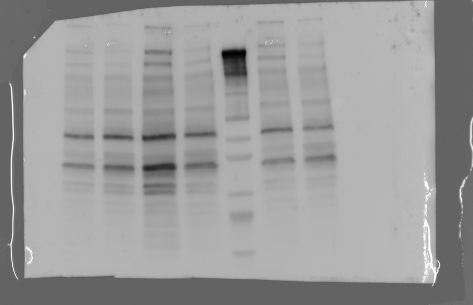
hala (Verified Customer) (01-27-2023) | there is 2 bands one at the specific size and the other one is higher
 |
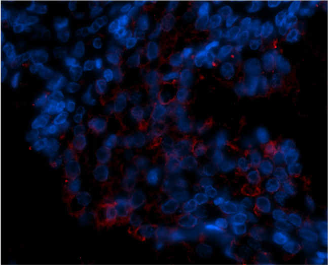
Emma (Verified Customer) (11-29-2021) | Used for IF on FFPE prostate tissue at 1:50 with Tris-EDTA antigen retrieval (pH 9.0).
 |
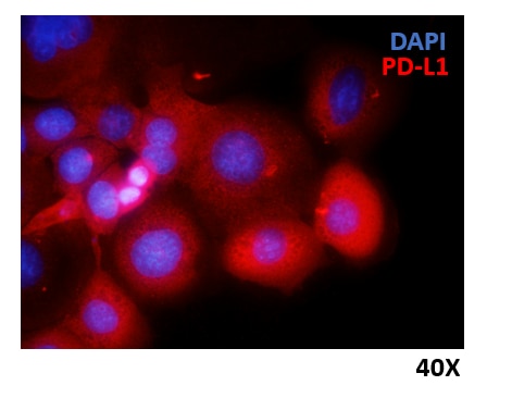
Marina (Verified Customer) (06-14-2021) | HCC1937 breast cancer cells were fixed with 4% PFA, permeabilized and stained overnight at 4ºC with anti-PD-L1 Ig diluted 1:300. Nuclei were stained with DAPI.
 |


























