

Tested Applications
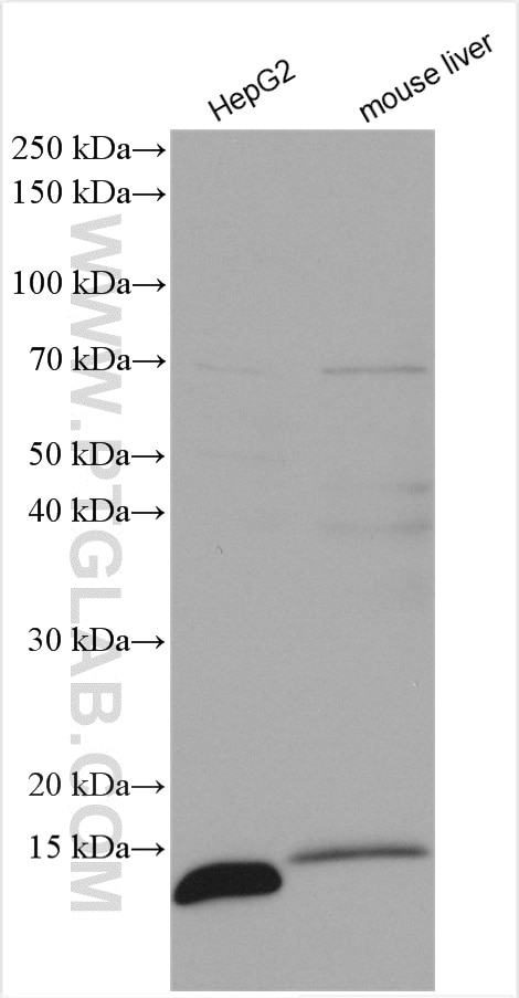
| Positive WB detected in | HepG2 cells, mouse liver tissue |
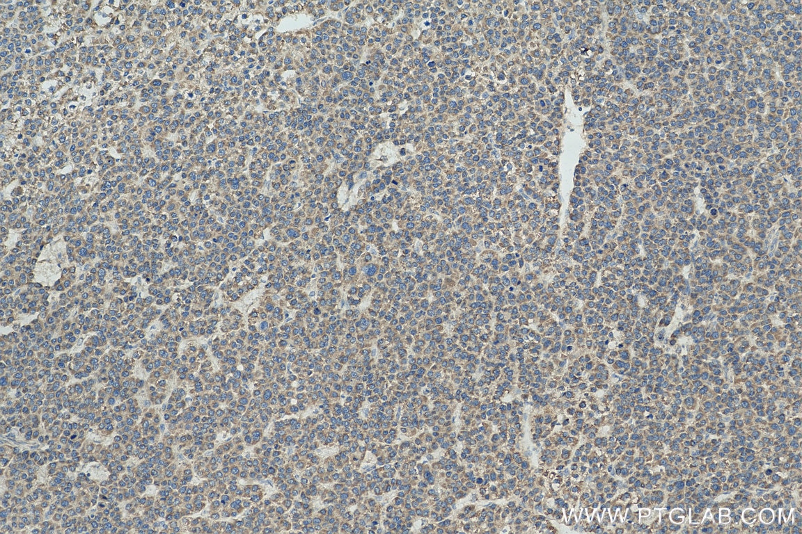
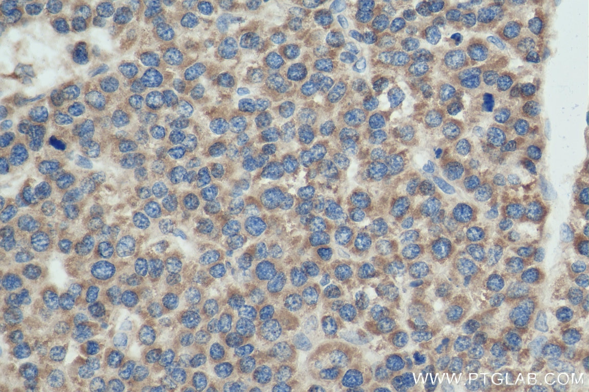
| Positive IHC detected in | human liver cancer tissue Note: suggested antigen retrieval with TE buffer pH 9.0; (*) Alternatively, antigen retrieval may be performed with citrate buffer pH 6.0 |
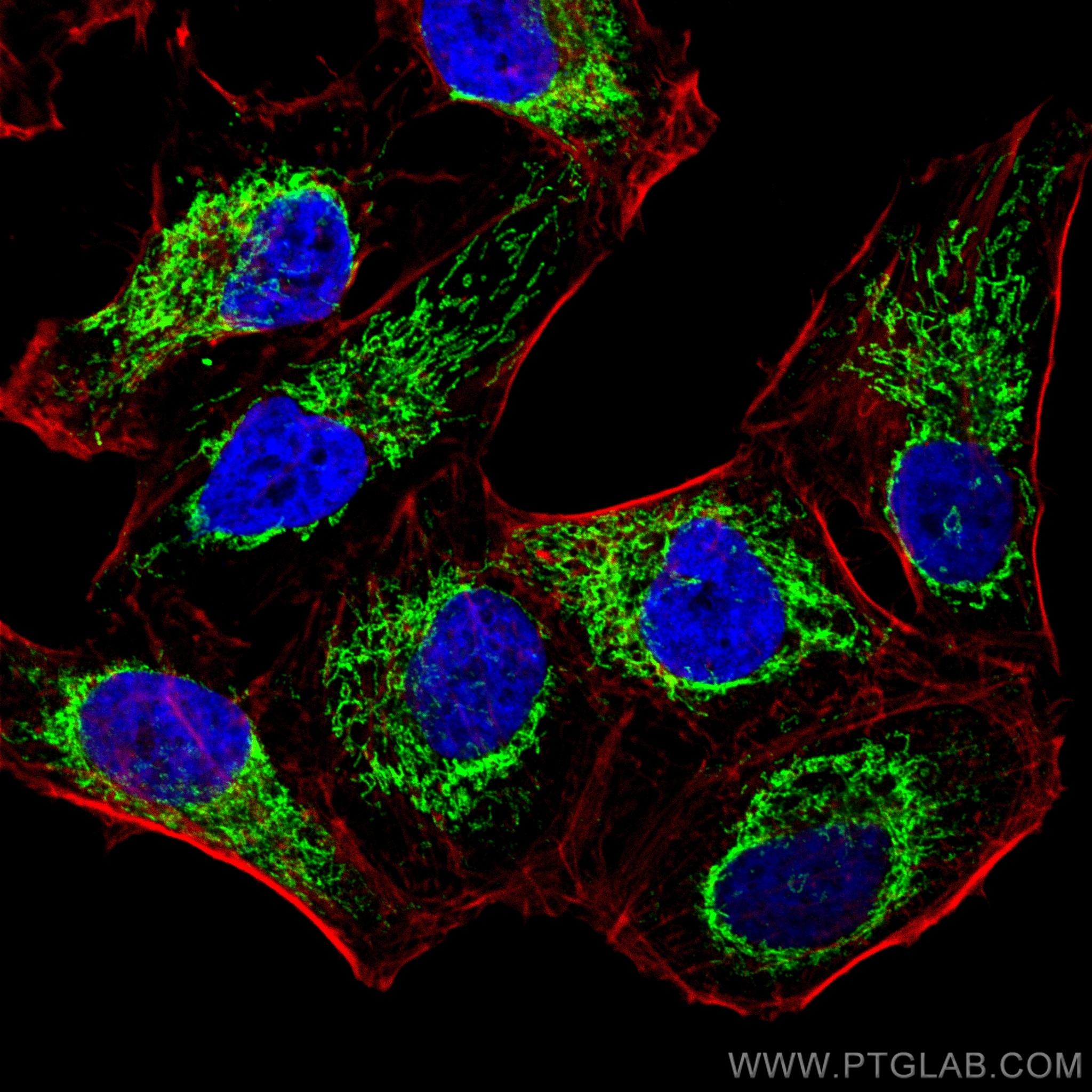
| Positive IF/ICC detected in | HeLa cells |
Recommended dilution
| Application | Dilution |
|---|---|
| Western Blot (WB) | WB : 1:500-1:2000 |
| Immunohistochemistry (IHC) | IHC : 1:50-1:500 |
| Immunofluorescence (IF)/ICC | IF/ICC : 1:50-1:500 |
| It is recommended that this reagent should be titrated in each testing system to obtain optimal results. | |
| Sample-dependent, Check data in validation data gallery. | |
Product Information
16483-1-AP targets ATP5I in WB, IHC, IF/ICC, ELISA applications and shows reactivity with human, mouse, rat samples.
| Tested Reactivity | human, mouse, rat |
| Cited Reactivity | human, mouse, chicken |
| Host / Isotype | Rabbit / IgG |
| Class | Polyclonal |
| Type | Antibody |
| Immunogen |
CatNo: Ag9605 Product name: Recombinant human ATP5I protein Source: e coli.-derived, PGEX-4T Tag: GST Domain: 1-69 aa of BC003679 Sequence: MVPPVQVSPLIKLGRYSALFLGVAYGATRYNYLKPRAEEERRIAAEEKKKQDELKRIARELAEDDSILK Predict reactive species |
| Full Name | ATP synthase, H+ transporting, mitochondrial F0 complex, subunit E |
| Calculated Molecular Weight | 69 aa, 8 kDa |
| Observed Molecular Weight | 8 kDa |
| GenBank Accession Number | BC003679 |
| Gene Symbol | ATP5I |
| Gene ID (NCBI) | 521 |
| RRID | AB_2062052 |
| Conjugate | Unconjugated |
| Form | Liquid |
| Purification Method | Antigen affinity purification |
| UNIPROT ID | P56385 |
| Storage Buffer | PBS with 0.02% sodium azide and 50% glycerol, pH 7.3. |
| Storage Conditions | Store at -20°C. Stable for one year after shipment. Aliquoting is unnecessary for -20oC storage. 20ul sizes contain 0.1% BSA. |
Background Information
ATP5I(ATP synthase subunit e) is also named as ATP5K and belongs to the ATPase e subunit family. The ATP5I gene encodes the e subunit of the mitochondrial ATP synthase Fo complex. Mitochondrial membrane ATP synthase(F1F0 ATP synthase or Complex V) produces ATP from ADP in the presence of a proton gradient across the membrane which is generated by electron transport complexes of the respiratory chain. Antisense ATP5I in a human HCC cell line inhibited cell growth suggesting that ATP5I acts through the MAP kinase pathway(PMID:11939412).
Protocols
| Product Specific Protocols | |
|---|---|
| IF protocol for ATP5I antibody 16483-1-AP | Download protocol |
| IHC protocol for ATP5I antibody 16483-1-AP | Download protocol |
| WB protocol for ATP5I antibody 16483-1-AP | Download protocol |
| Standard Protocols | |
|---|---|
| Click here to view our Standard Protocols |
Publications
What published studies show
The ATP5I Polyclonal antibody (Cat# 16483-1-AP) has 14 citations. These appear in journals including Nature, Cell Metabolism, Journal of Experimental Medicine, EBioMedicine, Environmental International, Frontiers in Aging Neuroscience, Journal of Inflammation Research, Genetics Selection Evolution, Biology (Basel), Journal of Cell Science, Life Science Alliance, Journal of Proteomics, and eLife. The associated research fields span mitochondrial biology and ATP synthase function, oxidative phosphorylation, neurocognitive disorders, neuroinflammation, cancer metabolism (metformin), hematopoietic stem cells, and brain ischemia.
| Species | Application | Title |
|---|---|---|
Cell Metab Single-nucleus transcriptomics identifies separate classes of UCP1 and futile cycle adipocytes | ||
EBioMedicine Mitochonic Acid 5 (MA-5) Facilitates ATP Synthase Oligomerization and Cell Survival in Various Mitochondrial Diseases. | ||
Environ Int Bisphenol S impairs mitochondrial function by targeting Myo19/oxidative phosphorylation pathway contributing to axonal and dendritic injury |
Reviews
What customers say
The single review for this product reports a 4-star rating. The user tested it at a 1:500 dilution on mouse embryonic tissue for Western Blot and stated it worked well. Overall, the feedback is positive, confirming reliable performance in WB applications on embryonic tissue.
The reviews below have been submitted by verified Proteintech customers who received an incentive for providing their feedback.
Yan (Verified Customer) (01-28-2020) | It worked well when I performed western blot in embryonic tissue.
|





