The Proteintech guarantee covers Proteintech antibodies in any species and any application, including those not listed on the datasheet. If the antibody doesn’t perform, you can receive a hassle-free refund or credit note.
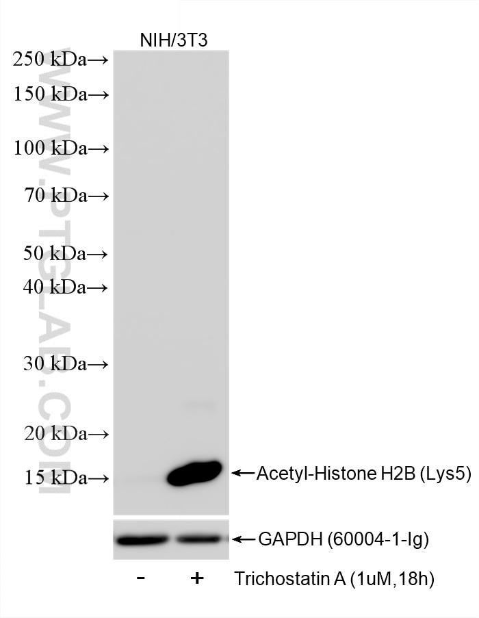
Trichostatin A treated NIH/3T3 cells were subjected to SDS PAGE followed by western blot with 86731-1-RR (Acetyl-Histone H2B (Lys12) antibody) at dilution of 1:10000 incubated at room temperature for 1.5 hours. The membrane was stripped and reblotted with GAPDH Monoclonal antibody (60004-1-Ig) as loading control.
This validation data has been generated in-house and reviewed by Proteintech scientists. Find out more on our antibody validation policy
Trichostatin A treated NIH/3T3 cells were subjected to SDS PAGE followed by western blot with 86731-1-RR (Acetyl-Histone H2B (Lys12) antibody) at dilution of 1:10000 incubated at room temperature for 1.5 hours. The membrane was stripped and reblotted with GAPDH Monoclonal antibody (60004-1-Ig) as loading control.
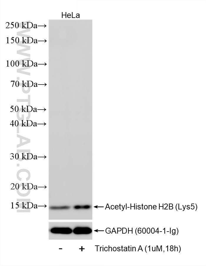
WB analysis of HeLa using 86731-1-RR
Trichostatin A treated HeLa cells were subjected to SDS PAGE followed by western blot with 86731-1-RR (Acetyl-Histone H2B (Lys12) antibody) at dilution of 1:10000 incubated at room temperature for 1.5 hours. The membrane was stripped and reblotted with GAPDH Monoclonal antibody (60004-1-Ig) as loading control.
This validation data has been generated in-house and reviewed by Proteintech scientists. Find out more on our antibody validation policy
Trichostatin A treated HeLa cells were subjected to SDS PAGE followed by western blot with 86731-1-RR (Acetyl-Histone H2B (Lys12) antibody) at dilution of 1:10000 incubated at room temperature for 1.5 hours. The membrane was stripped and reblotted with GAPDH Monoclonal antibody (60004-1-Ig) as loading control.
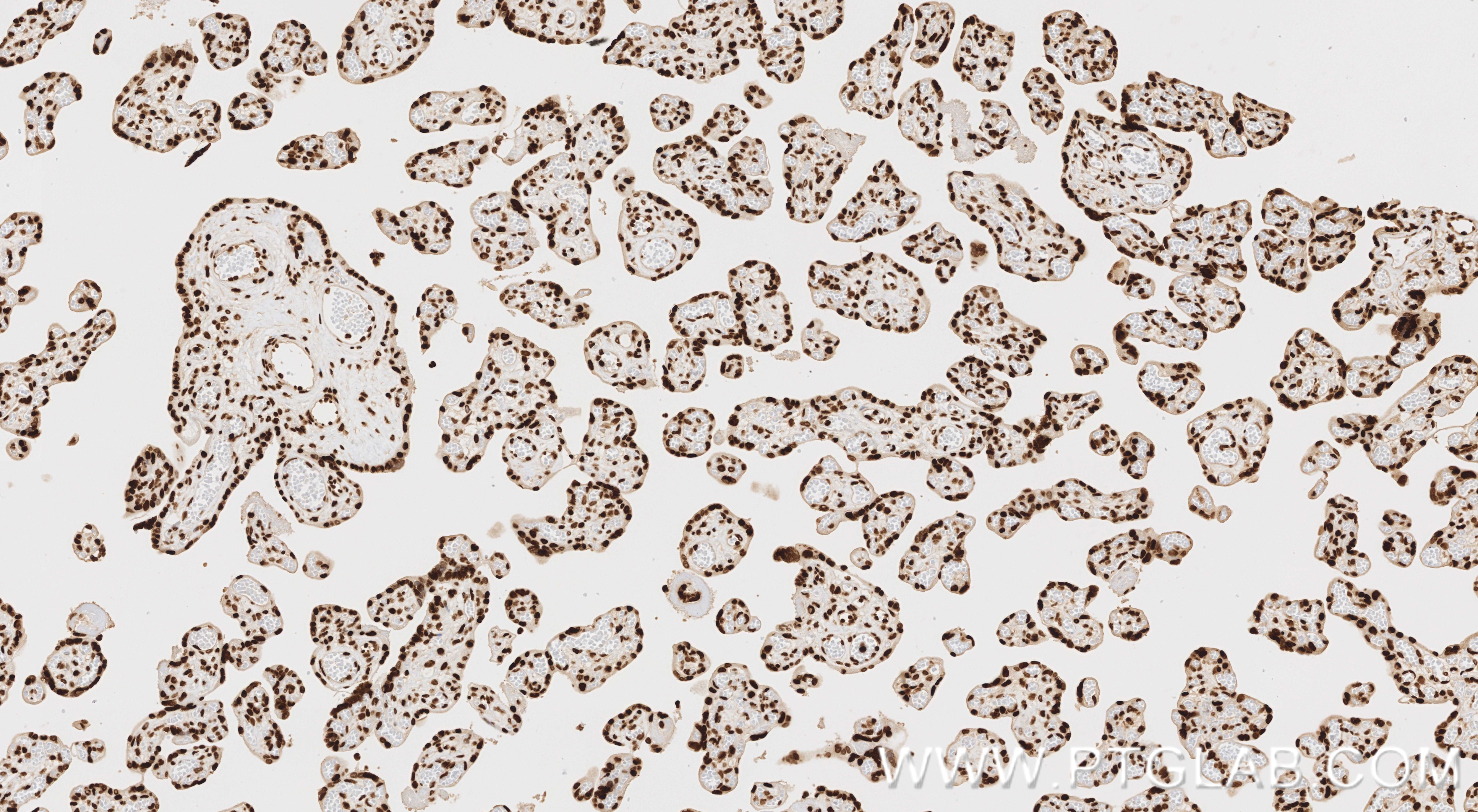
IHC staining of human placenta using 86731-1-RR
Immunohistochemical analysis of paraffin-embedded human placenta tissue slide using 86731-1-RR (Acetyl-Histone H2B (Lys12) antibody) at dilution of 1:1000 (under 10x lens). Heat mediated antigen retrieval with Tris-EDTA buffer (pH 9.0).
This validation data has been generated in-house and reviewed by Proteintech scientists. Find out more on our antibody validation policy
Immunohistochemical analysis of paraffin-embedded human placenta tissue slide using 86731-1-RR (Acetyl-Histone H2B (Lys12) antibody) at dilution of 1:1000 (under 10x lens). Heat mediated antigen retrieval with Tris-EDTA buffer (pH 9.0).
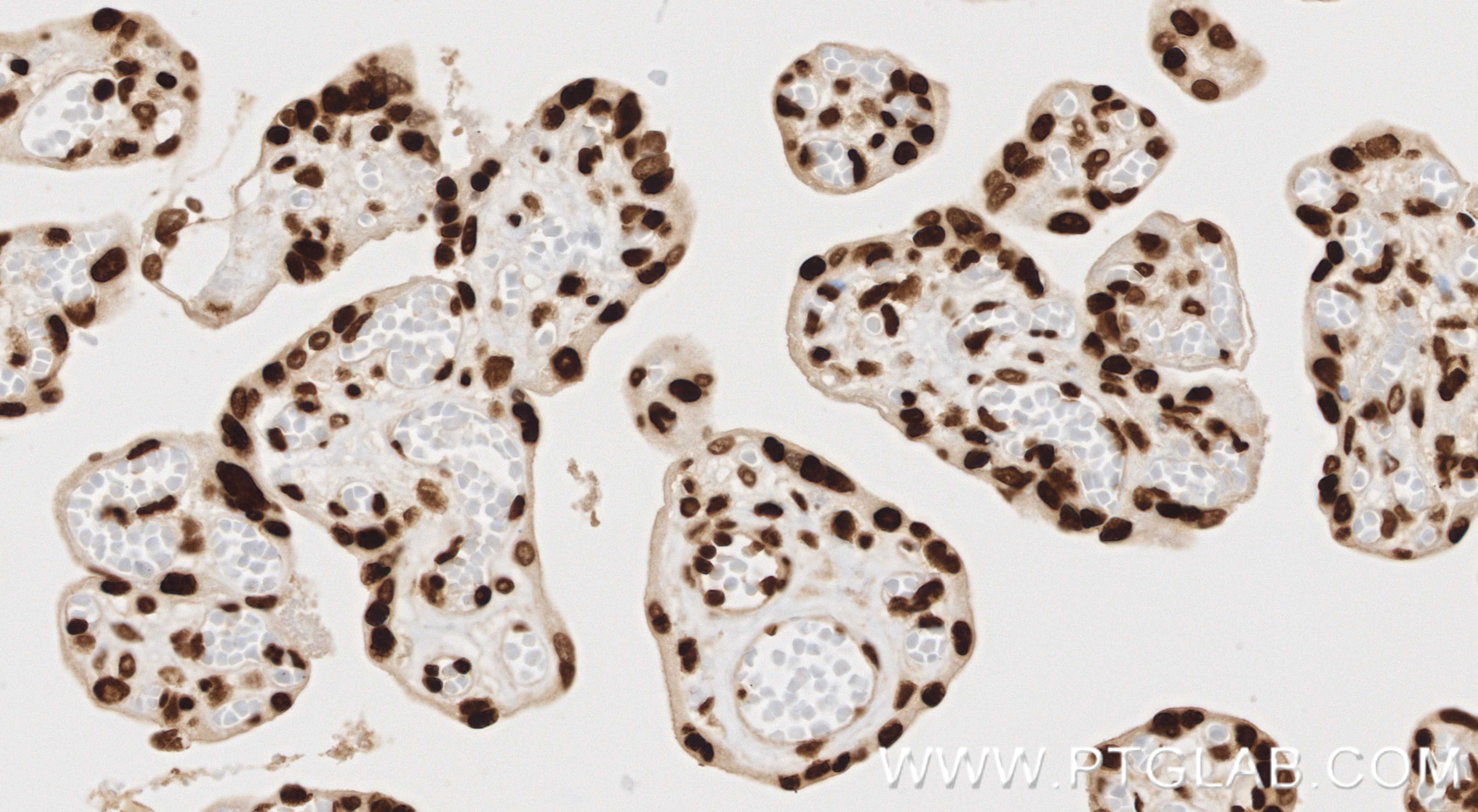
IHC staining of human placenta using 86731-1-RR
Immunohistochemical analysis of paraffin-embedded human placenta tissue slide using 86731-1-RR (Acetyl-Histone H2B (Lys12) antibody) at dilution of 1:1000 (under 40x lens). Heat mediated antigen retrieval with Tris-EDTA buffer (pH 9.0).
This validation data has been generated in-house and reviewed by Proteintech scientists. Find out more on our antibody validation policy
Immunohistochemical analysis of paraffin-embedded human placenta tissue slide using 86731-1-RR (Acetyl-Histone H2B (Lys12) antibody) at dilution of 1:1000 (under 40x lens). Heat mediated antigen retrieval with Tris-EDTA buffer (pH 9.0).
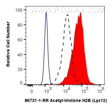
FC experiment of NIH/3T3 using 86731-1-RR
1x10^6 Trichostatin A treated NIH/3T3 cells were intracellularly stained with 0.13 ug Acetyl-Histone H2B (Lys12) Recombinant monoclonal antibody (86731-1-RR, Clone:250005E8) and CoraLite®488-Conjugated Goat Anti-Rabbit IgG(H+L) (SA00013-2)(red), or 0.13 ug Isotype Control (blue). Cells were fixed and permeabilized with Flow Cytometry Phosphorylated Protein Fixation/Permeabilization Kit (PF00026).
This validation data has been generated in-house and reviewed by Proteintech scientists. Find out more on our antibody validation policy
1x10^6 Trichostatin A treated NIH/3T3 cells were intracellularly stained with 0.13 ug Acetyl-Histone H2B (Lys12) Recombinant monoclonal antibody (86731-1-RR, Clone:250005E8) and CoraLite®488-Conjugated Goat Anti-Rabbit IgG(H+L) (SA00013-2)(red), or 0.13 ug Isotype Control (blue). Cells were fixed and permeabilized with Flow Cytometry Phosphorylated Protein Fixation/Permeabilization Kit (PF00026).
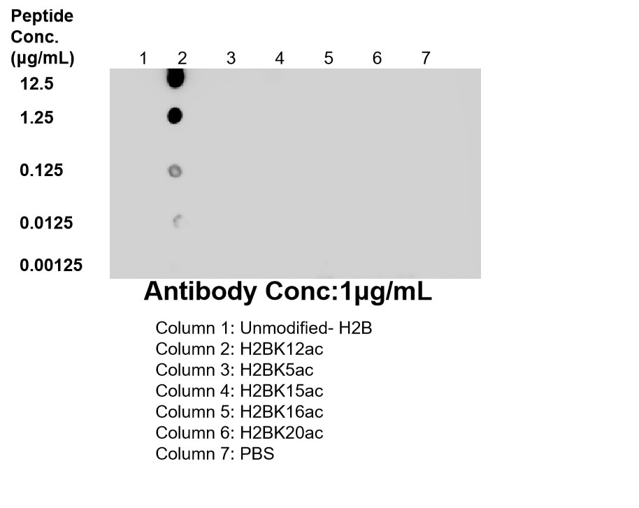
Dot Blot experiment of / using 86731-1-RR
Dot blot analysis was used to confirm the specificity of 86731-1-RR Acetyl-Histone H2B (Lys12) antibody. peptides were spotted onto NC and probed with antibody at 1 µg/ml.The amount of peptide (μg/mL) spotted is indicated next to each row.
This validation data has been generated in-house and reviewed by Proteintech scientists. Find out more on our antibody validation policy
Dot blot analysis was used to confirm the specificity of 86731-1-RR Acetyl-Histone H2B (Lys12) antibody. peptides were spotted onto NC and probed with antibody at 1 µg/ml.The amount of peptide (μg/mL) spotted is indicated next to each row.
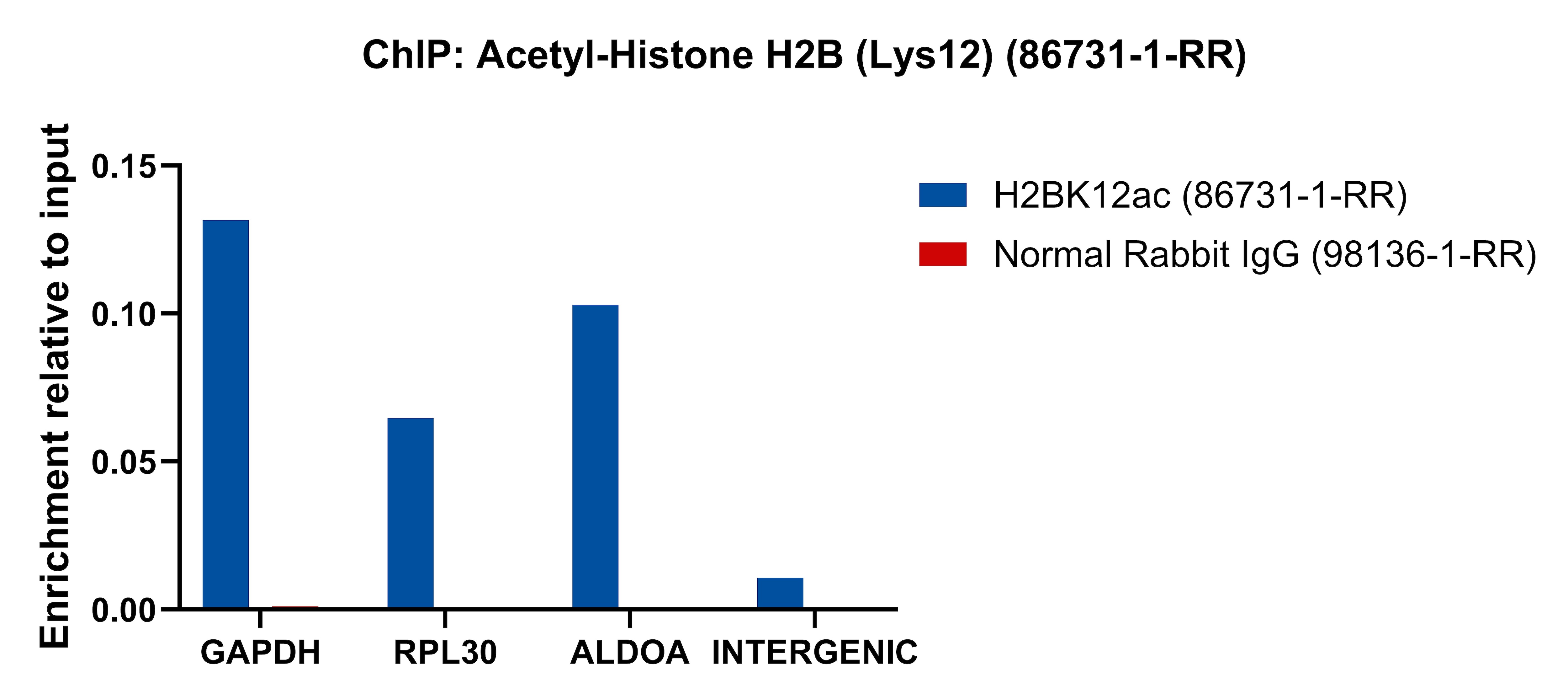
ChIP experiment of HeLa using 86731-1-RR
Chromatin was prepared from HeLa cells. Cells were fixed with formaldehyde for 10 minutes. The ChIP was performed with 15 µg of cross-linked chromatin, 5 µg of Acetyl-Histone H2B (Lys12) (86731-1-RR) or 5 ug of Normal Rabbit IgG (98136-1-RR), and 20 µl of Protein A Magarose Beads. The immunoprecipitated DNA was quantified by real-time PCR.
This validation data has been generated in-house and reviewed by Proteintech scientists. Find out more on our antibody validation policy
Chromatin was prepared from HeLa cells. Cells were fixed with formaldehyde for 10 minutes. The ChIP was performed with 15 µg of cross-linked chromatin, 5 µg of Acetyl-Histone H2B (Lys12) (86731-1-RR) or 5 ug of Normal Rabbit IgG (98136-1-RR), and 20 µl of Protein A Magarose Beads. The immunoprecipitated DNA was quantified by real-time PCR.
Trichostatin A treated NIH/3T3 cells, Trichostatin A treated HeLa cells
Positive IHC detected in
human placenta tissue Note: suggested antigen retrieval with TE buffer pH 9.0; (*) Alternatively, antigen retrieval may be performed with citrate buffer pH 6.0
Positive FC (Intra) detected in
Trichostatin A treated NIH/3T3 cells
Positive Dot blot detected in
/ /
Positive ChIP-qPCR detected in
HeLa cells
Recommended dilution
Application
Dilution
Western Blot (WB)
WB : 1:5000-1:50000
Immunohistochemistry (IHC)
IHC : 1:500-1:2000
Flow Cytometry (FC) (INTRA)
FC (INTRA) : 0.13 ug per 10^6 cells in a 100 µl suspension
DOT BLOT
DOT BLOT : 1:10-1:100
CHIP-QPCR
CHIP-QPCR : 1:10-1:100
It is recommended that this reagent should be titrated in each testing system to obtain optimal results.
Sample-dependent, Check data in validation data gallery.
Product Information
86731-1-RR targets Acetyl-Histone H2B (Lys12) in WB, IHC, FC (Intra), Dot blot, ELISA, ChIP-qPCR applications and shows reactivity with human, mouse samples.
PBS with 0.02% sodium azide and 50% glycerol, pH 7.3.
Storage Conditions
Store at -20°C. Stable for one year after shipment. Aliquoting is unnecessary for -20oC storage. 20ul sizes contain 0.1% BSA.
Background Information
Histones are small, highly basic proteins that consist of a globular domain with unstructured N- and C-terminal tails protruding from the main structure. Histone H3 is one of the five main histones that are responsible for the nucleosome structure of the chromosomal fiber in eukaryotes. Two molecules of each of the four core histones (H2A, H2B, H3, and H4) form an octamer, around which approximately 146 bp of DNA is wrapped in repeating units, called nucleosomes. In addition to their role in DNA compartmentalization, histones also play crucial roles in various biologic processes, including gene expression and regulation, DNA repair, chromatin condensation, cell cycle progression, chromosome segregation, and apoptosis. The ability of histones to regulate chromatin dynamics primarily originates from various posttranslational modifications carried out by histone-modifying enzymes.
Protocols
Product Specific Protocols
FC protocol for Acetyl-Histone H2B (Lys12) antibody 86731-1-RR
This validation data has been generated in-house and reviewed by Proteintech scientists.
Find out more on our antibody validation policy
WB Figures
WB analysis of NIH/3T3 using 86731-1-RR
Trichostatin A treated NIH/3T3 cells were subjected to SDS PAGE followed by western blot with 86731-1-RR (Acetyl-Histone H2B (Lys12) antibody) at dilution of 1:10000 incubated at room temperature for 1.5 hours. The membrane was stripped and reblotted with GAPDH Monoclonal antibody (60004-1-Ig) as loading control.
WB analysis of HeLa using 86731-1-RR
Trichostatin A treated HeLa cells were subjected to SDS PAGE followed by western blot with 86731-1-RR (Acetyl-Histone H2B (Lys12) antibody) at dilution of 1:10000 incubated at room temperature for 1.5 hours. The membrane was stripped and reblotted with GAPDH Monoclonal antibody (60004-1-Ig) as loading control.
IHC Figures
IHC staining of human placenta using 86731-1-RR
Immunohistochemical analysis of paraffin-embedded human placenta tissue slide using 86731-1-RR (Acetyl-Histone H2B (Lys12) antibody) at dilution of 1:1000 (under 10x lens). Heat mediated antigen retrieval with Tris-EDTA buffer (pH 9.0).
IHC staining of human placenta using 86731-1-RR
Immunohistochemical analysis of paraffin-embedded human placenta tissue slide using 86731-1-RR (Acetyl-Histone H2B (Lys12) antibody) at dilution of 1:1000 (under 40x lens). Heat mediated antigen retrieval with Tris-EDTA buffer (pH 9.0).
FC (INTRA) Figures
FC experiment of NIH/3T3 using 86731-1-RR
1x10^6 Trichostatin A treated NIH/3T3 cells were intracellularly stained with 0.13 ug Acetyl-Histone H2B (Lys12) Recombinant monoclonal antibody (86731-1-RR, Clone:250005E8) and CoraLite®488-Conjugated Goat Anti-Rabbit IgG(H+L) (SA00013-2)(red), or 0.13 ug Isotype Control (blue). Cells were fixed and permeabilized with Flow Cytometry Phosphorylated Protein Fixation/Permeabilization Kit (PF00026).
DOT BLOT Figures
Dot Blot experiment of / using 86731-1-RR
Dot blot analysis was used to confirm the specificity of 86731-1-RR Acetyl-Histone H2B (Lys12) antibody. peptides were spotted onto NC and probed with antibody at 1 µg/ml.The amount of peptide (μg/mL) spotted is indicated next to each row.
CHIP-QPCR Figures
ChIP experiment of HeLa using 86731-1-RR
Chromatin was prepared from HeLa cells. Cells were fixed with formaldehyde for 10 minutes. The ChIP was performed with 15 µg of cross-linked chromatin, 5 µg of Acetyl-Histone H2B (Lys12) (86731-1-RR) or 5 ug of Normal Rabbit IgG (98136-1-RR), and 20 µl of Protein A Magarose Beads. The immunoprecipitated DNA was quantified by real-time PCR.
The species listed in Tested Reactivity are in-house verified and applicable species. For unlisted species, please refer to the homology analysis of the immunogen sequence and related species. For rabbit polyclonal antibodies, homology >70% is recommended. For mouse monoclonal antibodies and rabbit recombinant antibodies, homology >90% is recommended. Generally, the higher the homology, the greater the applicability. However, there will be certain differences in protein expression in different species, tissues or cells. Therefore, the homology analysis results are for reference only and do not serve as a guarantee.
At Proteintech, we pride ourselves on our antibody quality, customer service and transparency. As such, we are comparing our antibodies with other vendors, enabling easy identification and comparisons of key data to help you choose the suitable antibody for your needs.
We have selected the top cited antibodies from these vendors for you to compare.
Proteintech
Acetyl-Histone H2B (Lys12) Recombinant antibody
Catalog Number
86731-1-RR
Citations
-
Dilutions
WB : 1:5000-1:50000 IHC : 1:500-1:2000 FC (INTRA) : 0.13 ug per 10^6 cells in a 100 µl suspension DOT BLOT : 1:10-1:100 CHIP-QPCR : 1:10-1:100
Applications
WB, IHC, FC (Intra), Dot blot, ELISA, ChIP-qPCR
Reactivity
human, mouse
Product Guarantee
Covers any species including not listed on datasheet
Covers any applications including not listed on datasheet