

Tested Applications
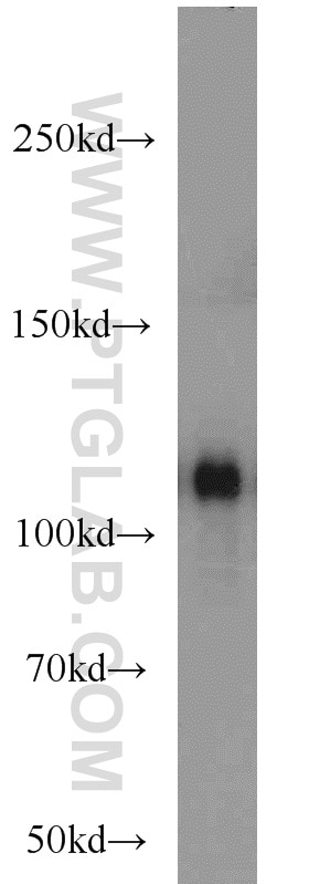
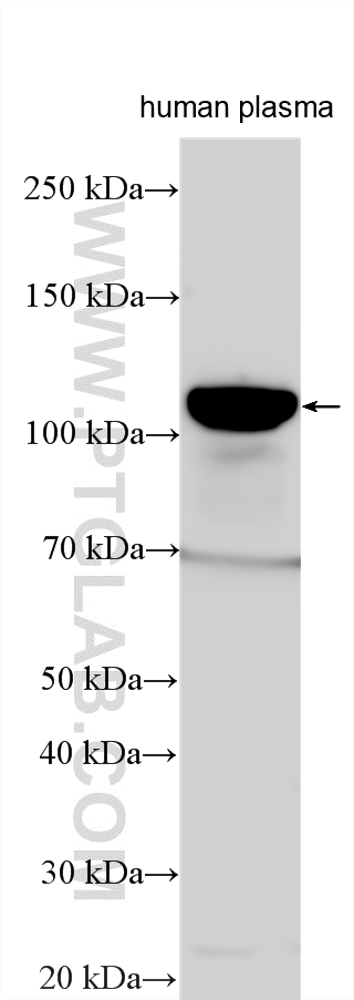
| Positive WB detected in | human plasma |
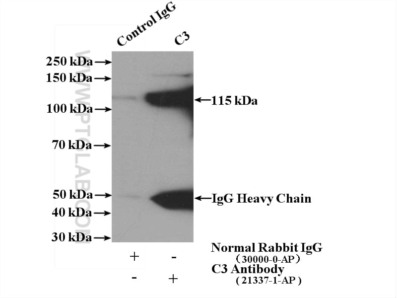
| Positive IP detected in | human plasma tissue |
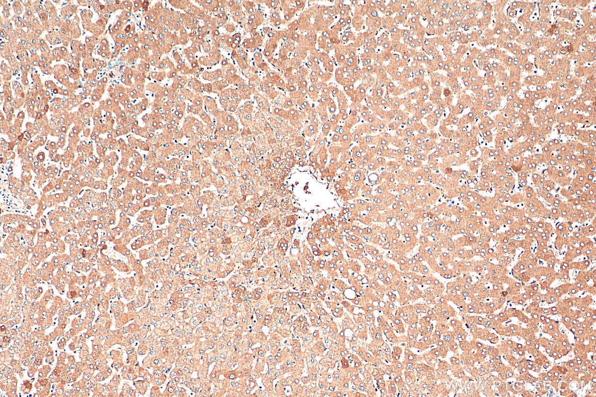
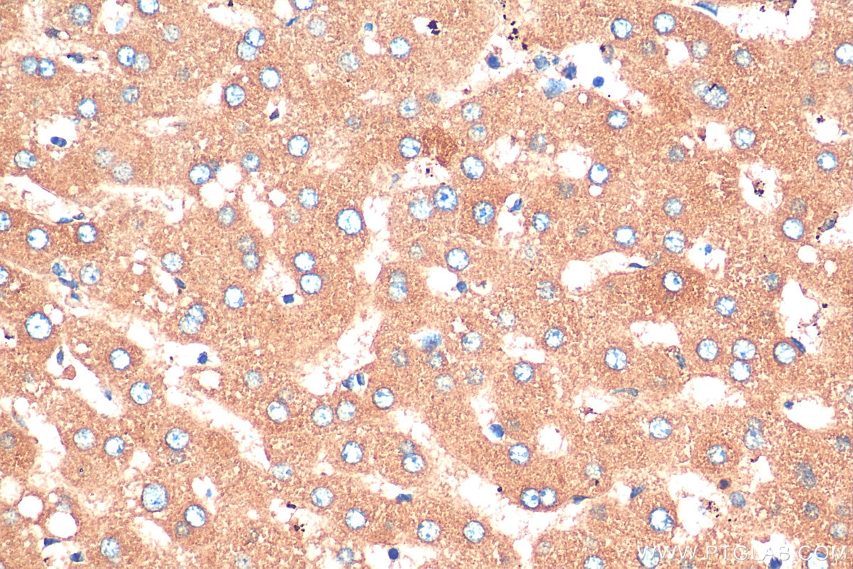
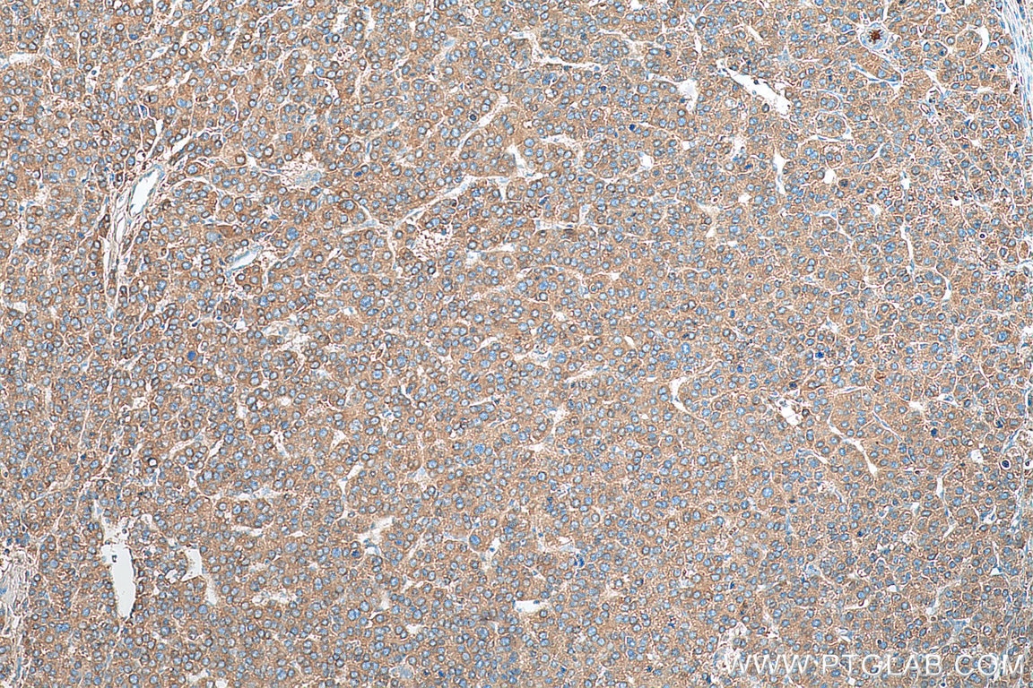
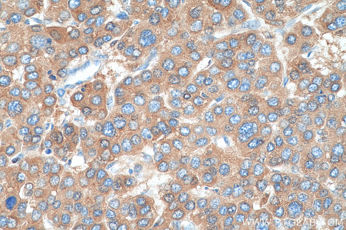
| Positive IHC detected in | human liver tissue, human liver cancer tissue Note: suggested antigen retrieval with TE buffer pH 9.0; (*) Alternatively, antigen retrieval may be performed with citrate buffer pH 6.0 |
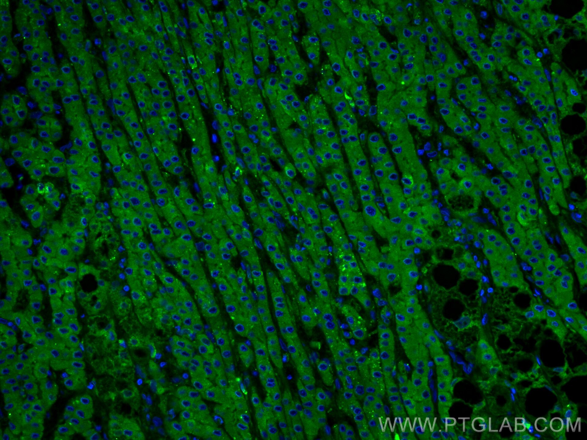
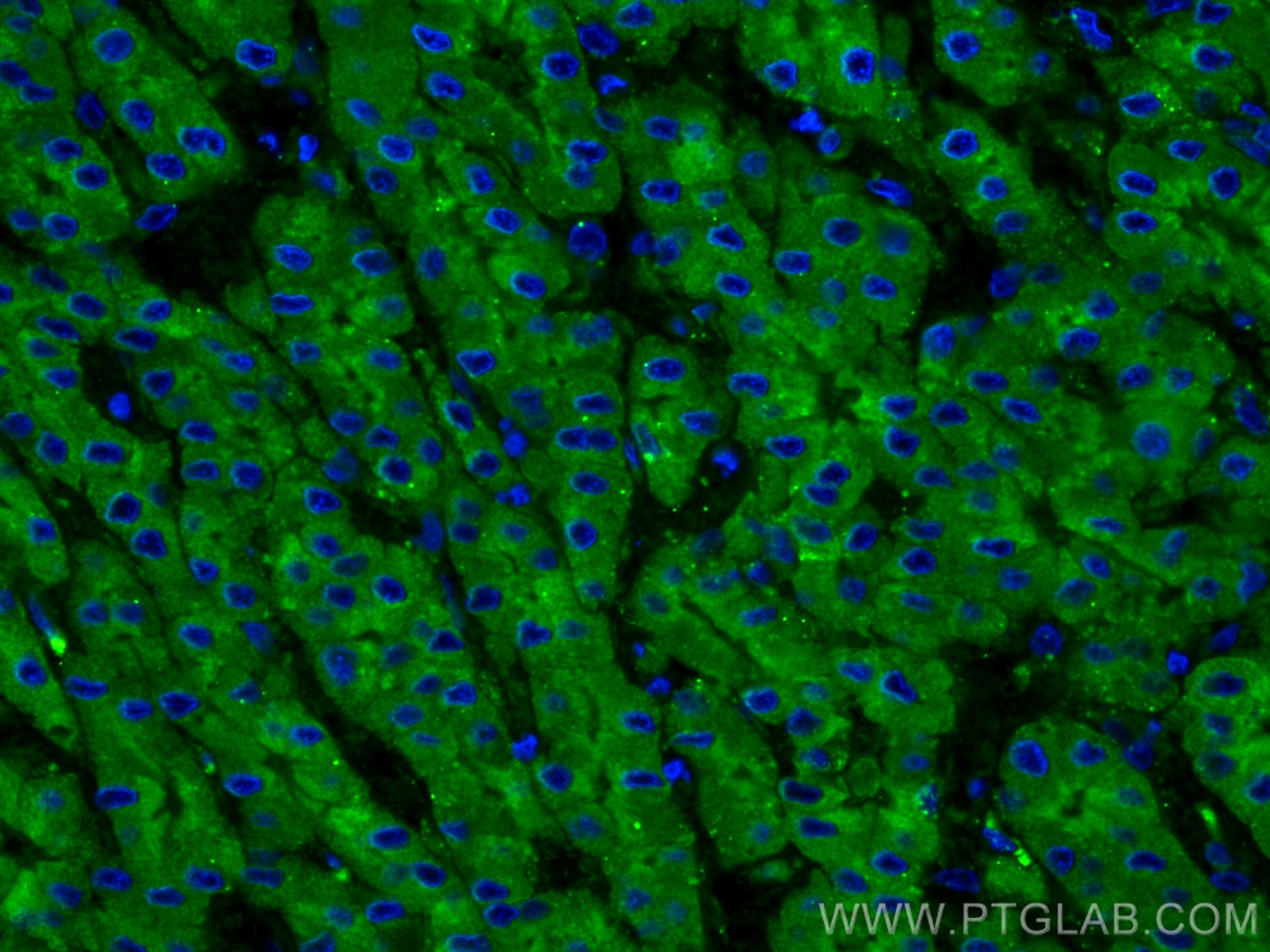
| Positive IF-P detected in | human liver cancer tissue |
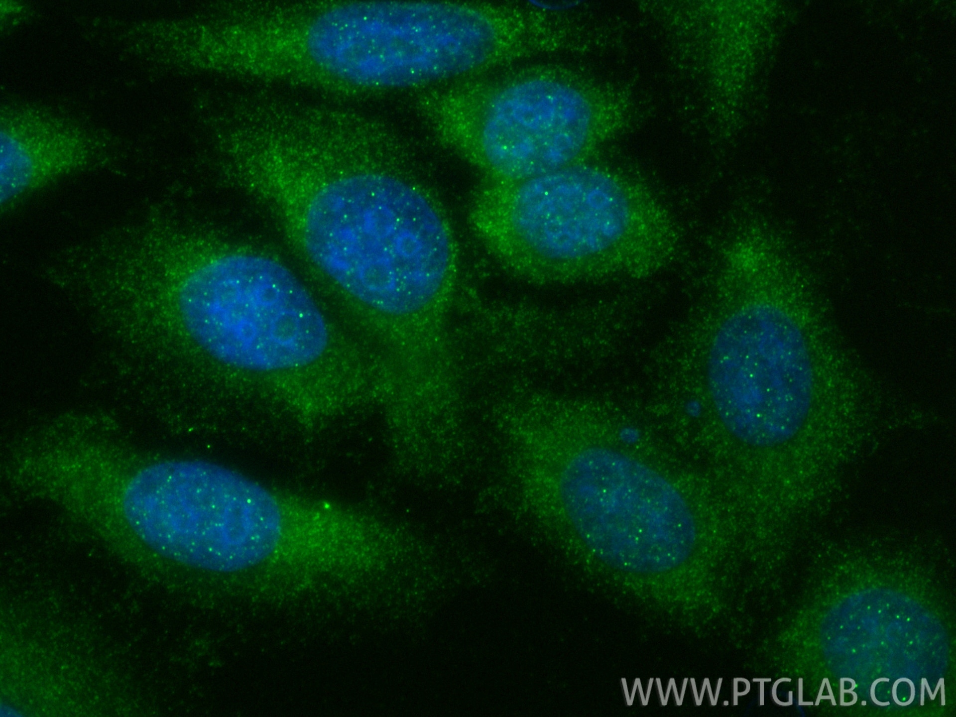
| Positive IF/ICC detected in | HepG2 cells |
Recommended dilution
| Application | Dilution |
|---|---|
| Western Blot (WB) | WB : 1:20000-1:100000 |
| Immunoprecipitation (IP) | IP : 0.5-4.0 ug for 1.0-3.0 mg of total protein lysate |
| Immunohistochemistry (IHC) | IHC : 1:1000-1:4000 |
| Immunofluorescence (IF)-P | IF-P : 1:50-1:500 |
| Immunofluorescence (IF)/ICC | IF/ICC : 1:50-1:500 |
| It is recommended that this reagent should be titrated in each testing system to obtain optimal results. | |
| Sample-dependent, Check data in validation data gallery. | |
Product Information
21337-1-AP targets C3/C3b/C3c in WB, IHC, IF/ICC, IF-P, IP, CoIP, ELISA, Blocking assay applications and shows reactivity with human samples.
| Tested Reactivity | human |
| Cited Reactivity | human, pig, monkey, bovine |
| Host / Isotype | Rabbit / IgG |
| Class | Polyclonal |
| Type | Antibody |
| Immunogen |
CatNo: Ag15537 Product name: Recombinant human C3 protein Source: e coli.-derived, PGEX-4T Tag: GST Domain: 1314-1663 aa of BC150179 Sequence: ESASLLRSEETKENEGFTVTAEGKGQGTLSVVTMYHAKAKDQLTCNKFDLKVTIKPAPETEKRPQDAKNTMILEICTRYRGDQDATMSILDISMMTGFAPDTDDLKQLANGVDRYISKYELDKAFSDRNTLIIYLDKVSHSEDDCLAFKVHQYFNVELIQPGAVKVYAYYNLEESCTRFYHPEKEDGKLNKLCRDELCRCAEENCFIQKSDDKVTLEERLDKACEPGVDYVYKTRLVKVQLSNDFDEYIMAIEQTIKSGSDEVQVGQQRTFISPIKCREALKLEEKKHYLMWGLSSDFWGEKPNLSYIIGKDTWVEHWPEEDECQDEENQKQCQDLGAFTESMVVFGCPN Predict reactive species |
| Full Name | complement component 3 |
| Calculated Molecular Weight | 1663 aa, 187 kDa |
| Observed Molecular Weight | 115 kDa, 70 kDa |
| GenBank Accession Number | BC150179 |
| Gene Symbol | C3 |
| Gene ID (NCBI) | 718 |
| RRID | AB_2878843 |
| Conjugate | Unconjugated |
| Form | Liquid |
| Purification Method | Antigen affinity purification |
| UNIPROT ID | P01024 |
| Storage Buffer | PBS with 0.02% sodium azide and 50% glycerol, pH 7.3. |
| Storage Conditions | Store at -20°C. Stable for one year after shipment. Aliquoting is unnecessary for -20oC storage. 20ul sizes contain 0.1% BSA. |
Background Information
The complement system is an important effector that bridges the innate and adaptive immune systems (PMID: 20010915). The third component of complement, C3, plays a central role in activating the complement system. Its processing by C3 convertase is the central reaction in both classical and alternative complement pathways (PMID: 11414361). Human C3 (190-195 kDa), composed of α and β chains (115-120 and 75 kDa, respectively), is cleaved into C3a and C3b by C3 convertase. C3b is composed of the α' chain and β chain (PMID: 27210597). Factor I cleaves the α′ chain of C3b to 68 kDa and 43 kDa degradation products (iC3b). Inactive iC3b stops convertase amplification but acts as an opsonin via CR3/CR4. Factor I and CR1 further cleave iC3b into C3d, linking innate recognition to B-cell activation via CR2 (PMID: 25395424; 14527961). This antibody raised against 1314-1663 aa of human C3 protein recognizes the C3 α chain (115-120 kDa), C3b α' chain (110-115 kDa), and C3c α' chain fragment 2 (43 kDa).
Protocols
| Product Specific Protocols | |
|---|---|
| IF protocol for C3/C3b/C3c antibody 21337-1-AP | Download protocol |
| IHC protocol for C3/C3b/C3c antibody 21337-1-AP | Download protocol |
| IP protocol for C3/C3b/C3c antibody 21337-1-AP | Download protocol |
| WB protocol for C3/C3b/C3c antibody 21337-1-AP | Download protocol |
| Standard Protocols | |
|---|---|
| Click here to view our Standard Protocols |
Publications
What published studies show
The Rabbit Polyclonal antibody C3/C3b/C3c (Cat# 21337-1-AP) has accumulated 179 citations across high-impact journals including Nature, Nat Cell Biol, Nat Neurosci, Nat Commun, Neuron, J Clin Invest, Adv Sci, Cell Death Differ, Brain, J Neuroinflammation, and FASEB J. Research fields span neuroinflammation, complement-mediated synaptic pruning, astrocyte reactivity, autoimmune diseases (SLE, lupus nephritis), neurological disorders (Alzheimer's, Parkinson's, epilepsy, stroke), renal pathology, cancer immunology, and infectious disease.
| Species | Application | Title |
|---|---|---|
Nat Cell Biol Autophagy enables microglia to engage amyloid plaques and prevents microglial senescence | ||
Nat Neurosci GABA-dependent microglial elimination of inhibitory synapses underlies neuronal hyperexcitability in epilepsy | ||
Nat Commun Increased circulating levels of Factor H-Related Protein 4 are strongly associated with age-related macular degeneration. | ||
ACS Nano Deciphering Apolipoprotein A4 in the Protein Corona of Ligand-Modified Liposomes for Tumor Targeting and Penetration. | ||
J Adv Res Tocilizumab-loaded nanoparticles block IL-6R and reduce edema after intracerebral hemorrhage. | ||
Neuron C5aR1+ microglia exacerbate neuroinflammation and cerebral edema in acute brain injury |
Reviews
What customers say
All three reviewers gave this antibody a 5-star rating. One user confirmed strong Western Blot performance on human samples, detecting two C3 bands at ~190 and 130 kDa. Two users successfully applied it for immunofluorescence—one on brain tissue at 1:100 dilution to visualize complement activation by confocal microscopy (noting mild background that improved with optimization), and another on rat kidney at 1:200 in an LPS induction model. Overall, the antibody performs well across WB and IF applications with clear, reliable signals.
The reviews below have been submitted by verified Proteintech customers who received an incentive for providing their feedback.
Boyan (Verified Customer) (08-27-2025) | good for WB, two bands at ~190 and 130 kd, both of which are C3 forms
|
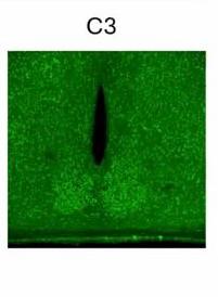
Marisa (Verified Customer) (07-31-2025) | We used the C3/C3b/C3c antibody in immunofluorescence and obtained a detectable signal, though some background was present. With slight optimization, it worked for visualizing complement activation by confocal microscopy.
 |
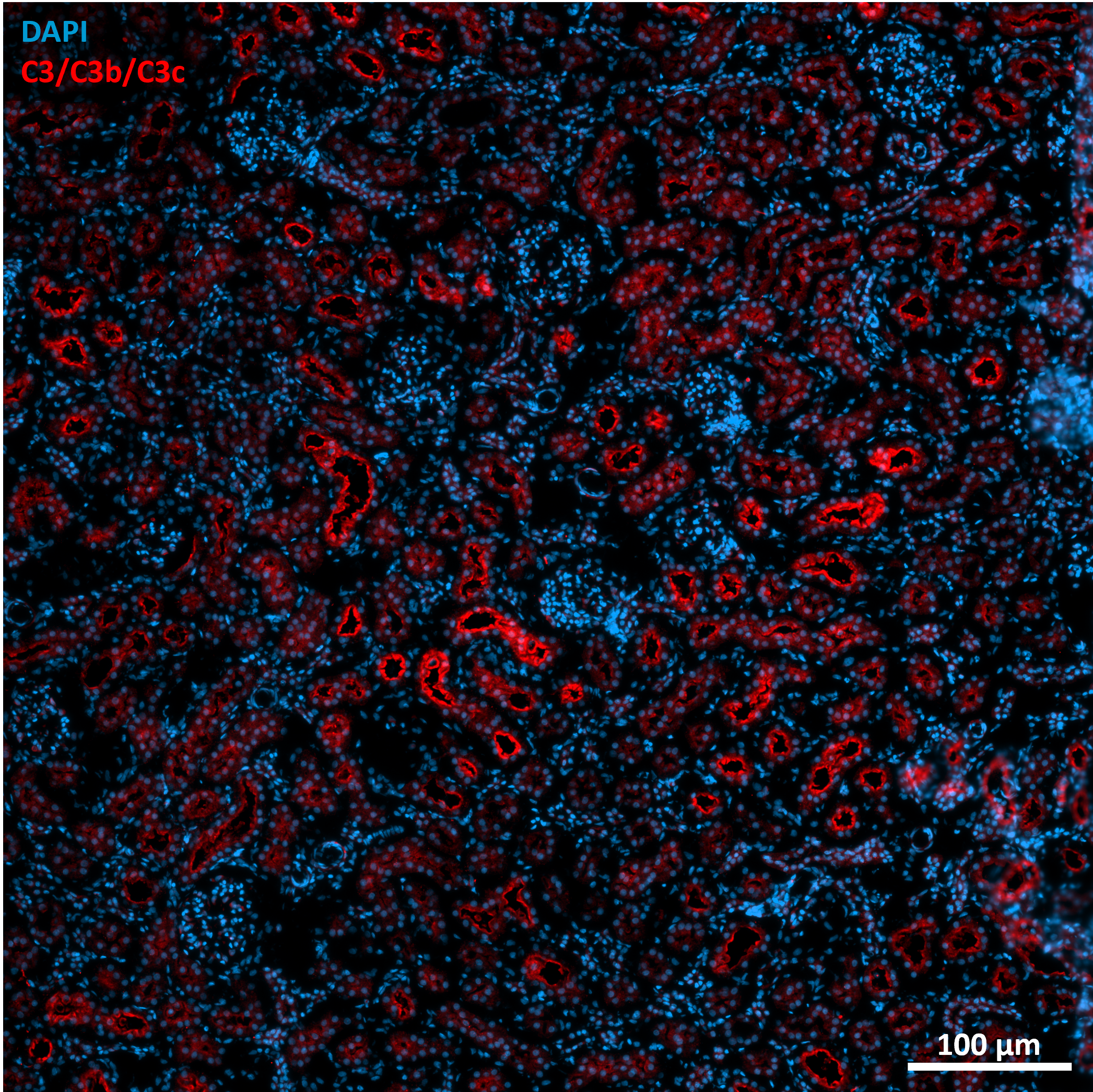
Marion (Verified Customer) (08-01-2024) | LPS induction model Fixative : Paraformaldehyde 4% Permeabilization: 10 min; RT; 0.2% Triton X-100 Blocking: 2 h; RT; 10% Normal donkey serum; 1% BSA Primary antibody: 1 night; 4°C Secondary antibody: ab150064 (Abcam); 1h30; RT Antibodies diluted in blocking solution
 |











