


Tested Applications
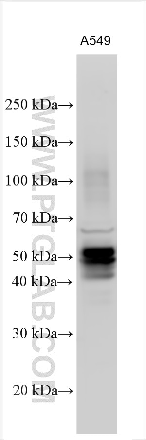
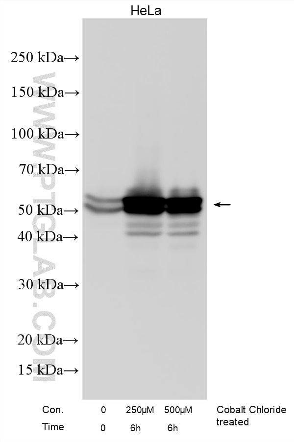
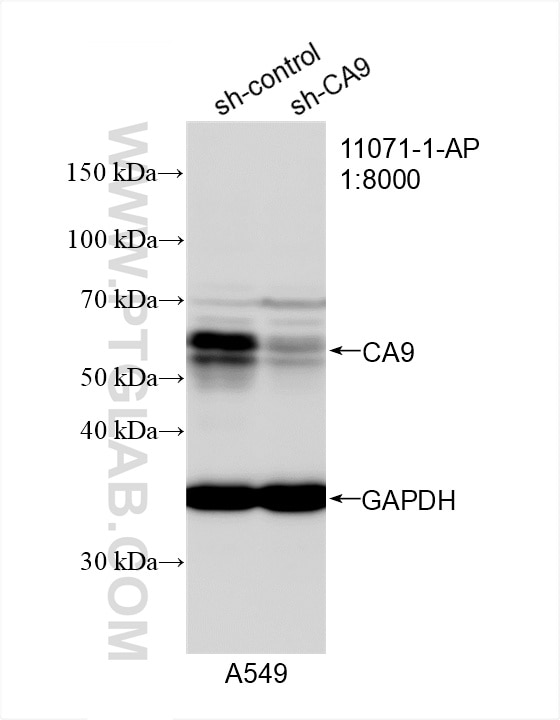
| Positive WB detected in | A549 cells, HeLa cells, Cobalt Chloride treated HeLa cells |
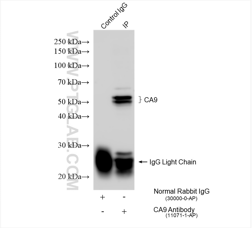
| Positive IP detected in | HeLa cells |
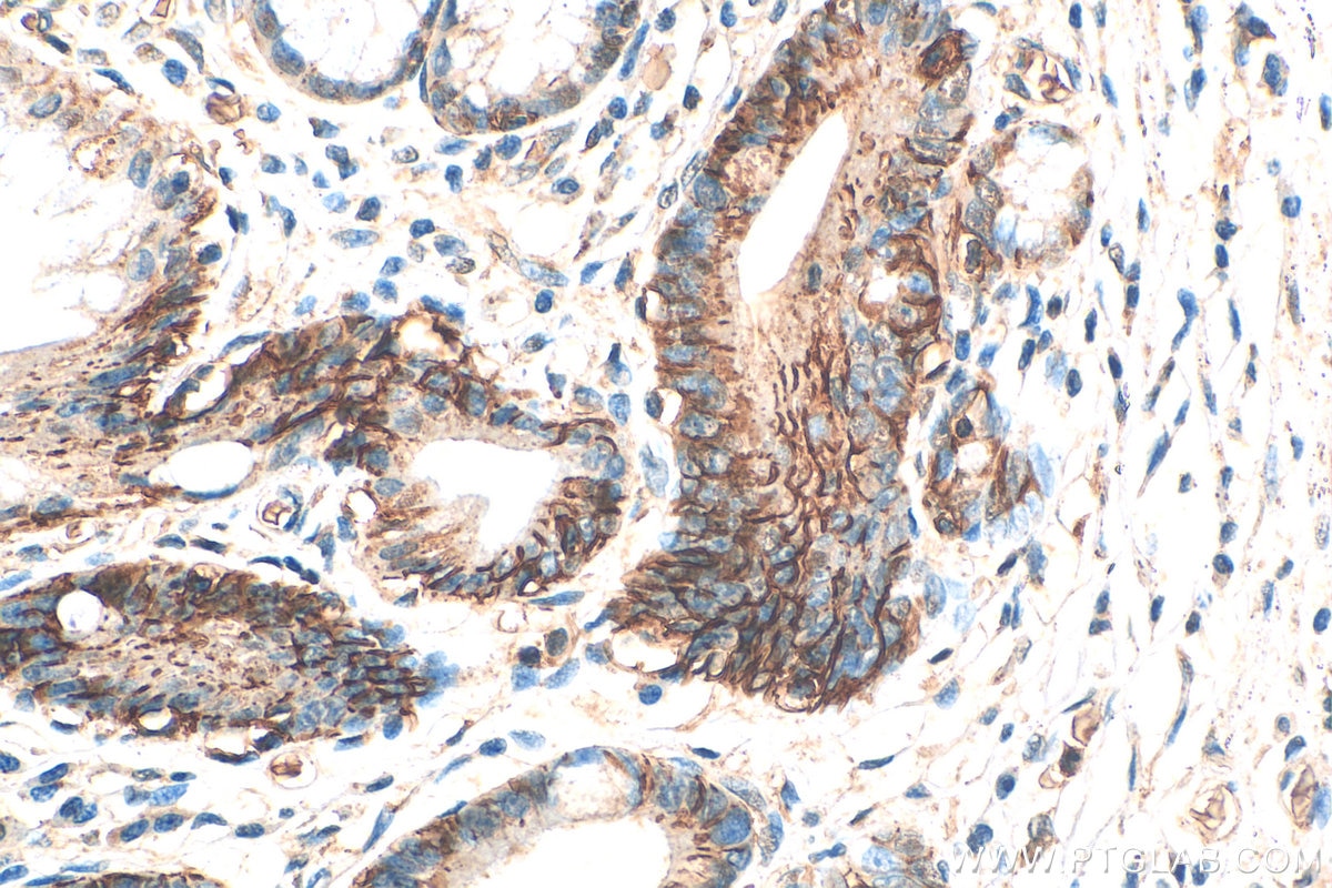
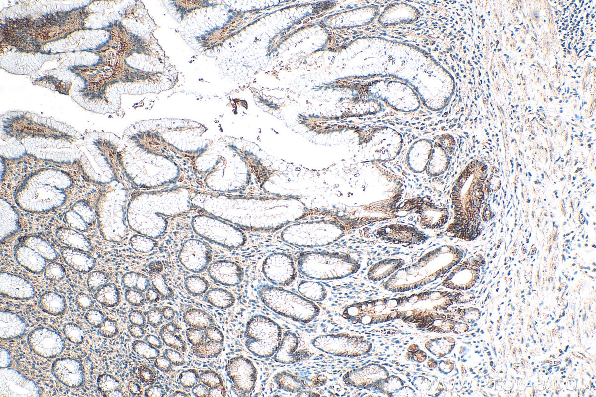
| Positive IHC detected in | human stomach cancer tissue Note: suggested antigen retrieval with TE buffer pH 9.0; (*) Alternatively, antigen retrieval may be performed with citrate buffer pH 6.0 |
| Positive IF/ICC detected in | HEK-293 cells |
Recommended dilution
| Application | Dilution |
|---|---|
| Western Blot (WB) | WB : 1:1000-1:6000 |
| Immunoprecipitation (IP) | IP : 0.5-4.0 ug for 1.0-3.0 mg of total protein lysate |
| Immunohistochemistry (IHC) | IHC : 1:50-1:500 |
| Immunofluorescence (IF)/ICC | IF/ICC : 1:200-1:800 |
| It is recommended that this reagent should be titrated in each testing system to obtain optimal results. | |
| Sample-dependent, Check data in validation data gallery. | |
Product Information
11071-1-AP targets Carbonic Anhydrase IX/CA9 in WB, IHC, IF/ICC, IP, ELISA applications and shows reactivity with human samples.
| Tested Reactivity | human |
| Cited Reactivity | human, mouse, rat |
| Host / Isotype | Rabbit / IgG |
| Class | Polyclonal |
| Type | Antibody |
| Immunogen |
CatNo: Ag1540 Product name: Recombinant human CA9 protein Source: e coli.-derived, PKG Tag: GST Domain: 32-408 aa of BC014950 Sequence: LMPVHPQRLPRMQEDSPLGGGSSGEDDPLGEEDLPSEEDSPREEDPPGEEDLPGEEDLPGEEDLPEVKPKSEEEGSLKLEDLPTVEAPGDPQEPQNNAHRDKEGDDQSHWRYGGDPPWPRVSPACAGRFQSPVDIRPQLAAFCPALRPLELLGFQLPPLPELRLRNNGHSVQLTLPPGLEMALGPGREYRALQLHLHWGAAGRPGSEHTVEGHRFPAEIHVVHLSTAFARVDEALGRPGGLAVLAAFLEEGPEENSAYEQLLSRLEEIAEEGSETQVPGLDISALLPSDFSRYFQYEGSLTTPPCAQGVIWTVFNQTVMLSAKQLHTLSDTLWGPGDSRLQLNFRATQPLNGRVIEASFPAGVDSSPRAAEPVQLNS Predict reactive species |
| Full Name | carbonic anhydrase IX |
| Calculated Molecular Weight | 459 aa, 50 kDa |
| Observed Molecular Weight | 50 kDa, 60-70 kDa |
| GenBank Accession Number | BC014950 |
| Gene Symbol | CA9 |
| Gene ID (NCBI) | 768 |
| RRID | AB_2066528 |
| Conjugate | Unconjugated |
| Form | Liquid |
| Purification Method | Antigen affinity purification |
| UNIPROT ID | Q16790 |
| Storage Buffer | PBS with 0.02% sodium azide and 50% glycerol, pH 7.3. |
| Storage Conditions | Store at -20°C. Stable for one year after shipment. Aliquoting is unnecessary for -20oC storage. 20ul sizes contain 0.1% BSA. |
Background Information
CA9 (Carbonic anhydrase 9) may be involved in the control of cell proliferation and transformation and appears to be a novel specific biomarker for a cervical neoplasia (PMID:18703501). It is a tumor-associated antigen that has been shown to have diagnostic utility in identifying cervical dysplasia and carcinoma. The protein is present in the cytoplasmic membrane and the nucleus, with a molecular weight of 50 kDa. The molecular weight of the glycosylated form is approximately 60-70 kDa. (PMID: 31142270, PMID: 31819036)
Protocols
| Product Specific Protocols | |
|---|---|
| IF protocol for Carbonic Anhydrase IX/CA9 antibody 11071-1-AP | Download protocol |
| IHC protocol for Carbonic Anhydrase IX/CA9 antibody 11071-1-AP | Download protocol |
| IP protocol for Carbonic Anhydrase IX/CA9 antibody 11071-1-AP | Download protocol |
| WB protocol for Carbonic Anhydrase IX/CA9 antibody 11071-1-AP | Download protocol |
| Standard Protocols | |
|---|---|
| Click here to view our Standard Protocols |
Publications
What published studies show
Anti-Carbonic Anhydrase 9 (CA9) antibody (Cat# 11071-1-AP) has 69 citations across journals including Nature Communications, Advanced Materials, PNAS, Advanced Science, Biomaterials, Cell Death & Disease, and Journal of Immunotherapy of Cancer. Research fields span tumor hypoxia, cancer biology (breast, lung, renal, colorectal, pancreatic, glioblastoma), angiogenesis, ferroptosis, drug resistance, tumor microenvironment, and radioligand imaging/therapy. Applications include WB, IHC, and IF in human, mouse, and rat models.
| Species | Application | Title |
|---|---|---|
Nanomicro Lett Targeting Hypoxic Tumors with Hybrid Nanobullets for Oxygen-Independent Synergistic Photothermal and Thermodynamic Therapy. | ||
Adv Mater GSH/Ph Dual Activatable Crosslinked And Fluorinated Pei for Cancer Gene Therapy Through Endogenous Iron De-Hijacking And in Situ Ros Amplification | ||
J Extracell Vesicles 124I-labelled BMSC-Derived Extracellular Vesicles Deliver CRISPR/Cas9 Ribonucleoproteins With a GFP-Reporter System to Inhibit Osteosarcoma Proliferation and Metastasis | ||
J Adv Res Targeting PLOD2 induces epithelioid differentiation and improves therapeutic response in sarcomatoid renal cell carcinoma. | ||
Adv Sci (Weinh) Understanding Glioblastoma Dynamics Using 3D Organoids and Engineered Extracellular Matrix. |
Reviews
What customers say
Across five reviews, the antibody performed well in Western Blot at dilutions ranging from 1:250 to 1:4000, detecting the expected band in renal cell carcinoma, HCC, and ccRCC cell lines. One user noted additional non-specific bands at 1:1000. For IHC on liver metastasis tissue, one reviewer reported loss of efficacy after a freeze-thaw cycle despite glycerol storage. Another user observed both 70 kDa and 25 kDa isoforms. Overall, the antibody is considered reliable for WB with good sensitivity, though users should be mindful of storage conditions for IHC applications.
The reviews below have been submitted by verified Proteintech customers who received an incentive for providing their feedback.
Aline Seiko (Verified Customer) (05-12-2025) | It was observed a 70kDa and 25kDa isoform.
|
Maelle (Verified Customer) (01-03-2025) | Lost efficacy after freeze-defreze even if it is in glycerol
|
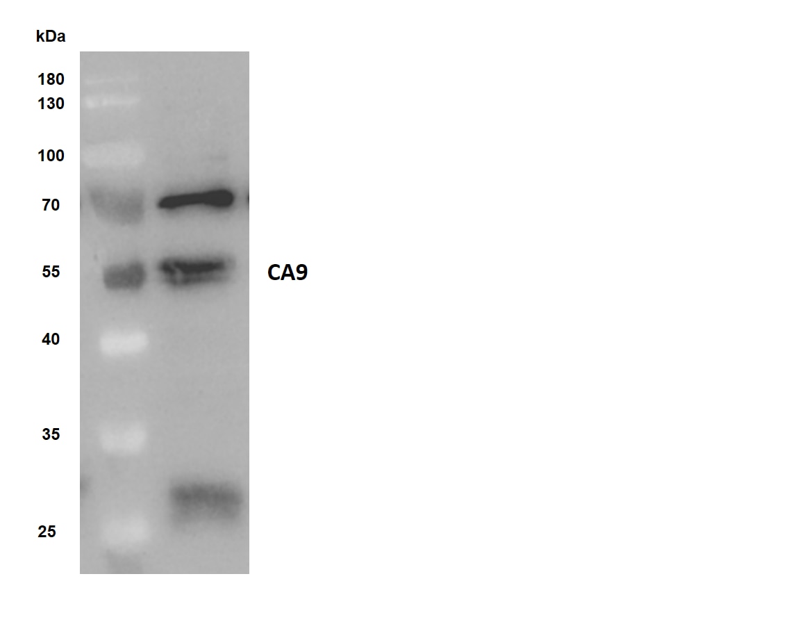
Josh (Verified Customer) (10-28-2021) | 15 ug of human clear cell renal cell carcinoma cell line RCC4 was resolved on 15% Tris/Glycine and transferred to PVDF membrane. Membrane was blocked in blocking buffer (2% BSA in TBS/0.1% Tween-20) for 1hr at room temperature, followed by overnight incubation with anti-CA9 (1:1000) in blocking buffer at 4oC. After 1hr incubation with anti-Rabbit secondary antibody, membrane was imaged with ECL. Expected band was detected along with several non-specific bands by western blot
 |
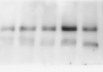
K (Verified Customer) (08-03-2021) | total cell lysate (50 ug of human HCC cell lines) was resolved on 10% Bis-Tris gel and transferred to nitrocellulose membrane. Membrane was incubated in blocking buffer (5% BSA in TBS/0.1% Tween-20) for 1h. Membrane was incubated with anti-CA9 in blocking buffer (1:500) at 40C overnight. After 1h incubation with appropriate secondary antibody (anti-Rabbit 1:5000) membranes were imaged with ECL and expected band was detected.
 |
Paulo (Verified Customer) (03-04-2019) | Total cell lysate (50 ug of human ccRCC cell lines) was resolved on 10% Bis-Tris gel and transferred to PVDF membrane. Membrane was incubated in blocking buffer (5% milk in PBS/0.1% Tween-20) for 1h. Membrane was incubated with anti-CAIX in blocking buffer (1:250) at 4C overnight. After 1h incubation with appropriate secondary antibody (DAKO anti-Rabbit 1:10000) membranes were imaged with ECL and expected band was detected
|








