Tested Applications
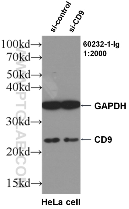
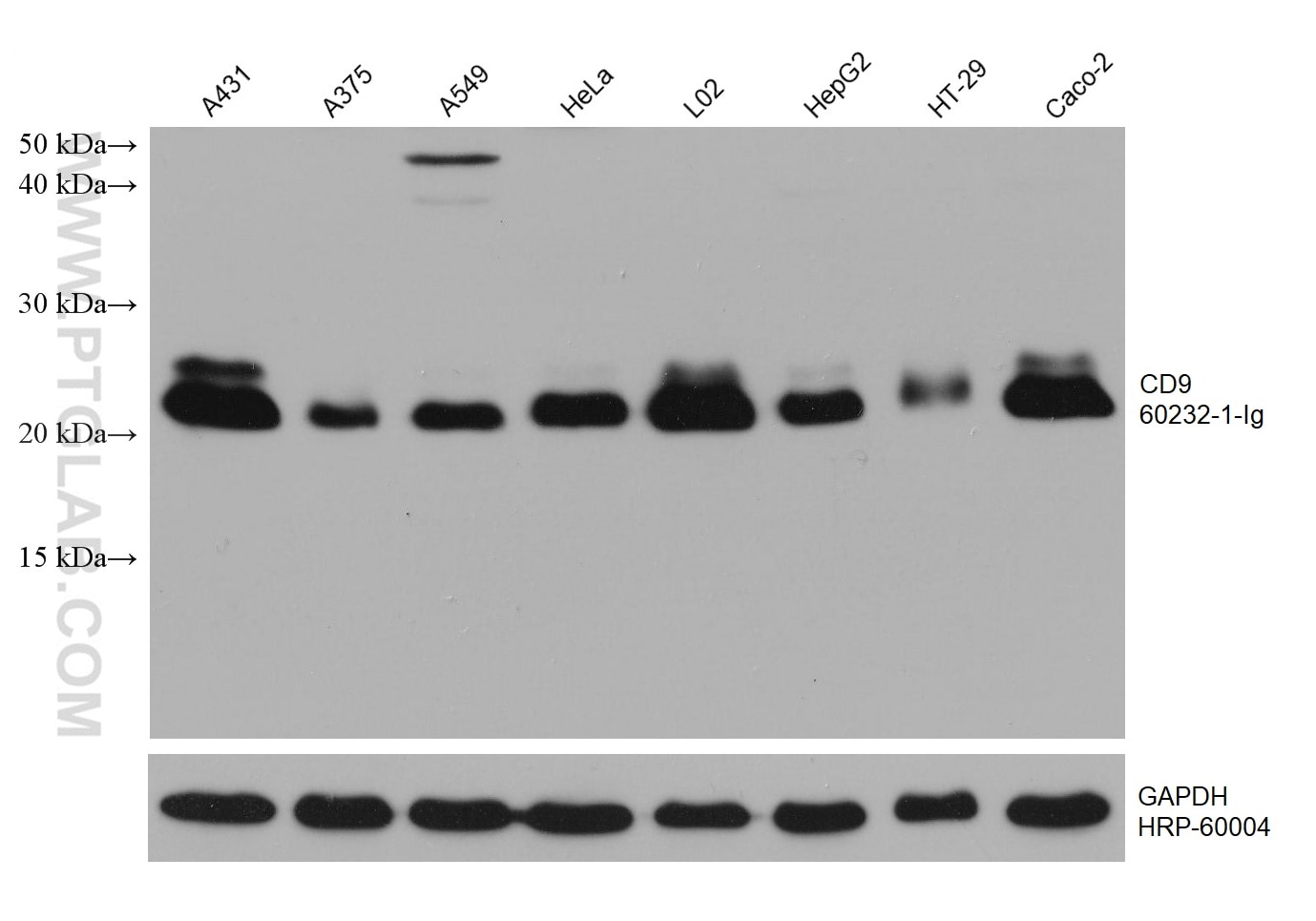
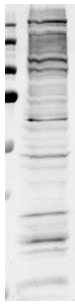
| Positive WB detected in | A431 cells, HeLa cells |
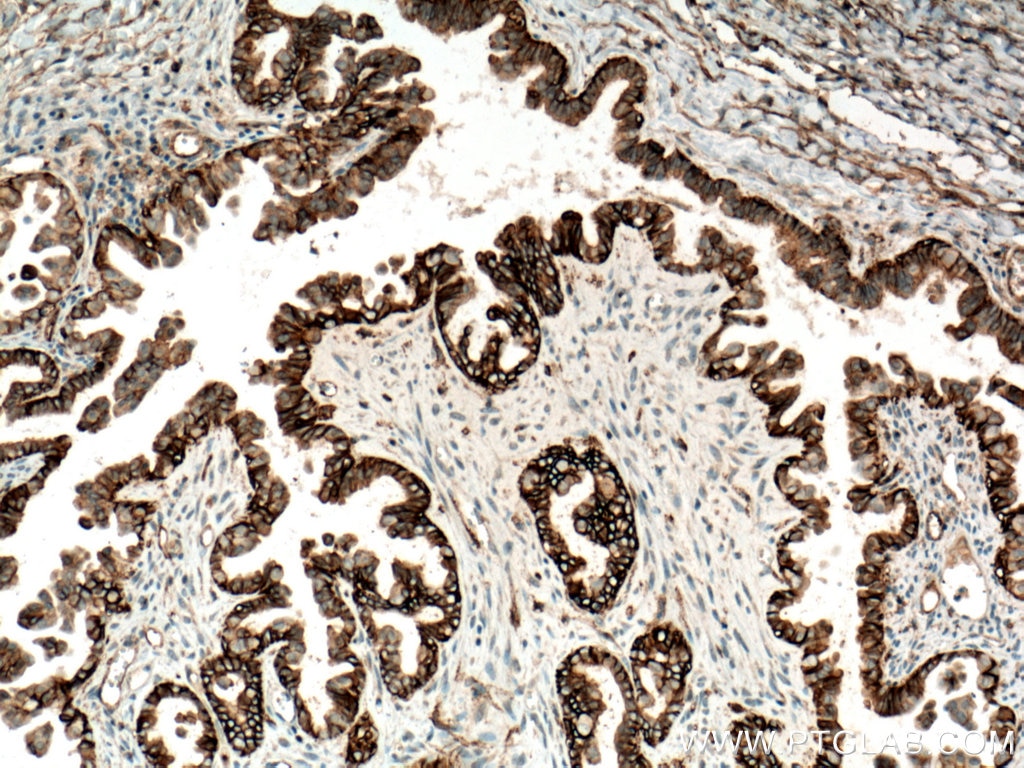
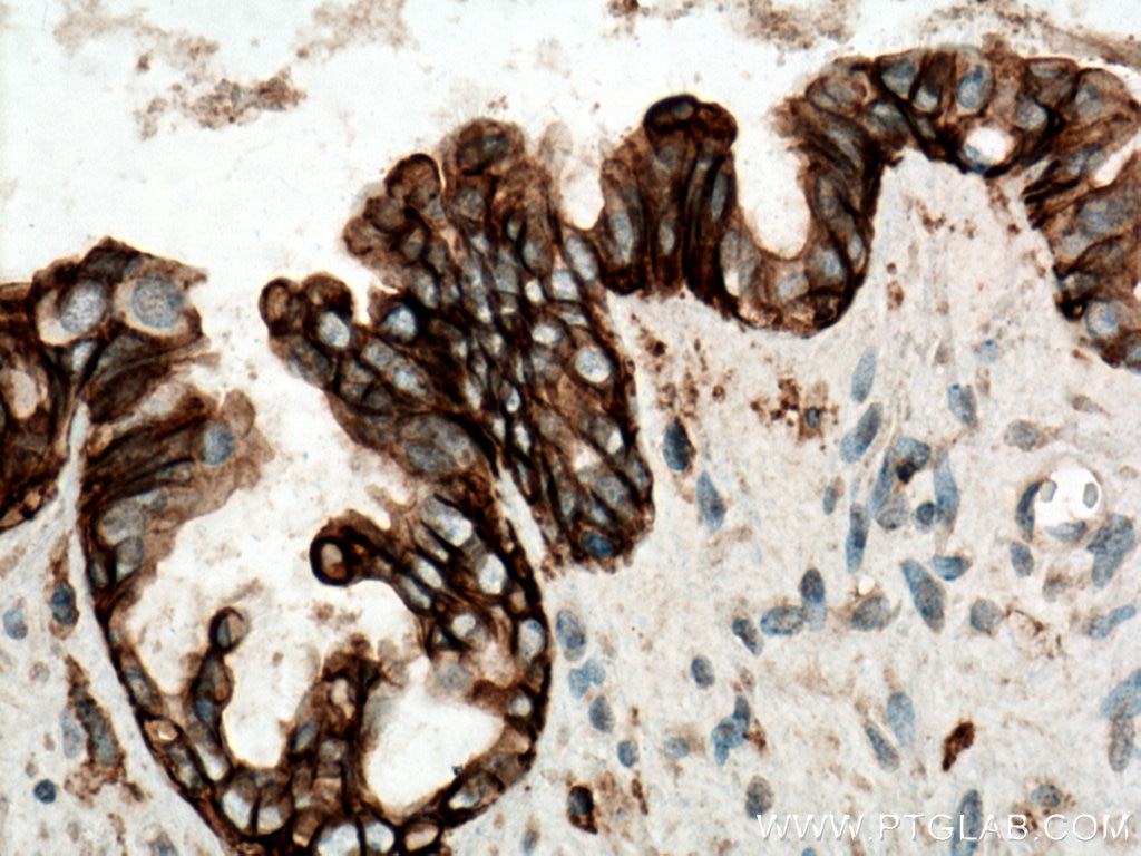
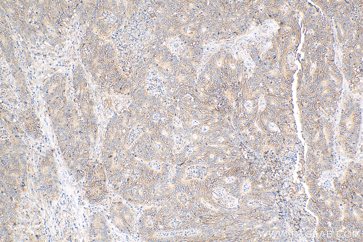
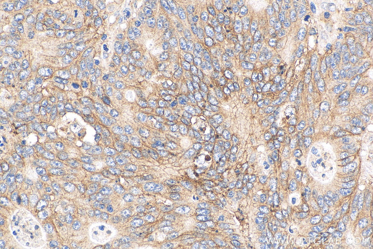
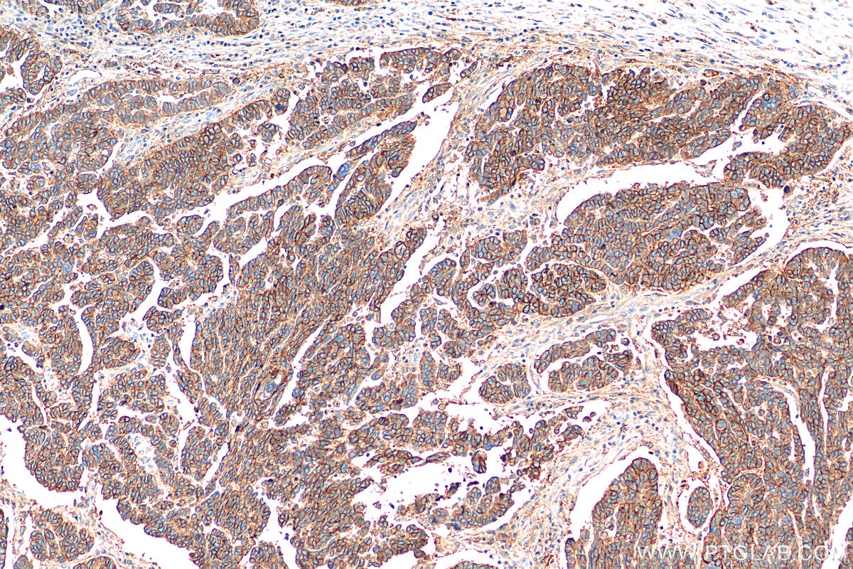
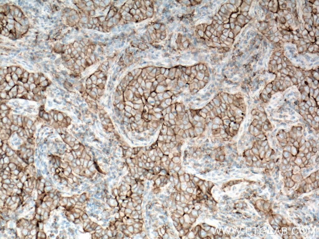
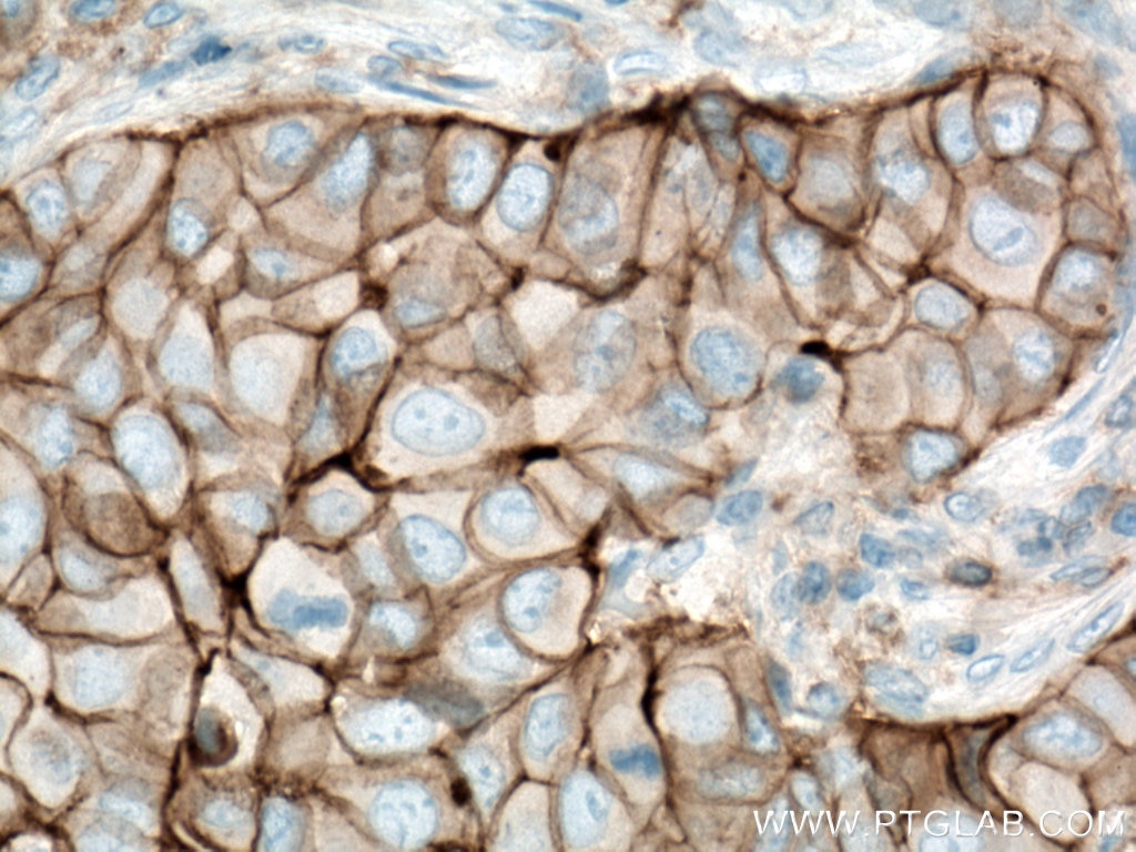
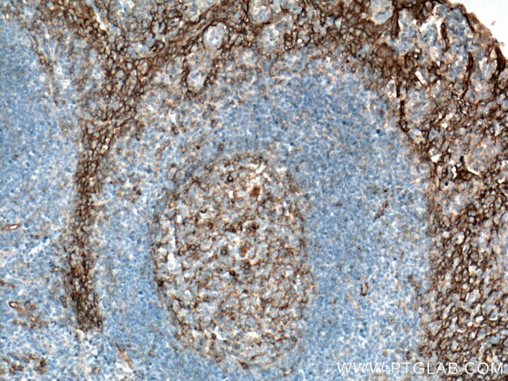
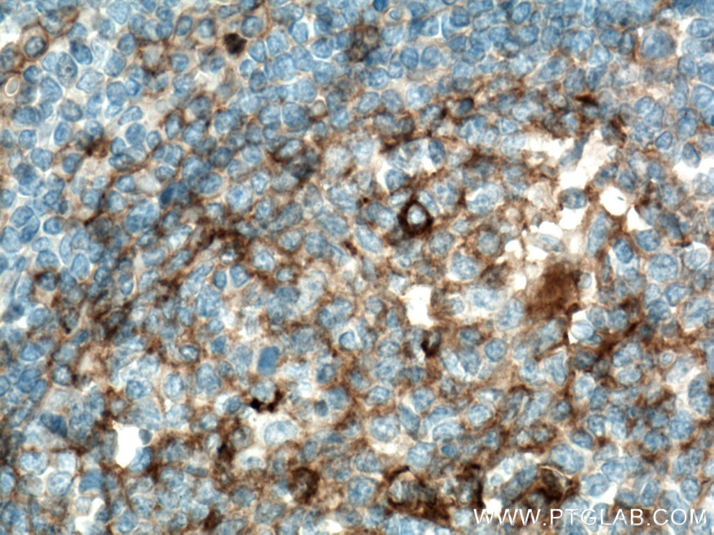
| Positive IHC detected in | human ovary tumor tissue, human breast cancer tissue, human colon cancer tissue, human tonsillitis tissue Note: suggested antigen retrieval with TE buffer pH 9.0; (*) Alternatively, antigen retrieval may be performed with citrate buffer pH 6.0 |
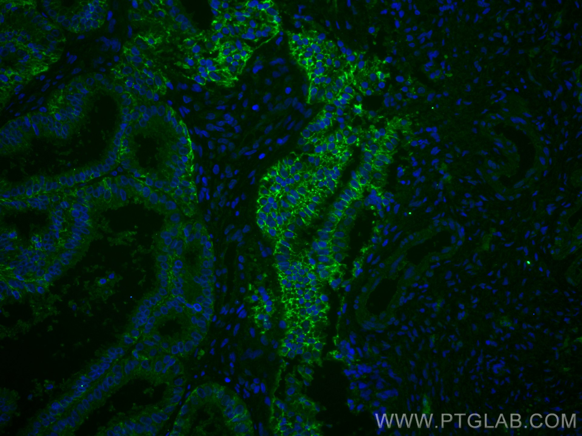
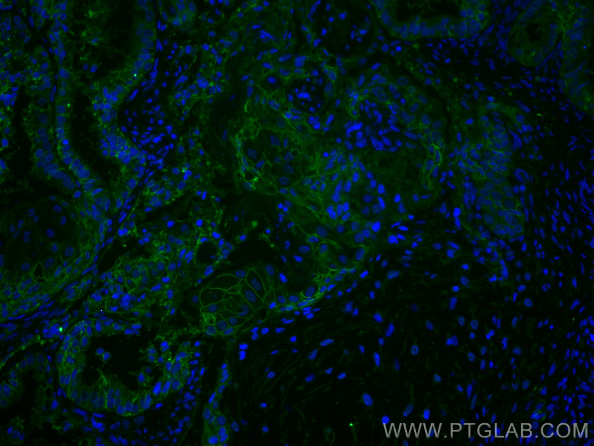
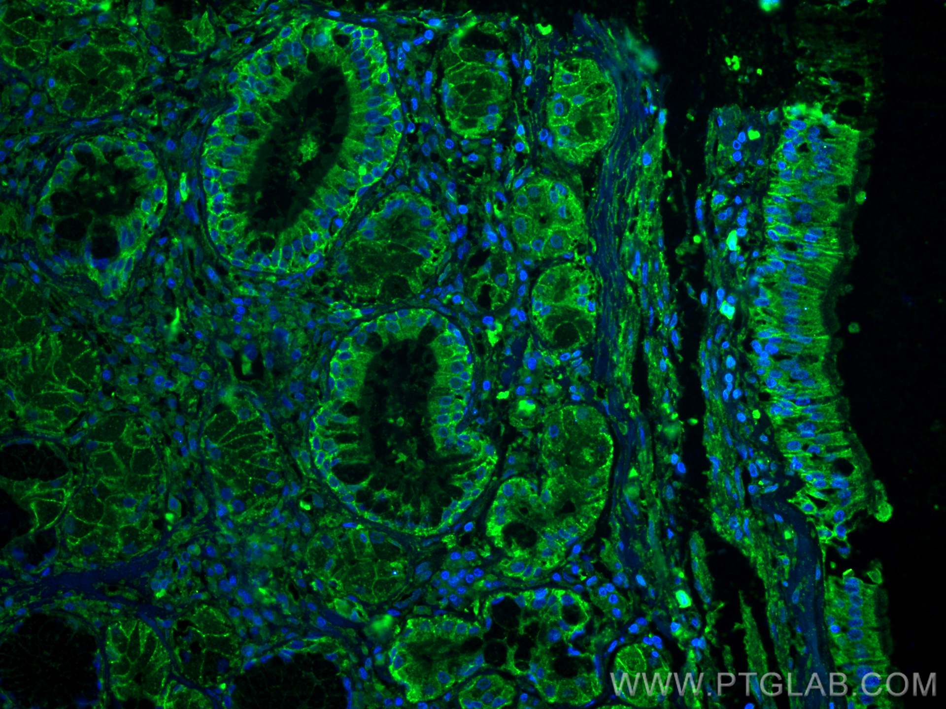
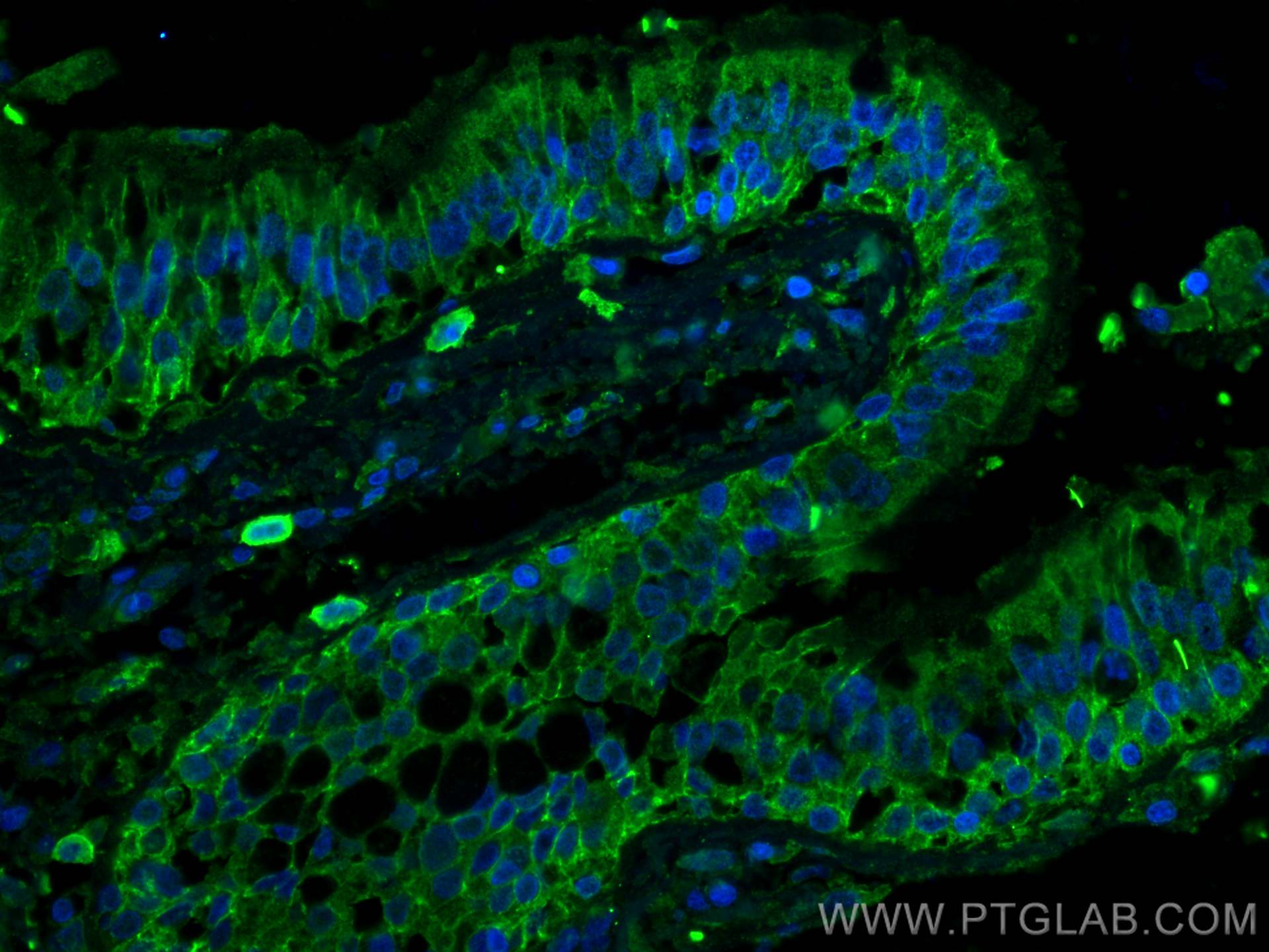
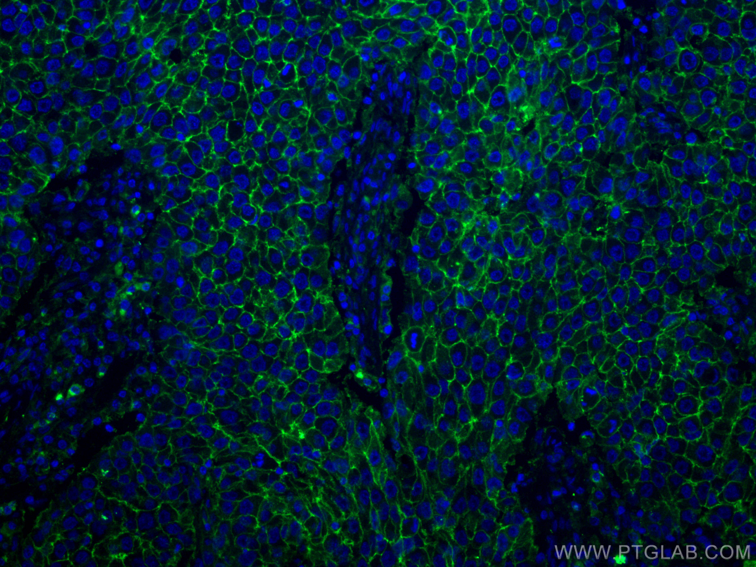
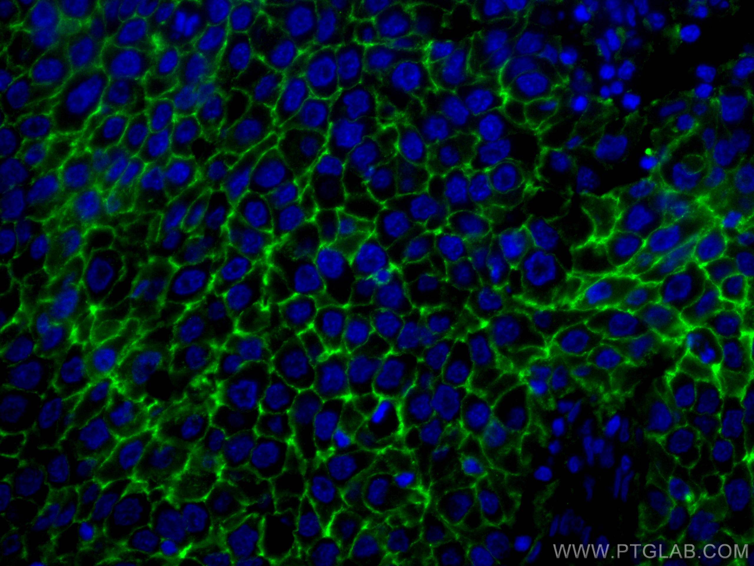
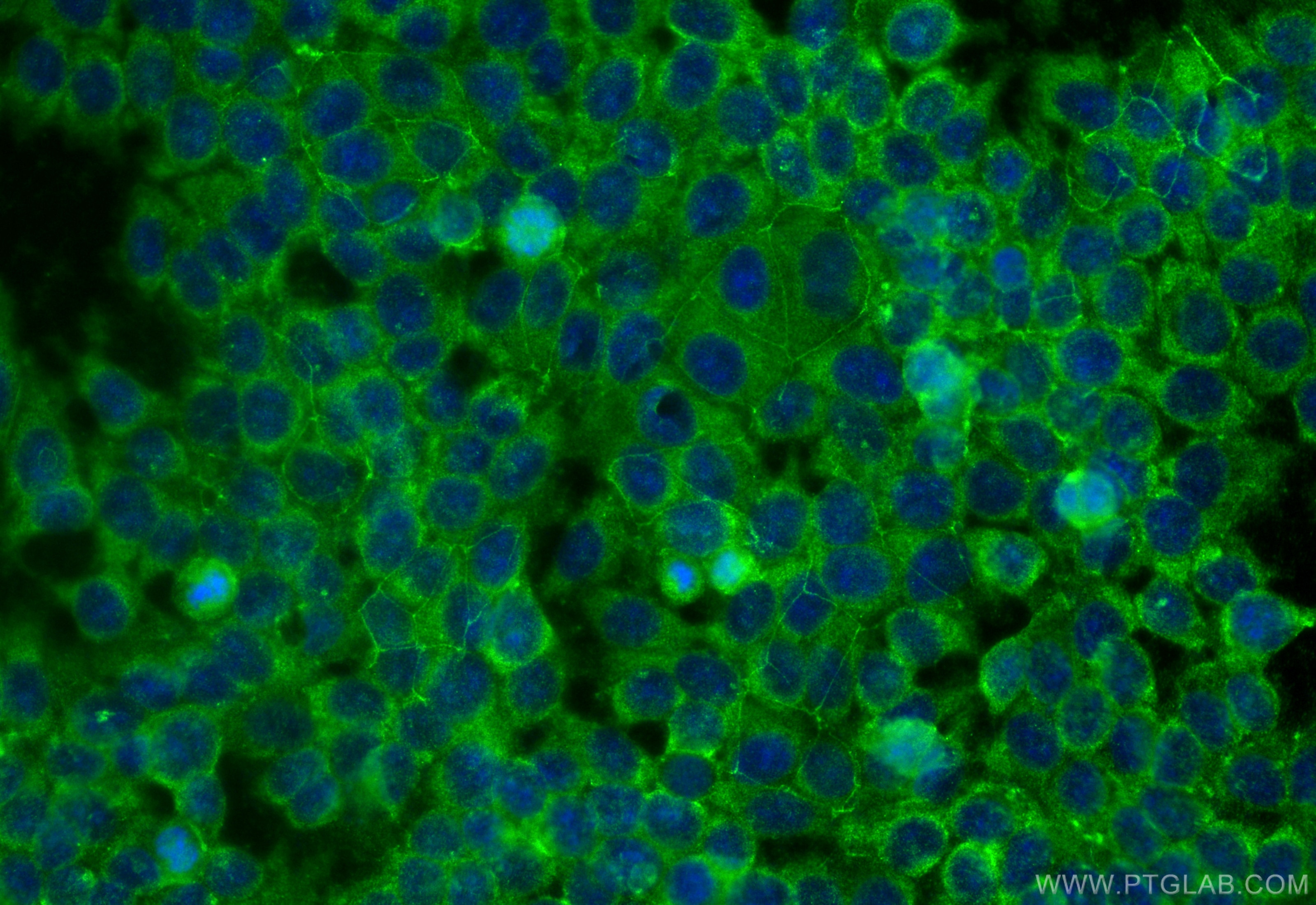
| Positive IF-P detected in | human breast cancer tissue, human ovary tumor tissue, human lung cancer tissue |
| Positive IF/ICC detected in | MCF-7 cells |
Recommended dilution
| Application | Dilution |
|---|---|
| Western Blot (WB) | WB : 1:5000-1:50000 |
| Immunohistochemistry (IHC) | IHC : 1:1000-1:4000 |
| Immunofluorescence (IF)-P | IF-P : 1:200-1:800 |
| Immunofluorescence (IF)/ICC | IF/ICC : 1:400-1:1600 |
| It is recommended that this reagent should be titrated in each testing system to obtain optimal results. | |
| Sample-dependent, Check data in validation data gallery. | |
Published Applications
| KD/KO | See 1 publications below |
| WB | See 140 publications below |
| IHC | See 4 publications below |
| IF | See 12 publications below |
| FC | See 1 publications below |
Product Information
60232-1-Ig targets CD9 in WB, IHC, IF/ICC, IF-P, ELISA, PLA applications and shows reactivity with human samples.
| Tested Reactivity | human |
| Cited Reactivity | human, rabbit |
| Host / Isotype | Mouse / IgG1 |
| Class | Monoclonal |
| Type | Antibody |
| Immunogen |
CatNo: Ag14529 Product name: Recombinant human CD9 protein Source: e coli.-derived, PET28a Tag: 6*His Domain: 100-208 aa of BC011988 Sequence: FAIEIAAAIWGYSHKDEVIKEVQEFYKDTYNKLKTKDEPQRETLKAIHYALNCCGLAGGVEQFISDICPKKDVLETFTVKSCPDAIKEVFDNKFHIIGAVGIGIAVVMI Predict reactive species |
| Full Name | CD9 molecule |
| Calculated Molecular Weight | 228 aa, 25 kDa |
| Observed Molecular Weight | 23-27 kDa |
| GenBank Accession Number | BC011988 |
| Gene Symbol | CD9 |
| Gene ID (NCBI) | 928 |
| RRID | AB_11232215 |
| Conjugate | Unconjugated |
| Form | Liquid |
| Purification Method | Protein G purification |
| UNIPROT ID | P21926 |
| Storage Buffer | PBS with 0.02% sodium azide and 50% glycerol, pH 7.3. |
| Storage Conditions | Store at -20°C. Stable for one year after shipment. Aliquoting is unnecessary for -20oC storage. 20ul sizes contain 0.1% BSA. |
Background Information
The cell-surface molecule CD9, a member of the transmembrane-4 superfamily, interacts with the integrin family and other membrane proteins, and is postulated to participate in cell migration and adhesion. Expression of CD9 enhances membrane fusion between muscle cells and promotes viral infection in some cells (PMID:10459022). It is often used as a mesenchymal stem cell marker (PMID:18005405). CD9 is also known as the p24 antigen besides MIC3, TSPAN29 because it is a protein of molecular weight 24 kD. The CD9 antigen appears to be a 227-amino acid molecule with 4 hydrophobic domains and 1 N-glycosylation site.
Protocols
| Product Specific Protocols | |
|---|---|
| IF protocol for CD9 antibody 60232-1-Ig | Download protocol |
| IHC protocol for CD9 antibody 60232-1-Ig | Download protocol |
| WB protocol for CD9 antibody 60232-1-Ig | Download protocol |
| Standard Protocols | |
|---|---|
| Click here to view our Standard Protocols |
Publications
| Species | Application | Title |
|---|---|---|
Nat Commun A functionally tunable magnetic nanochains platform for N-glycoproteomic analysis of extracellular vesicles from ultratrace biofluids | ||
Transl Neurodegener Abnormal α-synuclein binds to synaptotagmin 13, impairing extracellular vesicle release in synucleinopathies | ||
J Extracell Vesicles 124I-labelled BMSC-Derived Extracellular Vesicles Deliver CRISPR/Cas9 Ribonucleoproteins With a GFP-Reporter System to Inhibit Osteosarcoma Proliferation and Metastasis | ||
Adv Sci (Weinh) Cancer Cell-Derived Large Extracellular Vesicles Promote Venous Thromboembolism by Activating NETosis Through Delivering CYBA | ||
Adv Sci (Weinh) POSTN-Mediated Interplay of M1 Polarized Macrophage with Tendon-Derived Stem Cells to Drive Traumatic Heterotopic Ossification Formation through PTK7/ATK Signaling |
Reviews
The reviews below have been submitted by verified Proteintech customers who received an incentive for providing their feedback.
FH Guorong (Verified Customer) (03-22-2022) | Very weak bands with strong background were detected at 25 kDa
 |