

Tested Applications
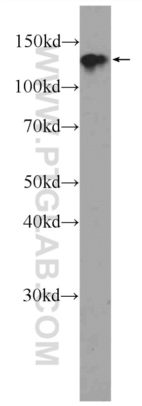
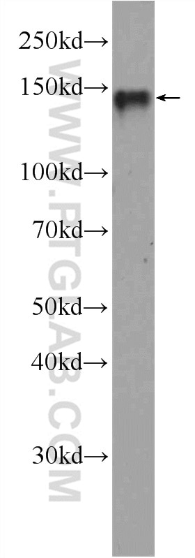
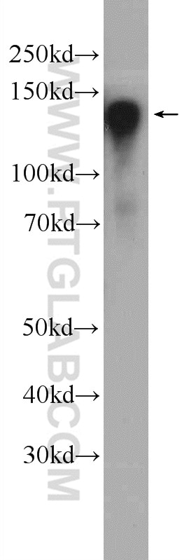
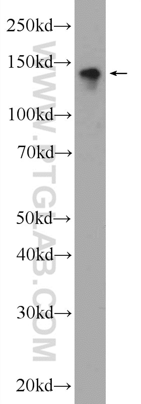
| Positive WB detected in | NIH/3T3 cells, HeLa cells, mouse brain tissue, Neuro-2a cells |
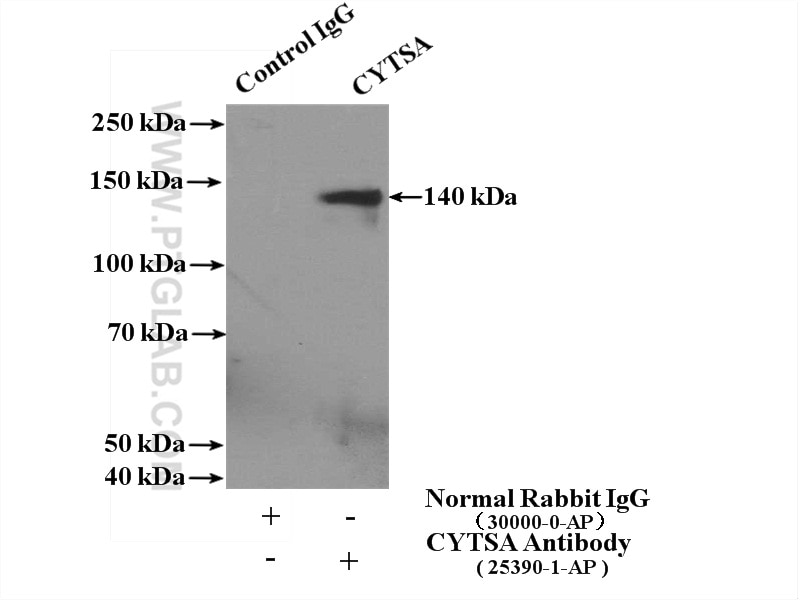
| Positive IP detected in | mouse brain tissue |
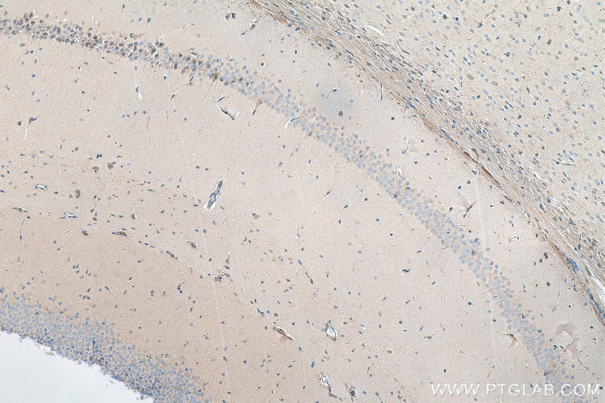
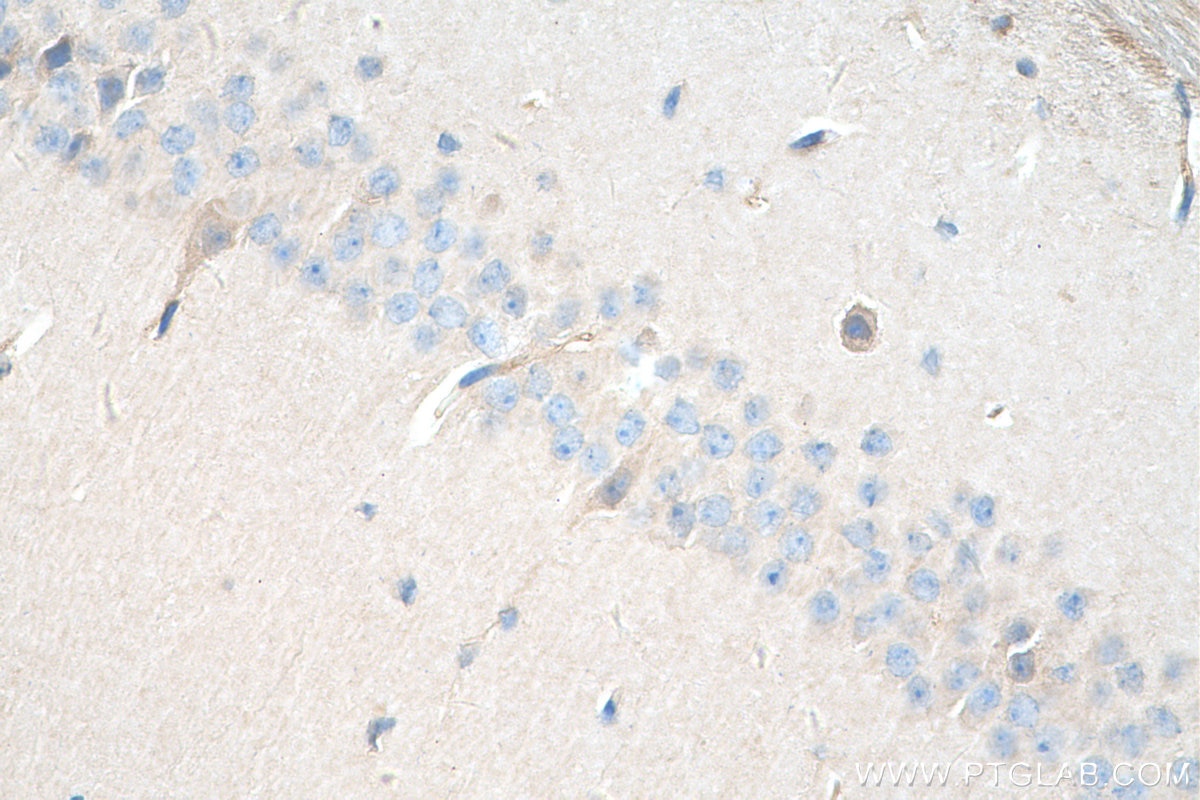
| Positive IHC detected in | mouse brain tissue Note: suggested antigen retrieval with TE buffer pH 9.0; (*) Alternatively, antigen retrieval may be performed with citrate buffer pH 6.0 |
Recommended dilution
| Application | Dilution |
|---|---|
| Western Blot (WB) | WB : 1:500-1:1000 |
| Immunoprecipitation (IP) | IP : 0.5-4.0 ug for 1.0-3.0 mg of total protein lysate |
| Immunohistochemistry (IHC) | IHC : 1:50-1:500 |
| It is recommended that this reagent should be titrated in each testing system to obtain optimal results. | |
| Sample-dependent, Check data in validation data gallery. | |
Product Information
25390-1-AP targets CYTSA in WB, IHC, IF, IP, ELISA applications and shows reactivity with human, mouse samples.
| Tested Reactivity | human, mouse |
| Cited Reactivity | human, mouse |
| Host / Isotype | Rabbit / IgG |
| Class | Polyclonal |
| Type | Antibody |
| Immunogen |
CatNo: Ag21896 Product name: Recombinant human CYTSA protein Source: e coli.-derived, PGEX-4T Tag: GST Domain: 1-352 aa of BC136471 Sequence: MKKASRSVGSVPKVSAISKTQTAEKIKPENSSSASTGGKLVKPGTAASLSKTKSSDDLLAGMAGGVTVTNGVKGKKSTCPSAAPSASAPAMTTVENKSKISTGTASSTKRSTSTGNKESSSTRERLRERTRLNQSKKLPSAGQGANDMALAKRSRSRTATECDVRMSKSKSDNQISDRAALEAKVKDLLTLAKTKDVEILHLRNELRDMRAQLGINEDHSEGDEKSEKETIMAHQPTDVESTLLQLQEQNTAIREELNQLKNENRMLKDRLNALGFSLEQRLDNSEKLFGYQSLSPEITPGNQSDGGGTLTSSVEGSAPGSVEDLLSQDENTLMDHQHSNSMDNLDSECSEV Predict reactive species |
| Full Name | cytospin A |
| Calculated Molecular Weight | 1117 aa, 125 kDa |
| Observed Molecular Weight | 130 kDa |
| GenBank Accession Number | BC136471 |
| Gene Symbol | CYTSA |
| Gene ID (NCBI) | 23384 |
| RRID | AB_2880056 |
| Conjugate | Unconjugated |
| Form | Liquid |
| Purification Method | Antigen affinity purification |
| UNIPROT ID | Q69YQ0 |
| Storage Buffer | PBS with 0.02% sodium azide and 50% glycerol, pH 7.3. |
| Storage Conditions | Store at -20°C. Stable for one year after shipment. Aliquoting is unnecessary for -20oC storage. 20ul sizes contain 0.1% BSA. |
Background Information
CYTSA, also named as SPECC1L or KIAA0376, is a 1117 amino acid protein, which contains one CH domain and belongs to the cytospin-A family. CYTSA localizes in the cytoplasm and is involved in cytokinesis and spindle organization. CYTSA may play a role in actin cytoskeleton organization and microtubule stabilization, hence it is required for proper cell adhesion and migration.
Protocols
| Product Specific Protocols | |
|---|---|
| IHC protocol for CYTSA antibody 25390-1-AP | Download protocol |
| IP protocol for CYTSA antibody 25390-1-AP | Download protocol |
| WB protocol for CYTSA antibody 25390-1-AP | Download protocol |
| Standard Protocols | |
|---|---|
| Click here to view our Standard Protocols |
Publications
What published studies show
The anti-SPECC1L antibody (Cat# 25390-1-AP) has 8 total citations. These appear in Current Biology, Frontiers in Physiology, Journal of Biological Chemistry, Human Molecular Genetics, and bioRxiv. Research fields include cytoskeletal biology (microtubule dynamics, actin regulation), developmental biology (cranial neural crest, palate development, frontonasal dysplasia), and signaling pathway regulation (Hedgehog signaling, IRF6 downstream effects).
| Species | Application | Title |
|---|---|---|
Curr Biol EML2-S constitutes a new class of proteins that recognizes and regulates the dynamics of tyrosinated microtubules | ||
Front Physiol Loss of SPECC1L in cranial neural crest cells results in increased hedgehog signaling and frontonasal dysplasia. | ||
J Biol Chem SPECC1L binds the myosin phosphatase complex MYPT1/PP1β and can regulate its distribution between microtubules and filamentous actin | ||
Hum Mol Genet In-frame deletion of SPECC1L microtubule association domain results in gain-of-function phenotypes affecting embryonic tissue movement and fusion events. | ||
bioRxiv Loss of SPECC1L in cranial neural crest cells results in increased hedgehog signaling and frontonasal dysplasia. |








