

Tested Applications
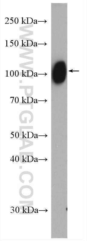
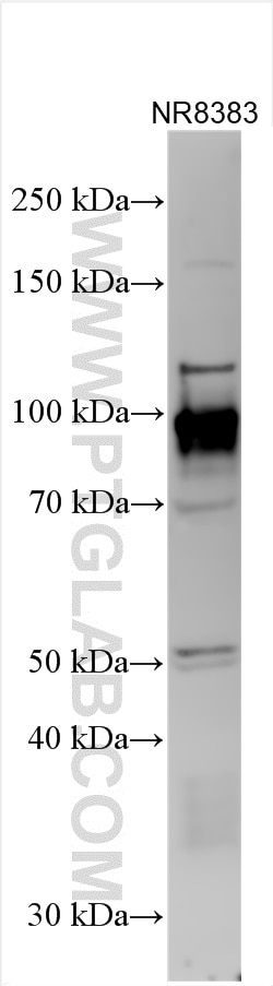
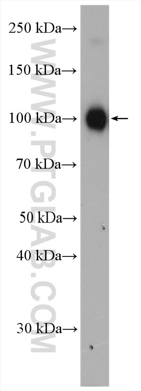
| Positive WB detected in | RAW 264.7 cells, mouse spleen tissue, NR8383 cells |
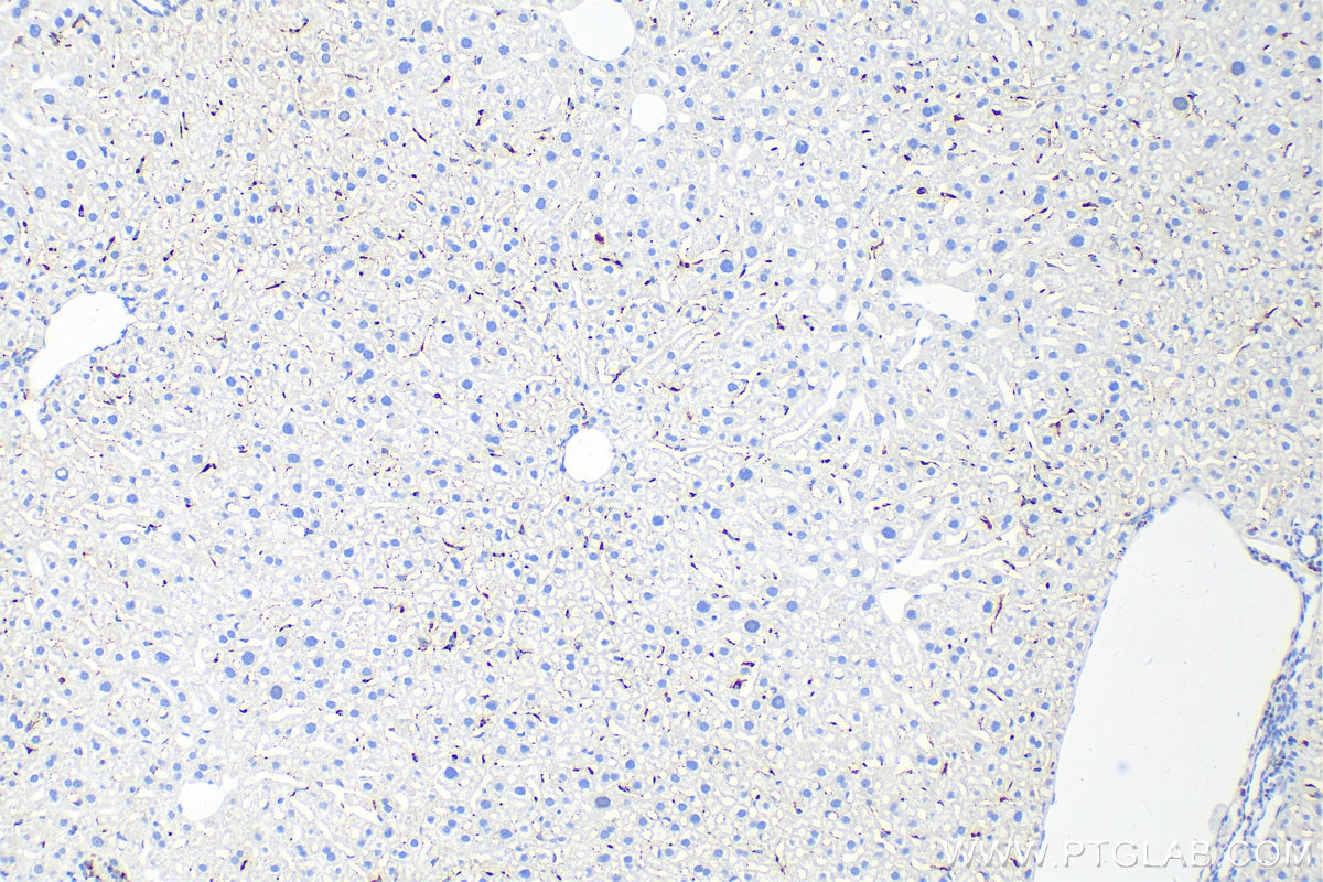
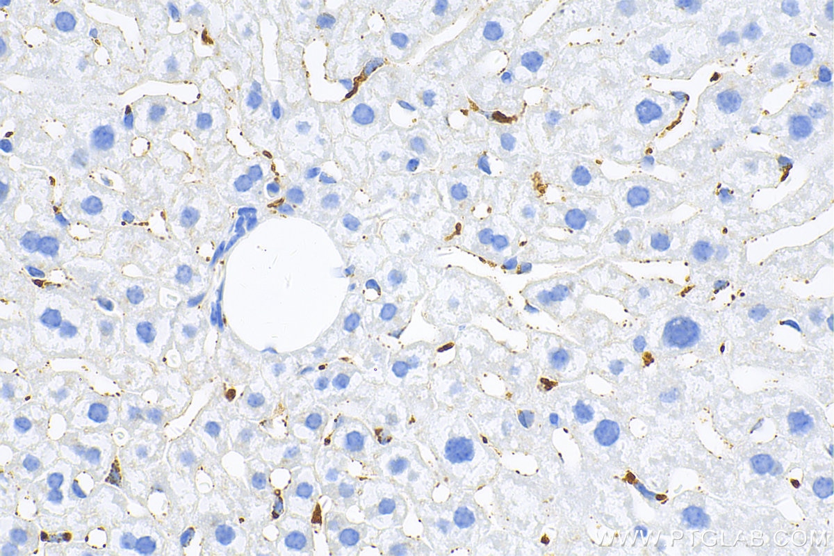
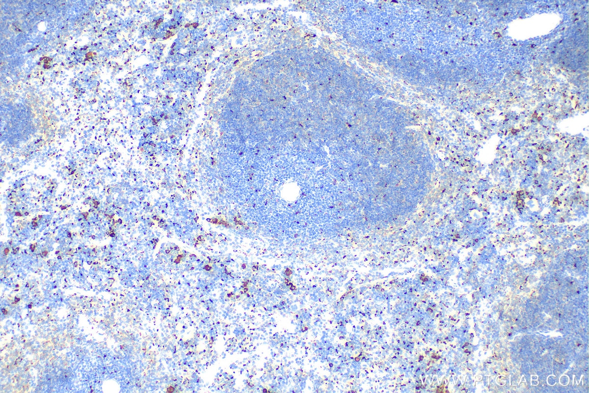
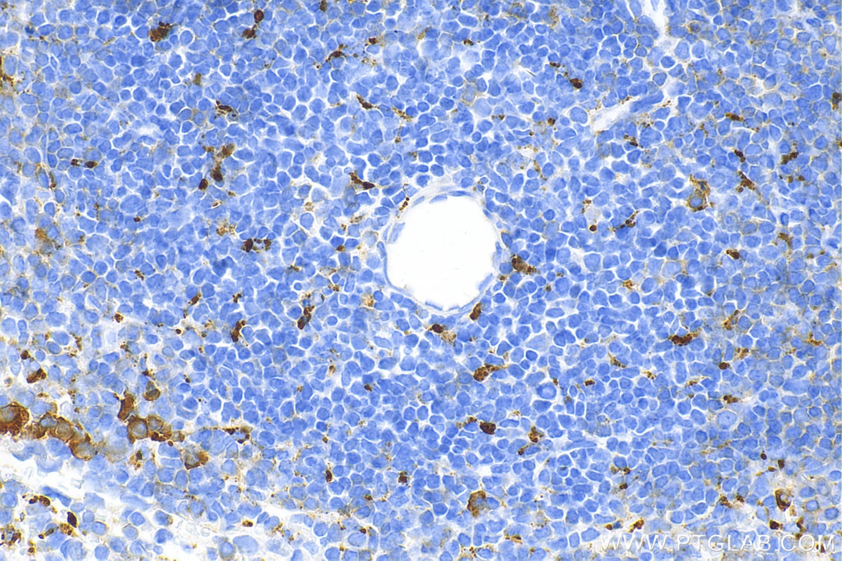
| Positive IHC detected in | mouse spleen tissue, mouse liver tissue Note: suggested antigen retrieval with TE buffer pH 9.0; (*) Alternatively, antigen retrieval may be performed with citrate buffer pH 6.0 |
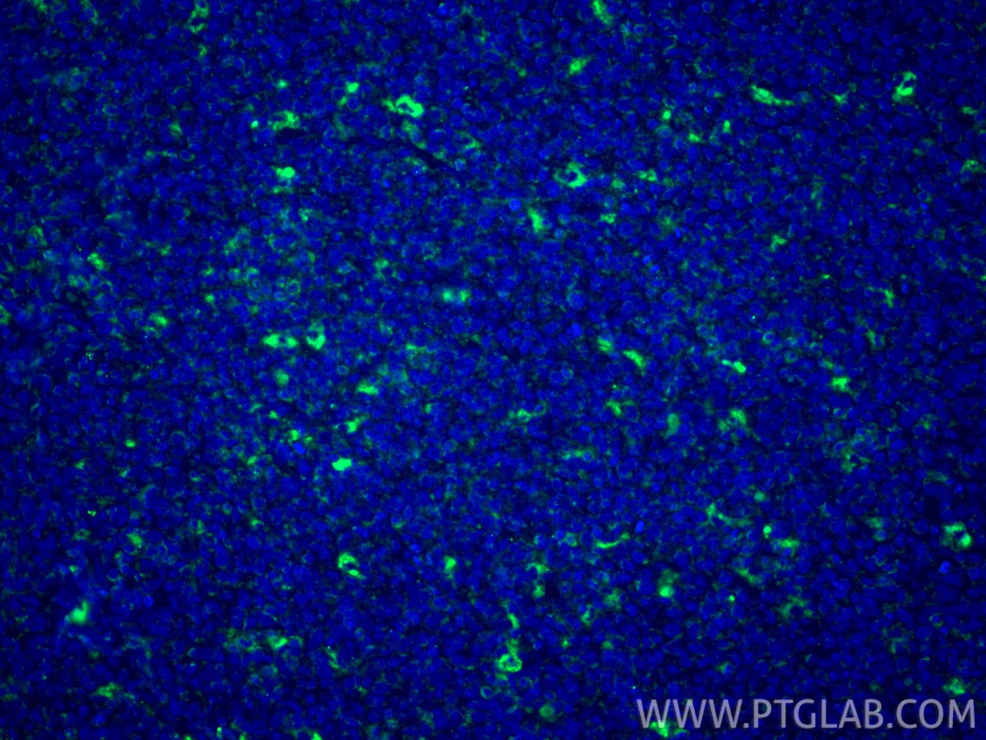
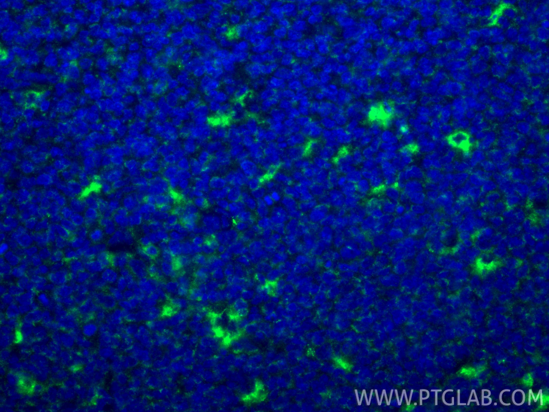
| Positive IF-P detected in | human tonsillitis tissue |
Recommended dilution
| Application | Dilution |
|---|---|
| Western Blot (WB) | WB : 1:600-1:2000 |
| Immunohistochemistry (IHC) | IHC : 1:1000-1:4000 |
| Immunofluorescence (IF)-P | IF-P : 1:50-1:500 |
| It is recommended that this reagent should be titrated in each testing system to obtain optimal results. | |
| Sample-dependent, Check data in validation data gallery. | |
Product Information
28058-1-AP targets CD68 in WB, IHC, IF-P, ELISA applications and shows reactivity with mouse, rat samples.
| Tested Reactivity | mouse, rat |
| Cited Reactivity | mouse, rat, pig, canine, sheep |
| Host / Isotype | Rabbit / IgG |
| Class | Polyclonal |
| Type | Antibody |
| Immunogen |
CatNo: Ag27827 Product name: Recombinant mouse Cd68 protein Source: e coli.-derived, PGEX-4T Tag: GST Domain: 119-291 aa of NM_001291058 Sequence: MPSPRSKGALGNYTWANGSQPCVQLQAQIQIRILYPIQGGRKAWGISVLNPNKTKVQGGCDGTHPHLSLSFPYGQLTFGFKQDLHQSPSTVYLDYMAVEYNVSFPQAAQWTFMAQNSSLRELQAPLGQSFCCGNASIVLSPAVHLDLLSLRLQAAQLPDKGHFGPCFSCNRDQS Predict reactive species |
| Full Name | CD68 antigen |
| Calculated Molecular Weight | 35 kDa |
| Observed Molecular Weight | 100 kDa |
| GenBank Accession Number | NM_001291058 |
| Gene Symbol | Cd68 |
| Gene ID (NCBI) | 12514 |
| RRID | AB_2881049 |
| Conjugate | Unconjugated |
| Form | Liquid |
| Purification Method | Antigen affinity purification |
| UNIPROT ID | P31996 |
| Storage Buffer | PBS with 0.02% sodium azide and 50% glycerol, pH 7.3. |
| Storage Conditions | Store at -20°C. Stable for one year after shipment. Aliquoting is unnecessary for -20oC storage. 20ul sizes contain 0.1% BSA. |
Background Information
Mouse CD68 (also known as macrosialin) is a type I transmembrane glycoprotein that is highly and specifically expressed by mouse tissue macrophages, and to a lesser extent by dendritic cells. It belongs to the lysosomal/endosomal-associated membrane glycoprotein (LAMP) family and primarily localizes to lysosomes and endosomes with a smaller fraction circulating to the cell surface. CD68 is also a member of the scavenger receptor family. It may play a role in phagocytic activities of tissue macrophages. Due to heavy glycosylation, the apparent molecular mass of CD68 detected by Western blot is generally observed between 80 and 140 kDa, far exceeding its unmodified core protein weight of ~34-37 kDa.
Protocols
| Product Specific Protocols | |
|---|---|
| IF protocol for CD68 antibody 28058-1-AP | Download protocol |
| IHC protocol for CD68 antibody 28058-1-AP | Download protocol |
| WB protocol for CD68 antibody 28058-1-AP | Download protocol |
| Standard Protocols | |
|---|---|
| Click here to view our Standard Protocols |
Publications
What published studies show
CD68 Polyclonal antibody (Cat# 28058-1-AP) has accumulated 479 citations in high-impact journals including Nature, Cancer Cell, Nature Communications, Advanced Science, Journal of Controlled Release, Biomaterials, Cell Death & Differentiation, ACS Nano, Acta Biomaterialia, and Journal of Nanobiotechnology. Key research fields include macrophage biology and polarization, neuroinflammation, atherosclerosis, tumor microenvironment, wound healing, ferroptosis, diabetic complications, liver fibrosis, and biomaterials/tissue engineering.
| Species | Application | Title |
|---|---|---|
Signal Transduct Target Ther C/EBPβ activation in vascular smooth muscle cells promotes hyperlipidemia-induced phenotypic transition and arterial stiffness | ||
Cancer Cell Spatial determinants of antibody-drug conjugate SHR-A1811 efficacy in neoadjuvant treatment for HER2-positive breast cancer | ||
Cancer Cell Multimodal spatial-omics reveal co-evolution of alveolar progenitors and proinflammatory niches in progression of lung precursor lesions. | ||
Immunity Expression of the readthrough transcript CiDRE in alveolar macrophages boosts SARS-CoV-2 susceptibility and promotes COVID-19 severity | ||
Nat Aging Activation of AMPK by GLP-1R agonists mitigates Alzheimer-related phenotypes in transgenic mice |
Reviews
What customers say
All three reviews are 5-star. Users report excellent performance across Western Blot, Immunohistochemistry, and Immunofluorescence applications. One user highlighted very bright WB bands and beautiful confocal images at 1:800 dilution. Another confirmed reliable IF results on mouse ventricle tissue. A third used it at 1:1000 for IF on mouse adrenal gland sections with heat-mediated antigen retrieval, achieving clear CD68 staining. Overall, reviewers consistently praise the antibody's strong signal and reliability.
The reviews below have been submitted by verified Proteintech customers who received an incentive for providing their feedback.
Mounika (Verified Customer) (01-08-2026) | Bands are very bright and cells under confocal microscope are very beautiful!
|
Alexandru (Verified Customer) (11-13-2023) | Great antibody, worked well in my experiments.
|
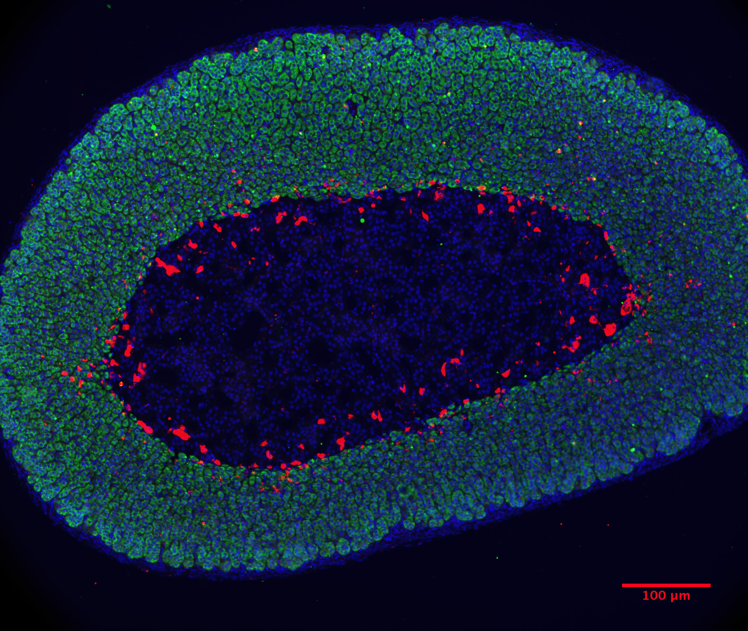
Amelie (Verified Customer) (09-09-2019) | sections were deparaffinized and subjected to heat mediated antigen retrieval using TE buffer ph9On the pictures CD68 in red
 |










