

Tested Applications
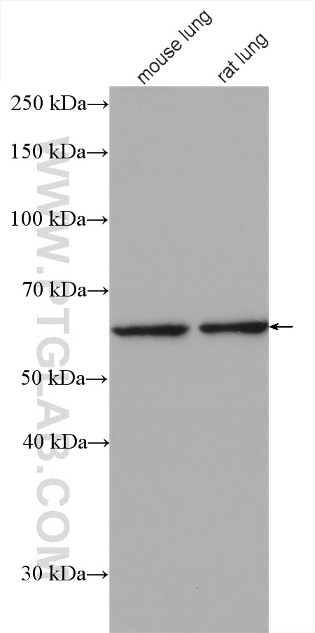
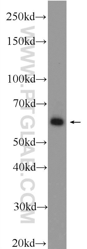
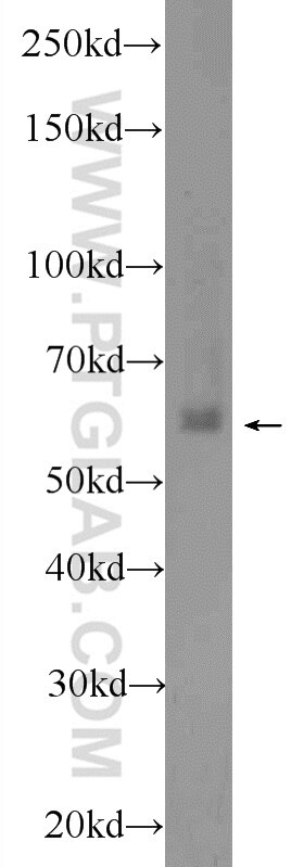
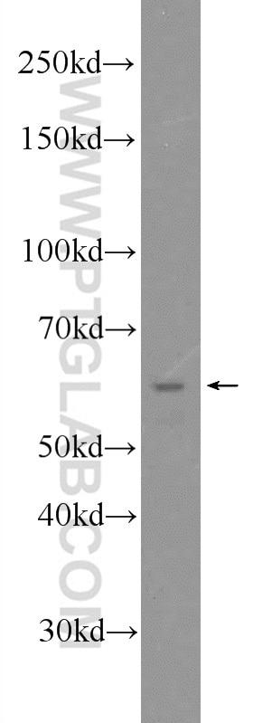
| Positive WB detected in | mouse lung tissue, mouse brain tissue, HeLa cells, mouse testis tissue, rat lung tissue |
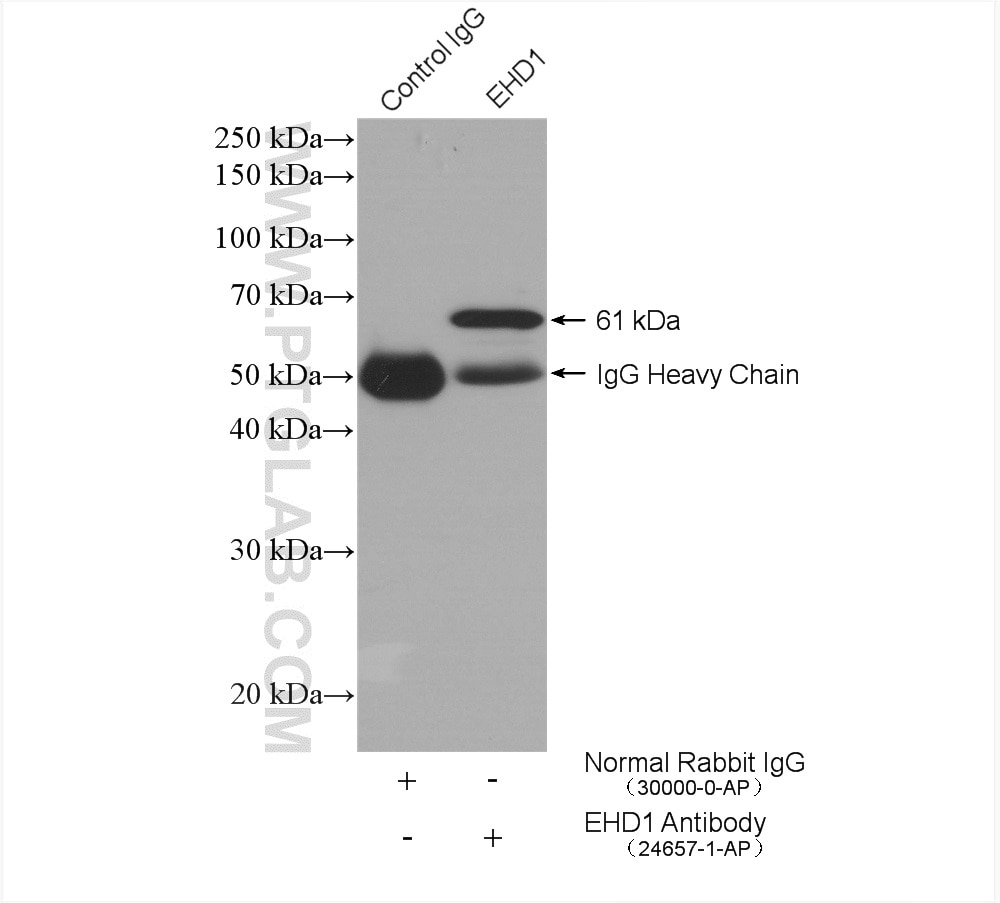
| Positive IP detected in | mouse testis tissue |
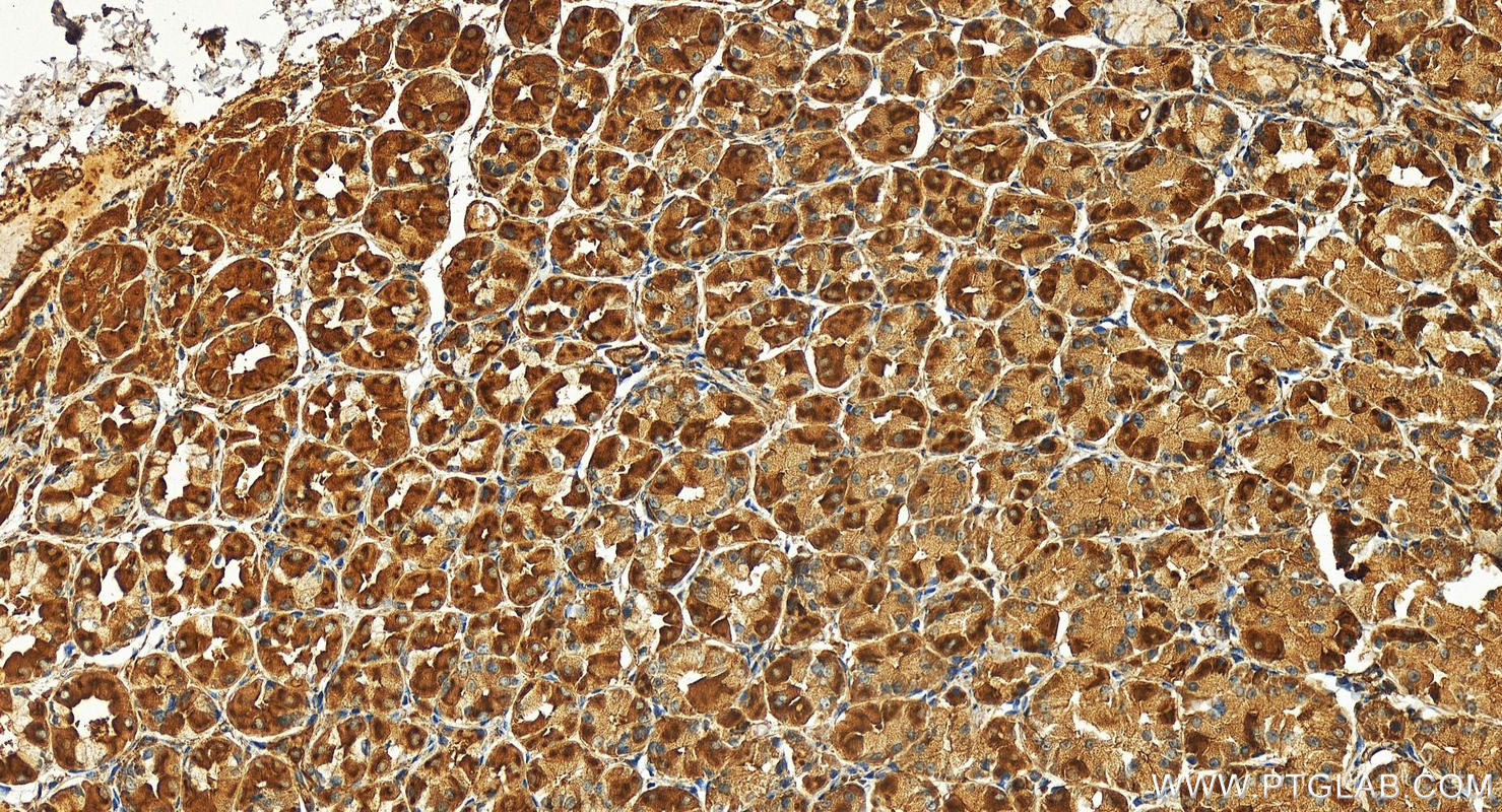
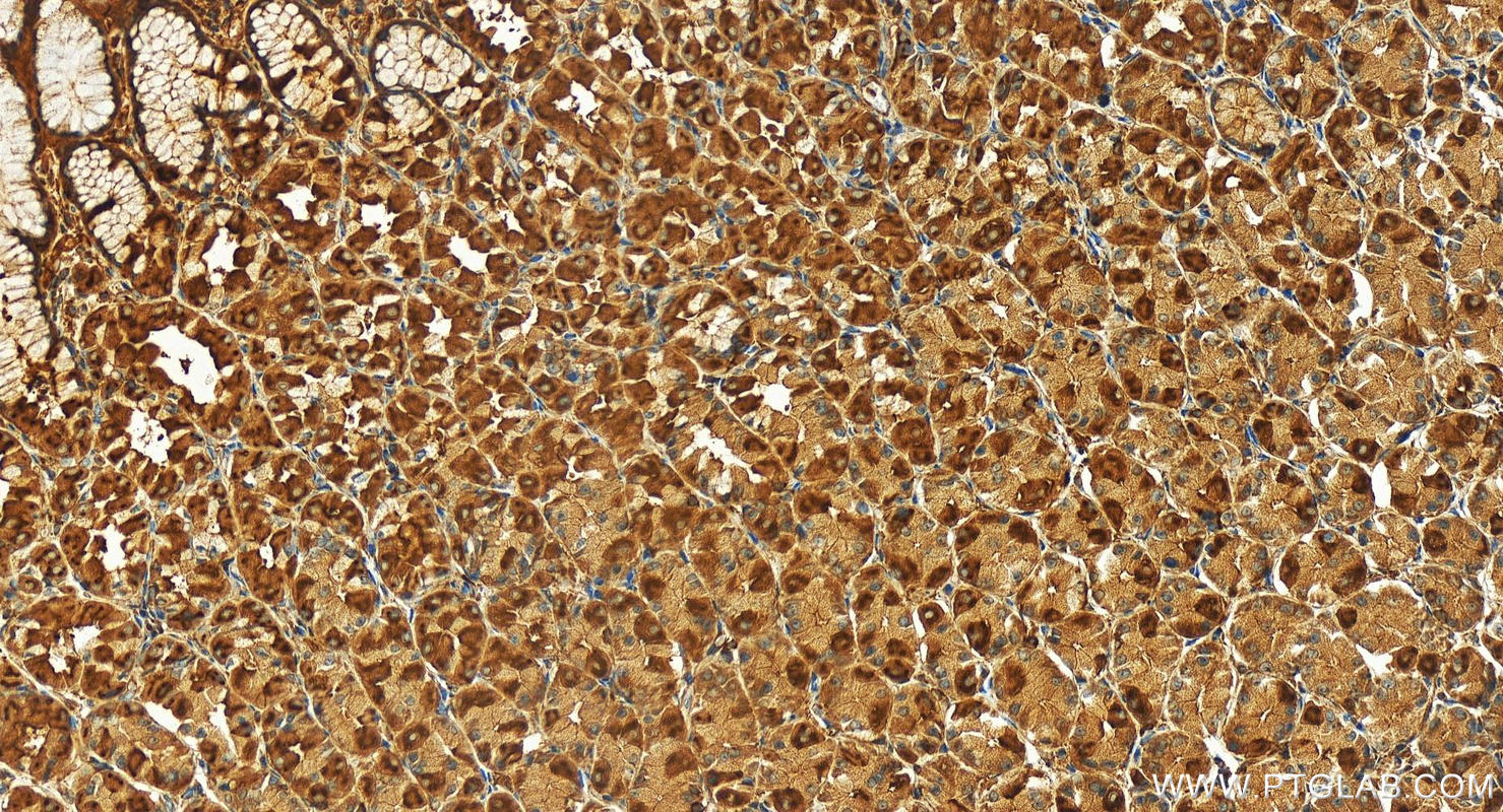
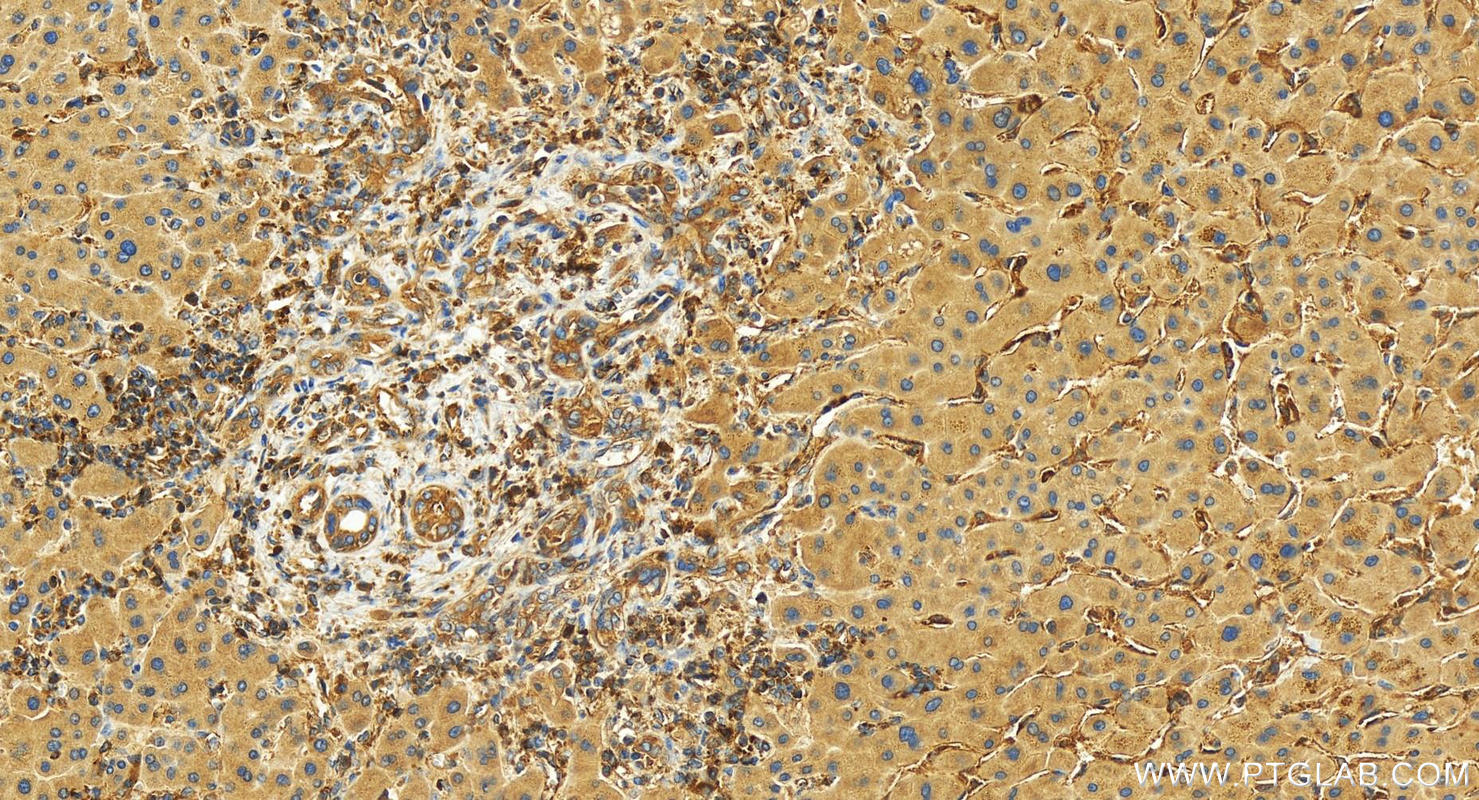
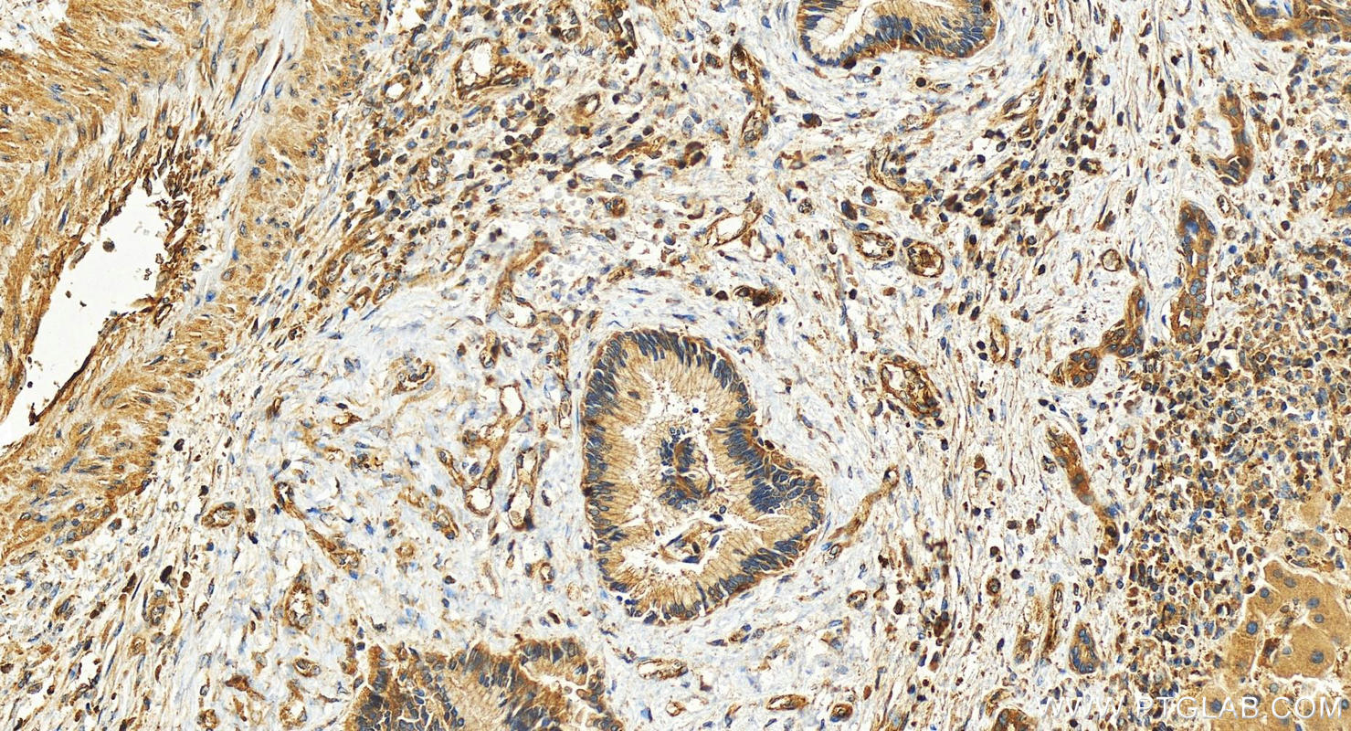
| Positive IHC detected in | human stomach cancer tissue, human intrahepatic cholangiocarcinoma tissue Note: suggested antigen retrieval with TE buffer pH 9.0; (*) Alternatively, antigen retrieval may be performed with citrate buffer pH 6.0 |
Recommended dilution
| Application | Dilution |
|---|---|
| Western Blot (WB) | WB : 1:1000-1:4000 |
| Immunoprecipitation (IP) | IP : 0.5-4.0 ug for 1.0-3.0 mg of total protein lysate |
| Immunohistochemistry (IHC) | IHC : 1:50-1:500 |
| It is recommended that this reagent should be titrated in each testing system to obtain optimal results. | |
| Sample-dependent, Check data in validation data gallery. | |
Product Information
24657-1-AP targets EHD1 in WB, IHC, IF, IP, ELISA applications and shows reactivity with human, mouse, rat samples.
| Tested Reactivity | human, mouse, rat |
| Cited Reactivity | human, mouse |
| Host / Isotype | Rabbit / IgG |
| Class | Polyclonal |
| Type | Antibody |
| Immunogen |
CatNo: Ag18400 Product name: Recombinant human EHD1 protein Source: e coli.-derived, PGEX-4T Tag: GST Domain: 353-422 aa of BC104799 Sequence: FPSLRKMQELLQTQDFSKFQALKPKLLDTVDDMLANDIARLMVMVRQEESLMPSQVVKGGAFDGTMNGPF Predict reactive species |
| Full Name | EH-domain containing 1 |
| Calculated Molecular Weight | 534 aa, 61 kDa |
| Observed Molecular Weight | 61 kDa |
| GenBank Accession Number | BC104799 |
| Gene Symbol | EHD1 |
| Gene ID (NCBI) | 10938 |
| RRID | AB_2879658 |
| Conjugate | Unconjugated |
| Form | Liquid |
| Purification Method | Antigen affinity purification |
| UNIPROT ID | Q9H4M9 |
| Storage Buffer | PBS with 0.02% sodium azide and 50% glycerol, pH 7.3. |
| Storage Conditions | Store at -20°C. Stable for one year after shipment. Aliquoting is unnecessary for -20oC storage. 20ul sizes contain 0.1% BSA. |
Background Information
EHD1 (Eps15 Homology Domain Containing 1) is a protein that plays a crucial role in endocytic recycling, which is the process by which cells recycle their membrane components. It is one of four paralogs in mammals (EHD1-4) and is involved in several membrane trafficking pathways. EHD1 is particularly well-studied and is known to regulate the recycling of various cell surface receptors back to the cell surface after they have been endocytosed. This process is essential for maintaining the proper distribution of receptors and other membrane proteins, which in turn affects cellular signaling and function. EHD1 has been implicated in the regulation of the transferrin receptor (TfR), major histocompatibility complex (MHC) class I proteins, β1 integrins, and other receptors. It has also been shown to interact with Rab11-FIP2 and is localized to peripheral endosomes, suggesting a role in the transport of receptors from early endosomes to the endocytic recycling compartment (ERC). Furthermore, EHD1 has been linked to dynein motors that drive transport from early endosomes to the ERC via a complex including MICAL-L1 and the collapsin response mediator protein-2 (Crmp2).
Protocols
| Product Specific Protocols | |
|---|---|
| IHC protocol for EHD1 antibody 24657-1-AP | Download protocol |
| IP protocol for EHD1 antibody 24657-1-AP | Download protocol |
| WB protocol for EHD1 antibody 24657-1-AP | Download protocol |
| Standard Protocols | |
|---|---|
| Click here to view our Standard Protocols |
Publications
What published studies show
The EHD1 Polyclonal antibody (Cat# 24657-1-AP) has 7 total citations. These appear in journals including Cancer Communications, Journal of Extracellular Vesicles, Developmental Cell, EMBO Journal, Scientific Reports, BMC Cancer, and Biochemical and Biophysical Research Communications. Research fields span lung adenocarcinoma and immune evasion, breast cancer extracellular vesicles, O-GlcNAcylation, Zellweger syndrome, neuronal tunneling nanotubes, cisplatin resistance in non-small cell lung cancer, and ciliogenesis.
| Species | Application | Title |
|---|---|---|
Cancer Commun (Lond) m6A-modified EHD1 controls PD-L1 endosomal trafficking to modulate immune evasion and immunotherapy responses in lung adenocarcinoma | ||
J Extracell Vesicles Breast Cancer-Derived Extracellular Vesicles Modulate the Cytoplasmic and Cytoskeletal Dynamics of Blood-Brain Barrier Endothelial Cells | ||
Dev Cell A pathological role of O-GlcNAcylation-driven TR11B production and function in lung adenocarcinoma.
| ||
EMBO J Insufficiency of ciliary cholesterol in hereditary Zellweger syndrome.
| ||
Sci Rep Rab35 and its effectors promote formation of tunneling nanotubes in neuronal cells.
| ||
Reviews
What customers say
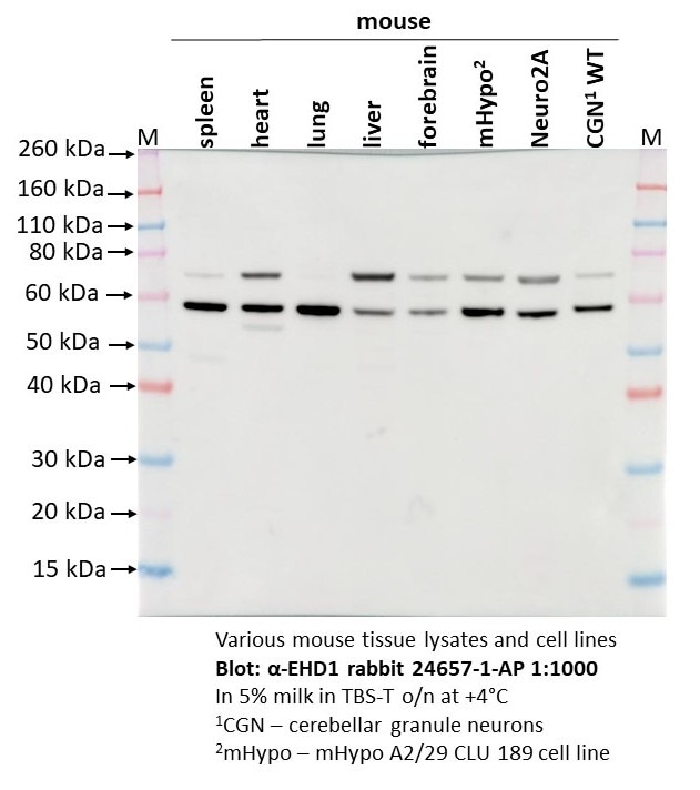
Customers report that this antibody performs well in Western blot across multiple applications and species. It produces clear, visible bands in mouse cell lines (N2a, N115, Neuro2A, mHypo), mouse tissues (spleen, heart, lung, liver, forebrain), mouse platelets, and HeLa cells, typically at a 1:1000 dilution. Most reviewers rate it 4–5 stars, praising its reliability for transfection studies and thrombosis research. One user noted some confusion from observing two bands in certain mouse cell lines and tissues, but overall found the blots clear. The antibody is generally described as working as expected with strong, single-band results.
The reviews below have been submitted by verified Proteintech customers who received an incentive for providing their feedback.
Krikor (Verified Customer) (09-17-2026) | Works as expected! We are using them with N2a and N115 cell lines and the bands are clearly visible when running westerns after transfection.
|
Olga (Verified Customer) (09-12-2025) | The blot is quite clear but a bit confusing that there are two bands in mouse cell lines and tissues.
 |
Kyosuke (Verified Customer) (06-12-2019) | I am working on thrombosis study. I use this for mouse platelet Western blot and it works very well.
|
Juan (Verified Customer) (05-02-2019) | It works well with a single band
|










