

Tested Applications
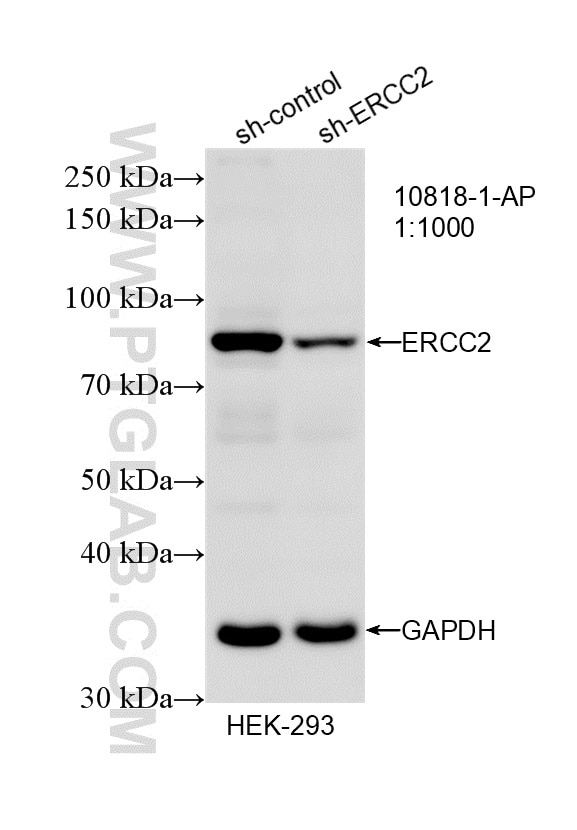
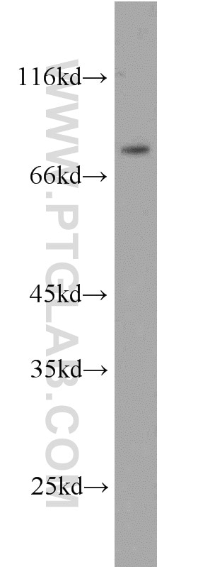
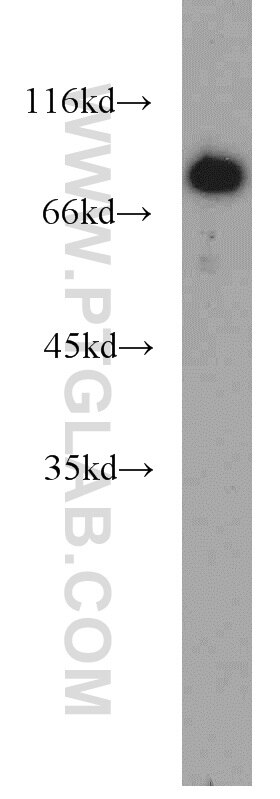
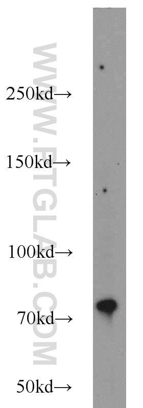
| Positive WB detected in | HeLa cells, HEK-293 cells, K-562 cells |
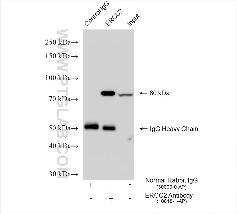
| Positive IP detected in | HEK-293 cells |
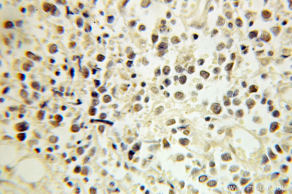
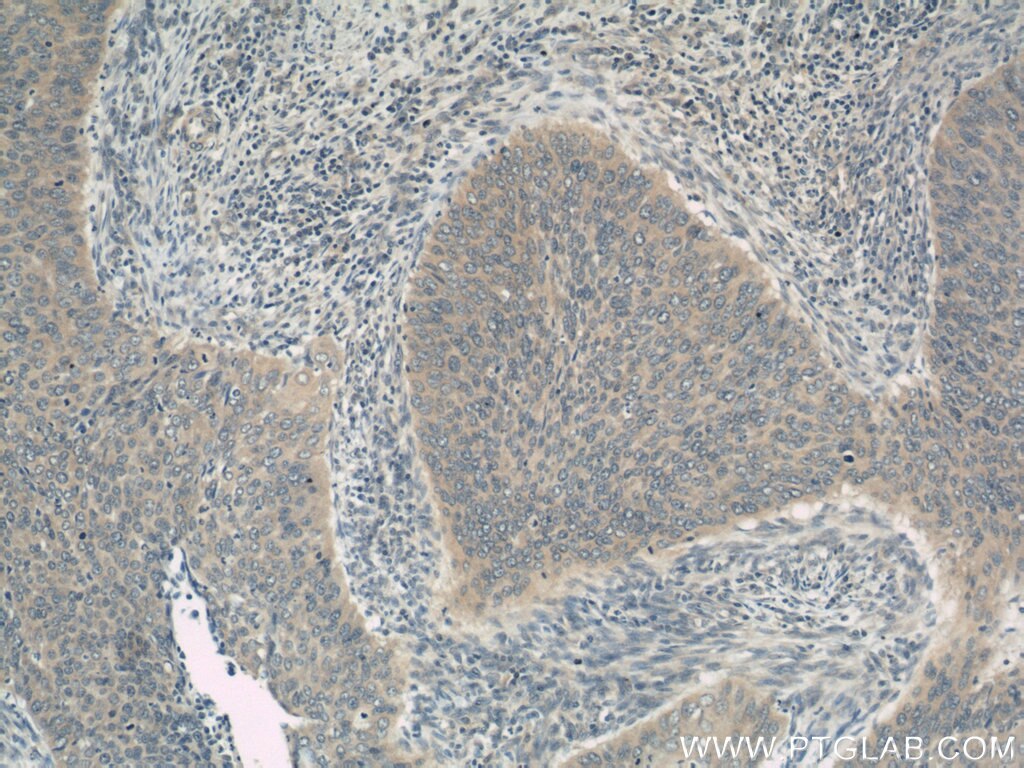
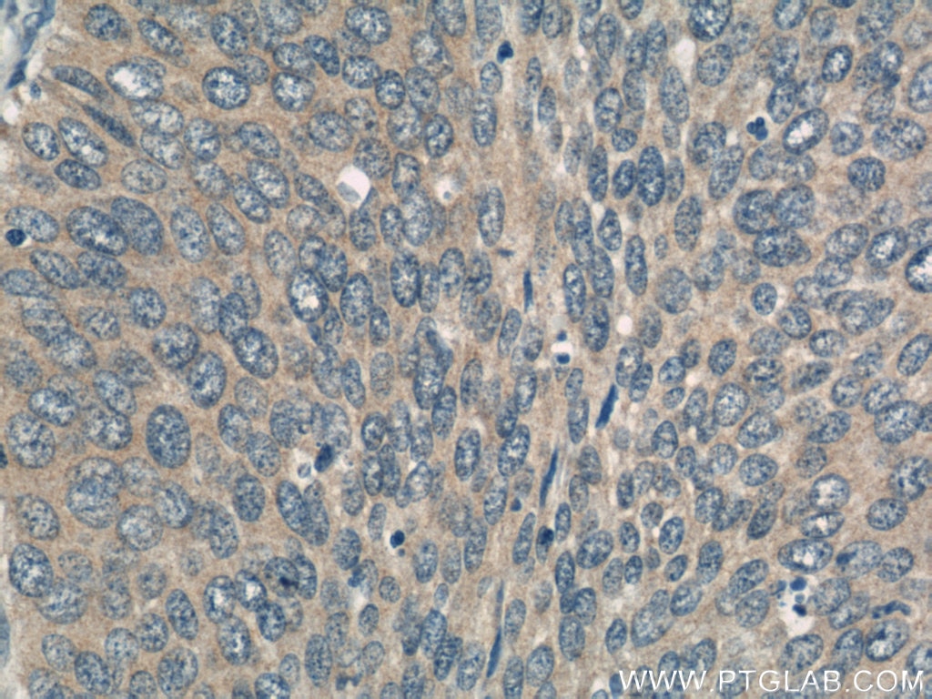
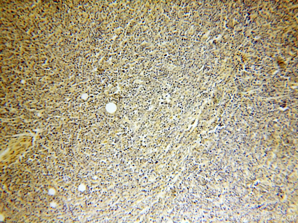
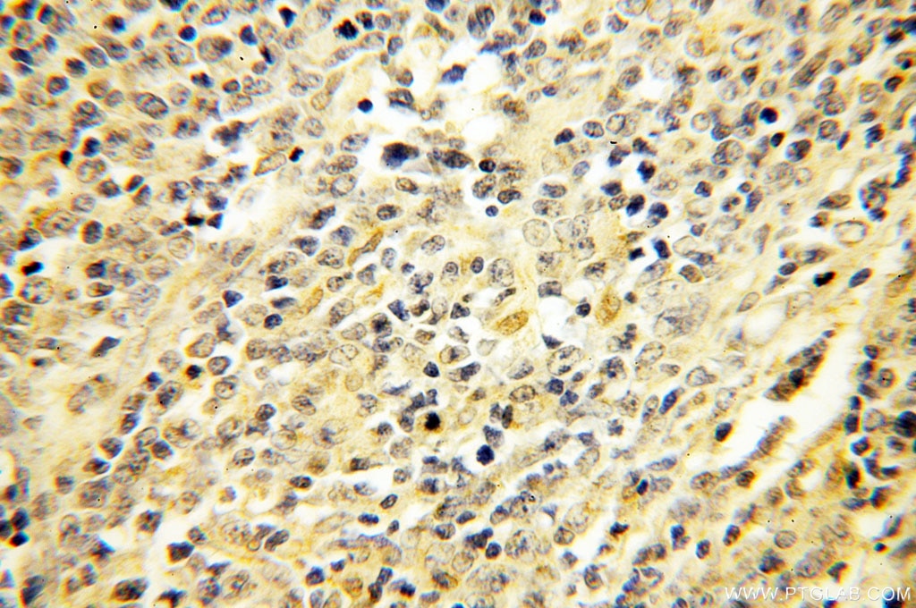
| Positive IHC detected in | human lymphoma tissue, human cervical cancer tissue Note: suggested antigen retrieval with TE buffer pH 9.0; (*) Alternatively, antigen retrieval may be performed with citrate buffer pH 6.0 |
Recommended dilution
| Application | Dilution |
|---|---|
| Western Blot (WB) | WB : 1:500-1:1000 |
| Immunoprecipitation (IP) | IP : 0.5-4.0 ug for 1.0-3.0 mg of total protein lysate |
| Immunohistochemistry (IHC) | IHC : 1:20-1:200 |
| It is recommended that this reagent should be titrated in each testing system to obtain optimal results. | |
| Sample-dependent, Check data in validation data gallery. | |
Product Information
10818-1-AP targets ERCC2 in WB, IHC, IF, IP, ELISA applications and shows reactivity with human samples.
| Tested Reactivity | human |
| Cited Reactivity | human, mouse, rat |
| Host / Isotype | Rabbit / IgG |
| Class | Polyclonal |
| Type | Antibody |
| Immunogen |
CatNo: Ag1038 Product name: Recombinant human ERCC2 protein Source: e coli.-derived, PGEX-4T Tag: GST Domain: 1-301 aa of BC008346 Sequence: MRELKRTLDAKGHGVLEMPSGTGKTVSLLALIMAYQRAYPLEVTKLIYCSRTVPEIEKVIEELRKLLNFYEKQEGEKLPFLGLALSSRKNLCIHPEVTPLRFGKDVDGKCHSLTASYVRAQYQHDTSLPHCRFYEEFDAHGREVPLPAGIYNLDDLKALGRRQGWCPYFLARYSILHANVVVYSYHYLLDPKIADLVSKELARKAVVVFDEAHNIDNVCIDSMSVNLTRRTLDRCQGNLETLQKTVLRIKETDEQRLRDEYRRLVEGLREASAARETDAHLANPVLPDEVLQEAVPGSIRT Predict reactive species |
| Full Name | excision repair cross-complementing rodent repair deficiency, complementation group 2 |
| Calculated Molecular Weight | 87 kDa |
| Observed Molecular Weight | 80 kDa |
| GenBank Accession Number | BC008346 |
| Gene Symbol | ERCC2 |
| Gene ID (NCBI) | 2068 |
| RRID | AB_2231330 |
| Conjugate | Unconjugated |
| Form | Liquid |
| Purification Method | Antigen affinity purification |
| UNIPROT ID | P18074 |
| Storage Buffer | PBS with 0.02% sodium azide and 50% glycerol, pH 7.3. |
| Storage Conditions | Store at -20°C. Stable for one year after shipment. Aliquoting is unnecessary for -20oC storage. 20ul sizes contain 0.1% BSA. |
Background Information
Excision-repair complementing defective in Chinese hamster 2(ERCC2), belongs to the nucleotide excision repair pathway, is a component of the TFIIH transcription complex and required for the association of the CDK-activating kinase(CAK) subcomplex with TFIIH. Since TFIIH is involved in both DNA repair and cell cycle progression, ERCC2 is implicated to play an integral role in the nucleotide excision repair pathway. Also it involves in the regulation of vitamin-D receptor activity. As a part of the mitotic spindle-associated MMXD complex, ERCC2 has a role in chromosome segregation.
Protocols
| Product Specific Protocols | |
|---|---|
| IHC protocol for ERCC2 antibody 10818-1-AP | Download protocol |
| IP protocol for ERCC2 antibody 10818-1-AP | Download protocol |
| WB protocol for ERCC2 antibody 10818-1-AP | Download protocol |
| Standard Protocols | |
|---|---|
| Click here to view our Standard Protocols |
Publications
What published studies show
The anti-XPD (ERCC2) antibody (Cat# 10818-1-AP) has 18 citations spanning journals including Molecular Cell, JCI, Advanced Science, FASEB Journal, Journal of Biological Chemistry, Cancer Science, Oncotarget, and others. Research fields cover nucleotide excision repair (NER), UV-induced DNA damage, cancer (glioblastoma, melanoma, hepatocellular, ovarian, pancreatic, colorectal), iron-sulfur cluster biology, mitochondrial dysfunction, cytoskeletal organization, and epigenetic regulation. Applications include WB and IHC across human, mouse, and rat species.
| Species | Application | Title |
|---|---|---|
Mol Cell The ribotoxic stress response drives acute inflammation, cell death, and epidermal thickening in UV-irradiated skin in vivo | ||
J Clin Invest CIAO1 loss of function causes a neuromuscular disorder with compromise of nucleocytoplasmic Fe-S enzymes | ||
Adv Sci (Weinh) Impaired Iron-Sulfur Cluster Synthesis Induces Mitochondrial PARthanatos in Diabetic Cardiomyopathy | ||
Adv Sci (Weinh) Arid1a Deficiency Drives Aristolochic Acid-Induced Liver Tumorigenesis through Ctnnb1 Mutation and Defective Nucleotide Excision Repair. | ||
J Transl Med Proteomic analysis predicts anti-angiogenic resistance in recurred glioblastoma | ||
Reprod Biol Endocrinol Map-1a regulates Sertoli cell BTB dynamics through the cytoskeletal organization of microtubule and F-actin |
Reviews
What customers say
One user rated this product 4 out of 5 stars. The reviewer used it at a 1:1000 dilution on human keratinocytes for Western Blot, Immunofluorescence, and Immunoprecipitation. No additional written comments were provided. Overall, the single review suggests satisfactory performance across multiple applications with no reported issues.
The reviews below have been submitted by verified Proteintech customers who received an incentive for providing their feedback.
Zizhao (Verified Customer) (08-15-2018) |
|











