

Tested Applications
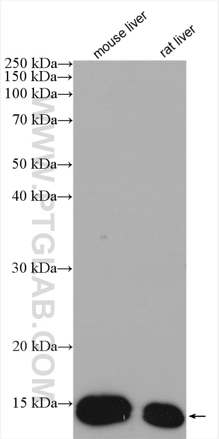
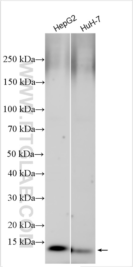
| Positive WB detected in | mouse liver tissue, HepG2 cells, HuH-7 cells, rat liver tissue |
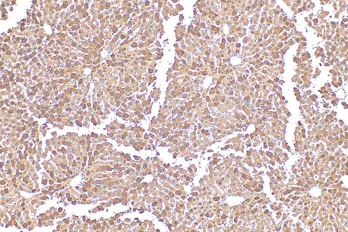
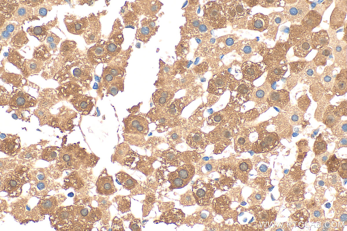
| Positive IHC detected in | mouse liver tissue Note: suggested antigen retrieval with TE buffer pH 9.0; (*) Alternatively, antigen retrieval may be performed with citrate buffer pH 6.0 |
Recommended dilution
| Application | Dilution |
|---|---|
| Western Blot (WB) | WB : 1:2000-1:16000 |
| Immunohistochemistry (IHC) | IHC : 1:300-1:1200 |
| It is recommended that this reagent should be titrated in each testing system to obtain optimal results. | |
| Sample-dependent, Check data in validation data gallery. | |
Product Information
13626-1-AP targets FABP1 in WB, IHC, IF, IP, ELISA applications and shows reactivity with human, mouse, rat samples.
| Tested Reactivity | human, mouse, rat |
| Cited Reactivity | human, mouse, rat, bovine |
| Host / Isotype | Rabbit / IgG |
| Class | Polyclonal |
| Type | Antibody |
| Immunogen |
CatNo: Ag4540 Product name: Recombinant human FABP1 protein Source: e coli.-derived, PGEX-4T Tag: GST Domain: 1-127 aa of BC032801 Sequence: MSFSGKYQLQSQENFEAFMKAIGLPEELIQKGKDIKGVSEIVQNGKHFKFTITAGSKVIQNEFTVGEECELETMTGEKVKTVVQLEGDNKLVTTFKNIKSVTELNGDIITNTMTLGDIVFKRISKRI Predict reactive species |
| Full Name | fatty acid binding protein 1, liver |
| Calculated Molecular Weight | 127 aa, 14 kDa |
| Observed Molecular Weight | 14 kDa |
| GenBank Accession Number | BC032801 |
| Gene Symbol | FABP1 |
| Gene ID (NCBI) | 2168 |
| RRID | AB_2102017 |
| Conjugate | Unconjugated |
| Form | Liquid |
| Purification Method | Antigen affinity purification |
| UNIPROT ID | P07148 |
| Storage Buffer | PBS with 0.02% sodium azide and 50% glycerol, pH 7.3. |
| Storage Conditions | Store at -20°C. Stable for one year after shipment. Aliquoting is unnecessary for -20oC storage. 20ul sizes contain 0.1% BSA. |
Background Information
FABP1, liver fatty acid-binding protein, is abundant in cytoplasm that regulates lipid transport and metabolism. It plays a role in lipoprotein-mediated cholesterol uptake in hepatocytes (PMID:25732850). FABP1 is mainly expressed in hepatocytes, enterocytes and to a lesser degree in renal tubular cells, associated with liver injury (PMID: 15653098). Levels of FABP1 have been found to be elevated in patients with hepatocyte injury secondary to alcohol or drug toxicity (PMID: 14563446).
Protocols
| Product Specific Protocols | |
|---|---|
| IHC protocol for FABP1 antibody 13626-1-AP | Download protocol |
| WB protocol for FABP1 antibody 13626-1-AP | Download protocol |
| Standard Protocols | |
|---|---|
| Click here to view our Standard Protocols |
Publications
What published studies show
The FABP1 Polyclonal antibody (Cat# 13626-1-AP) has 46 citations. It appears in high-impact journals including Nature Communications, Advanced Science, Biomaterials, Cell Death & Disease, Developmental Cell, EMBO Reports, Free Radical Biology & Medicine, and iScience, among others. Research fields center on lipid metabolism and fatty acid binding, with strong emphasis on liver disease (NAFLD/NASH, hepatocellular carcinoma), metabolic syndrome, obesity, diabetes, and renal injury. Studies also explore PPARα/PPARγ signaling, ferroptosis, inflammation, and lipid homeostasis, primarily using Western blot, IHC, and IF in human, mouse, and rat models.
| Species | Application | Title |
|---|---|---|
Nat Commun N1-methyladenosine methylation in tRNA drives liver tumourigenesis by regulating cholesterol metabolism. | ||
Nat Commun Hyodeoxycholic acid ameliorates nonalcoholic fatty liver disease by inhibiting RAN-mediated PPARα nucleus-cytoplasm shuttling | ||
Adv Sci (Weinh) Hepatocellular Carcinoma LINC01116 Outcompetes T Cells for Linoleic Acid and Accelerates Tumor Progression | ||
Biomaterials MMP-12 siRNA improves the homeostasis of the small intestine and metabolic dysfunction in high-fat diet feeding-induced obese mice. | ||
Cell Death Dis The natural compound GL22, isolated from Ganoderma mushrooms, suppresses tumor growth by altering lipid metabolism and triggering cell death. |
Reviews
What customers say
All three reviews rate this antibody 5 stars for Western Blot. Users report strong specificity and sensitivity with clean, background-free results. It performed well on mouse tissue, colon tissue, and colon cancer cell lines at a 1:1000 dilution. Reviewers consistently recommend it for WB applications, describing it as reliable and effective.
The reviews below have been submitted by verified Proteintech customers who received an incentive for providing their feedback.
Meixin (Verified Customer) (03-10-2026) | this antibody shows good specificity and sensitivity, and I would recommend it for Western blot applications.
|
MALLIKARJUNA (Verified Customer) (10-17-2025) | GOOD FOR WESTERN BLOT
|
Balawant (Verified Customer) (08-24-2022) | this antibody is working great. it has no background.
|





