Tested Applications
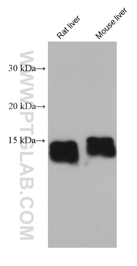
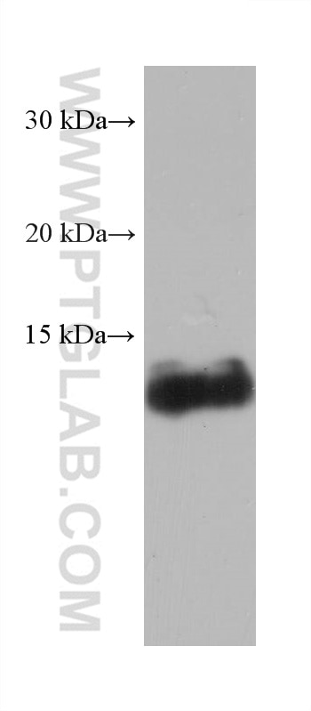
| Positive WB detected in | rat liver tissue, pig liver tissue, mouse liver tissue |
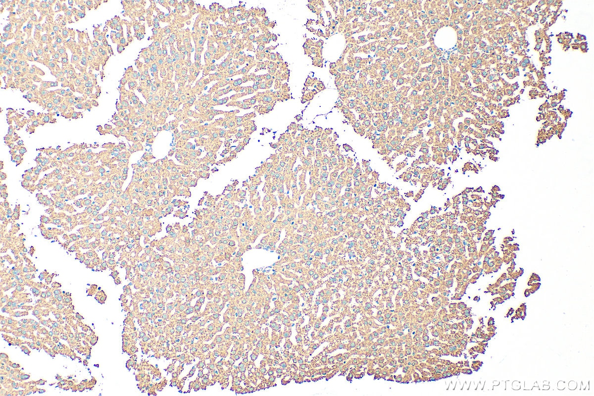
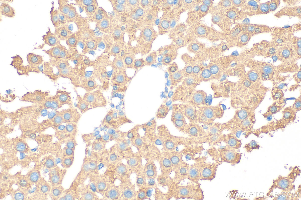
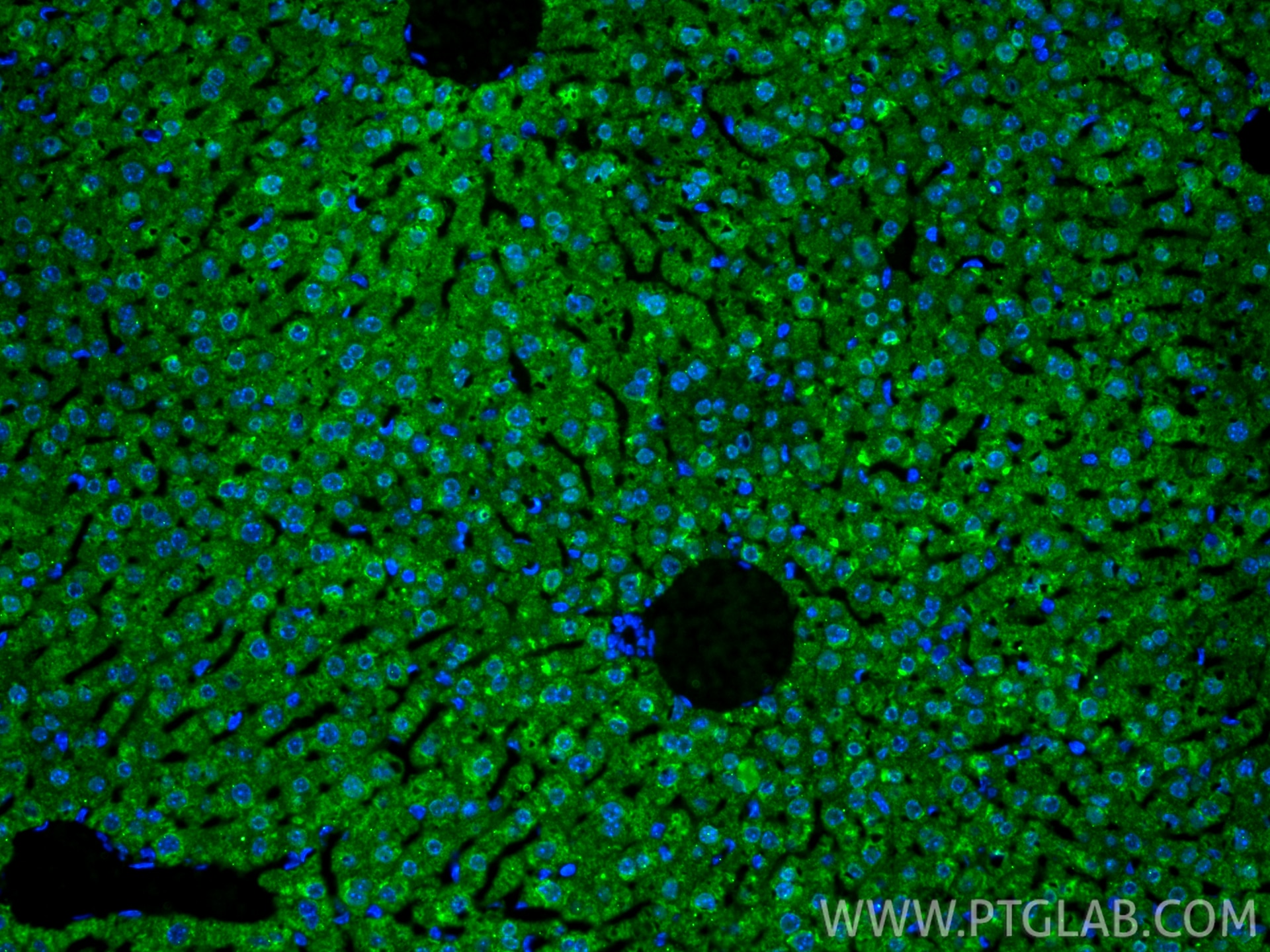
| Positive IHC detected in | mouse liver tissue Note: suggested antigen retrieval with TE buffer pH 9.0; (*) Alternatively, antigen retrieval may be performed with citrate buffer pH 6.0 |
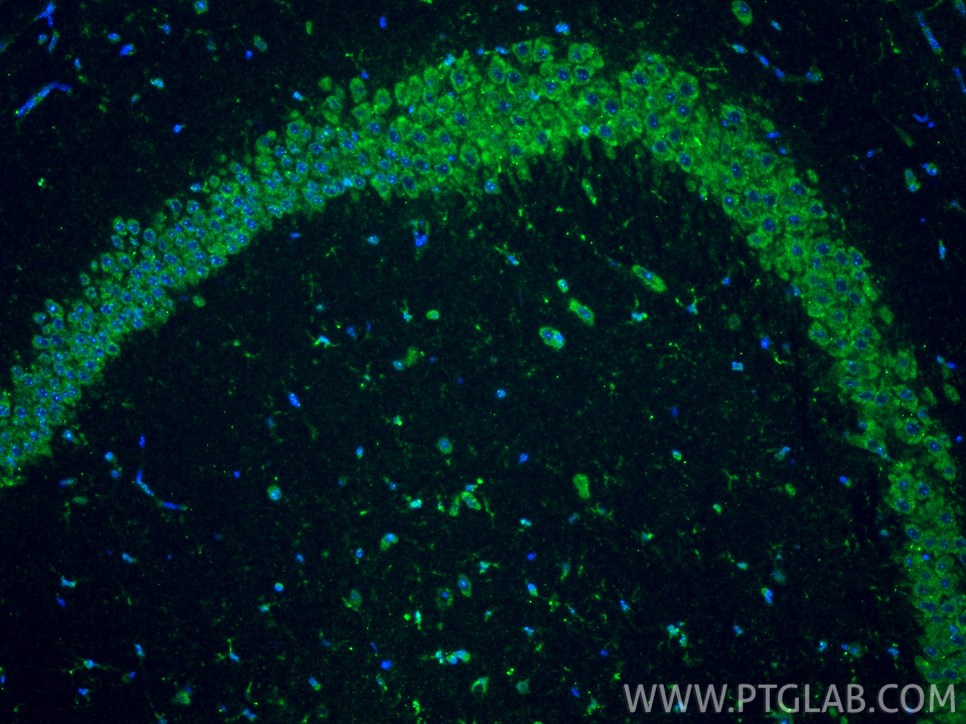
| Positive IF-P detected in | mouse liver tissue, mouse brain tissue |
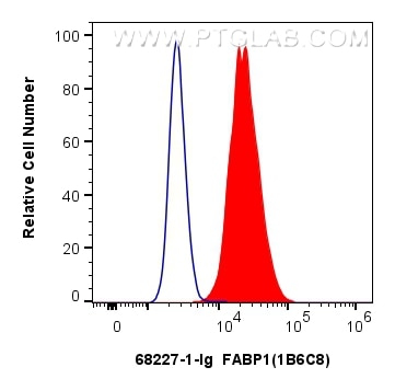
| Positive FC (Intra) detected in | HepG2 cells |
Recommended dilution
| Application | Dilution |
|---|---|
| Western Blot (WB) | WB : 1:5000-1:50000 |
| Immunohistochemistry (IHC) | IHC : 1:500-1:2000 |
| Immunofluorescence (IF)-P | IF-P : 1:500-1:2000 |
| Flow Cytometry (FC) (INTRA) | FC (INTRA) : 0.80 ug per 10^6 cells in a 100 µl suspension |
| It is recommended that this reagent should be titrated in each testing system to obtain optimal results. | |
| Sample-dependent, Check data in validation data gallery. | |
Product Information
68227-1-Ig targets FABP1 in WB, IHC, IF-P, FC (Intra), ELISA applications and shows reactivity with human, mouse, rat, pig samples.
| Tested Reactivity | human, mouse, rat, pig |
| Host / Isotype | Mouse / IgG1 |
| Class | Monoclonal |
| Type | Antibody |
| Immunogen |
CatNo: Ag32836 Product name: Recombinant human FABP1 protein Source: e coli.-derived, PET28a Tag: 6*His Domain: 1-127 aa of BC032801 Sequence: MSFSGKYQLQSQENFEAFMKAIGLPEELIQKGKDIKGVSEIVQNGKHFKFTITAGSKVIQNEFTVGEECELETMTGEKVKTVVQLEGDNKLVTTFKNIKSVTELNGDIITNTMTLGDIVFKRISKRI Predict reactive species |
| Full Name | fatty acid binding protein 1, liver |
| Calculated Molecular Weight | 127 aa, 14 kDa |
| Observed Molecular Weight | 14 kDa |
| GenBank Accession Number | BC032801 |
| Gene Symbol | FABP1 |
| Gene ID (NCBI) | 2168 |
| RRID | AB_2935315 |
| Conjugate | Unconjugated |
| Form | Liquid |
| Purification Method | Protein G purification |
| UNIPROT ID | P07148 |
| Storage Buffer | PBS with 0.02% sodium azide and 50% glycerol, pH 7.3. |
| Storage Conditions | Store at -20°C. Stable for one year after shipment. Aliquoting is unnecessary for -20oC storage. 20ul sizes contain 0.1% BSA. |
Background Information
FABP1, liver fatty acid-binding protein, is abundant in cytoplasm that regulates lipid transport and metabolism. It plays a role in lipoprotein-mediated cholesterol uptake in hepatocytes (PMID:25732850). FABP1 is mainly expressed in hepatocytes, enterocytes and to a lesser degree in renal tubular cells, associated with liver injury (PMID: 15653098). Levels of FABP1 was elevated in patients with hepatocyte injury secondary to alcohol or drug toxicity (PMID: 14563446).
Protocols
| Product Specific Protocols | |
|---|---|
| FC protocol for FABP1 antibody 68227-1-Ig | Download protocol |
| IF protocol for FABP1 antibody 68227-1-Ig | Download protocol |
| IHC protocol for FABP1 antibody 68227-1-Ig | Download protocol |
| WB protocol for FABP1 antibody 68227-1-Ig | Download protocol |
| Standard Protocols | |
|---|---|
| Click here to view our Standard Protocols |