

Tested Applications
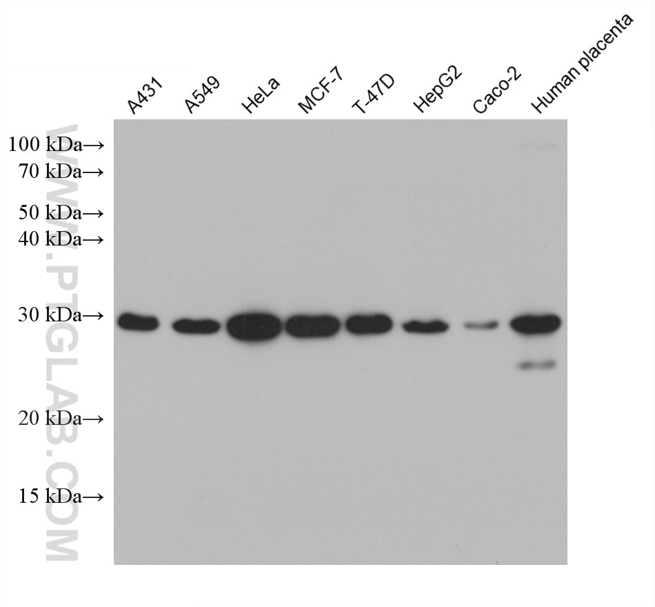
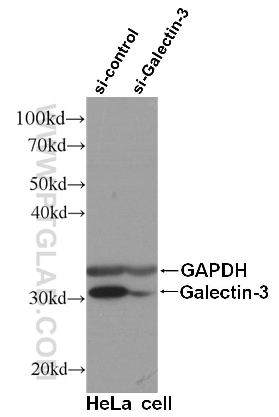
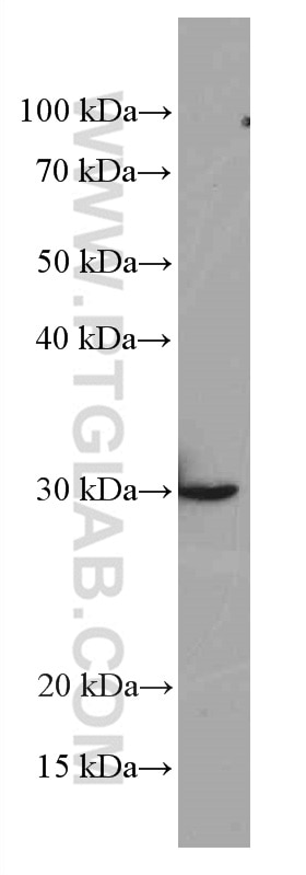
| Positive WB detected in | A431 cells, HSC-T6 cells, HeLa cells, A549 cells, MCF-7 cells, T-47D cells, HepG2 cells, Caco-2 cells, human placenta tissue |
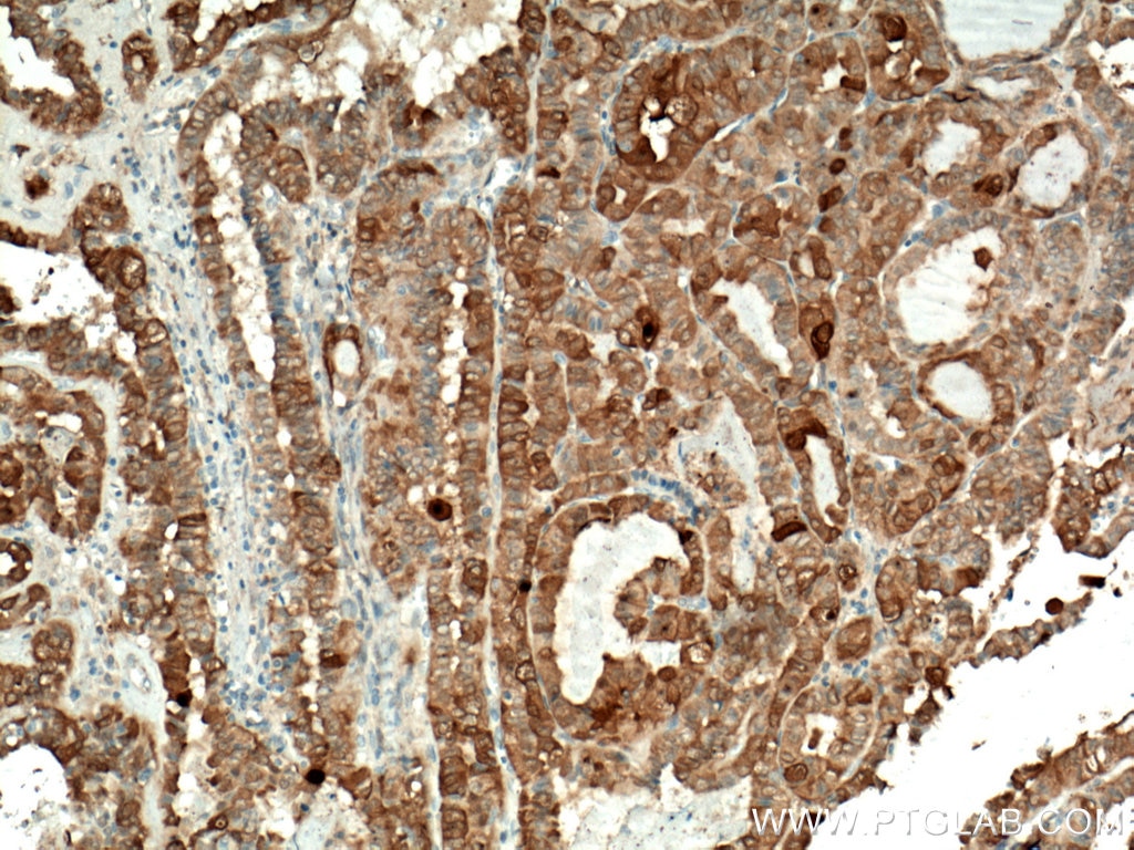
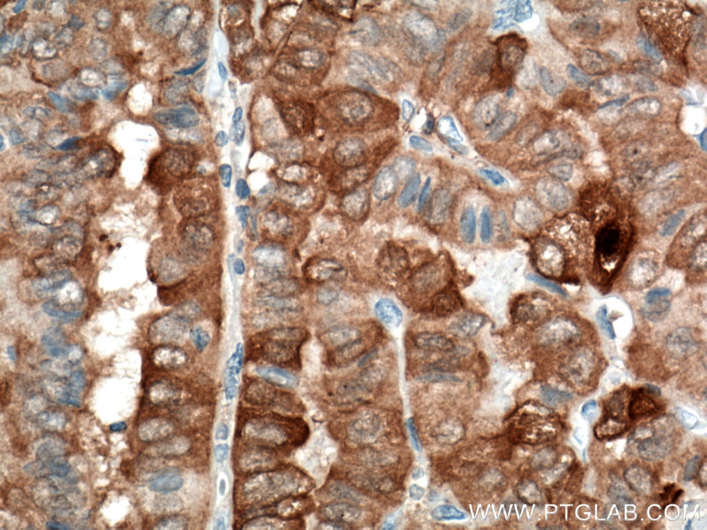
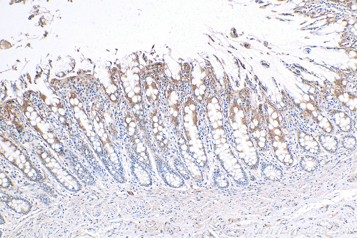
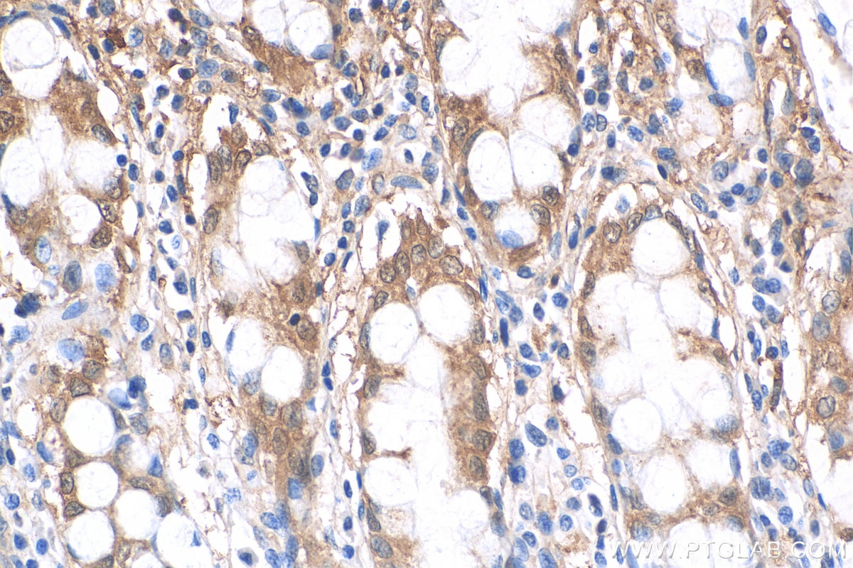
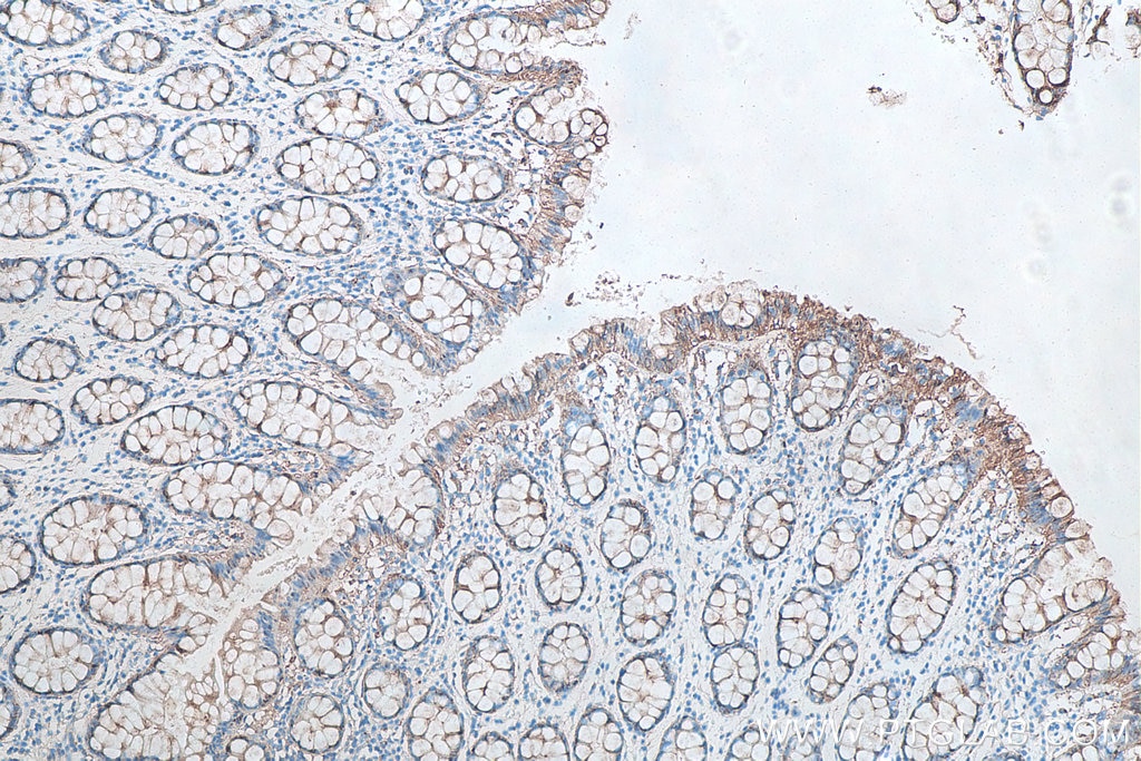
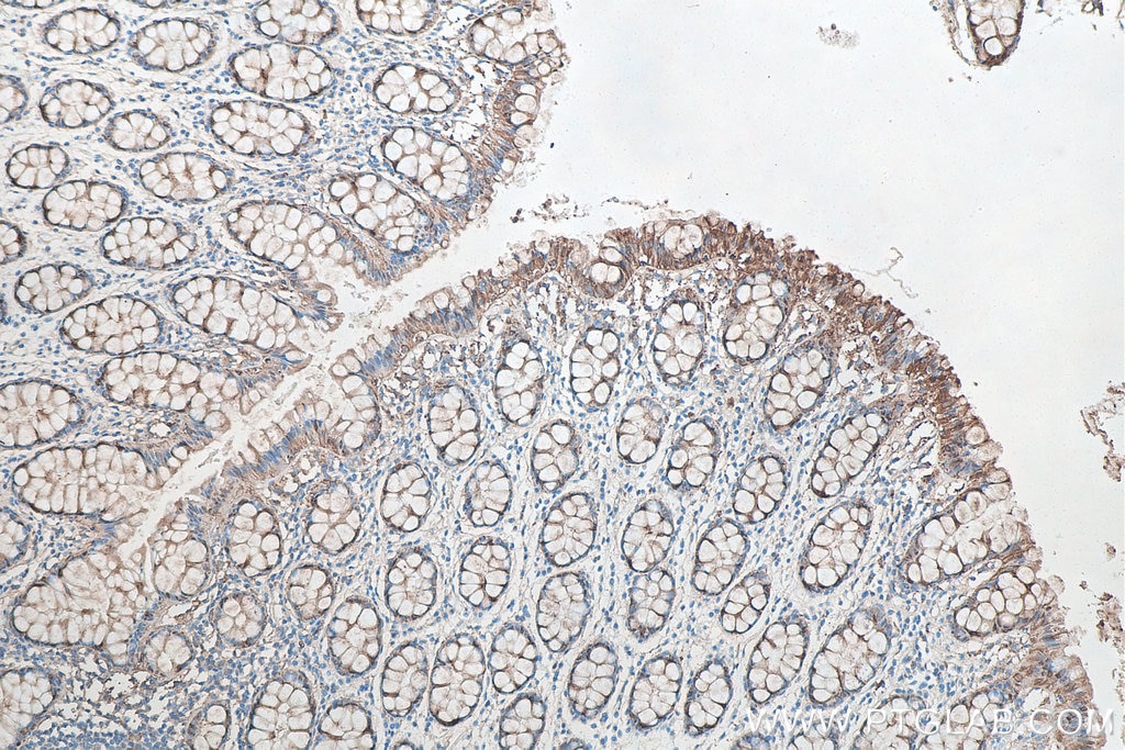
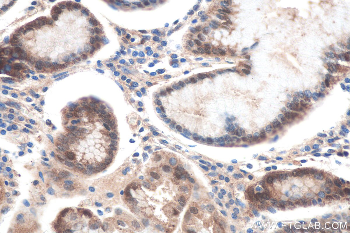
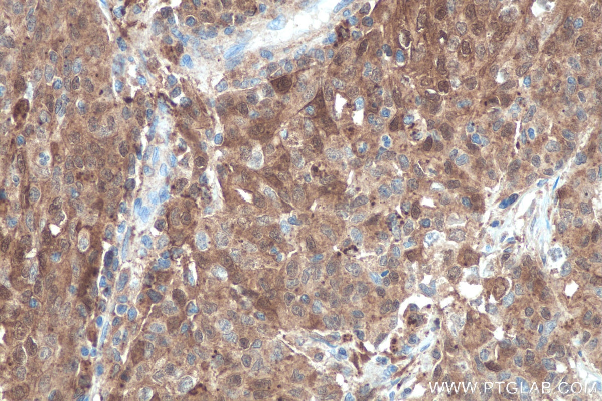
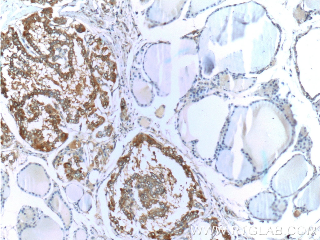
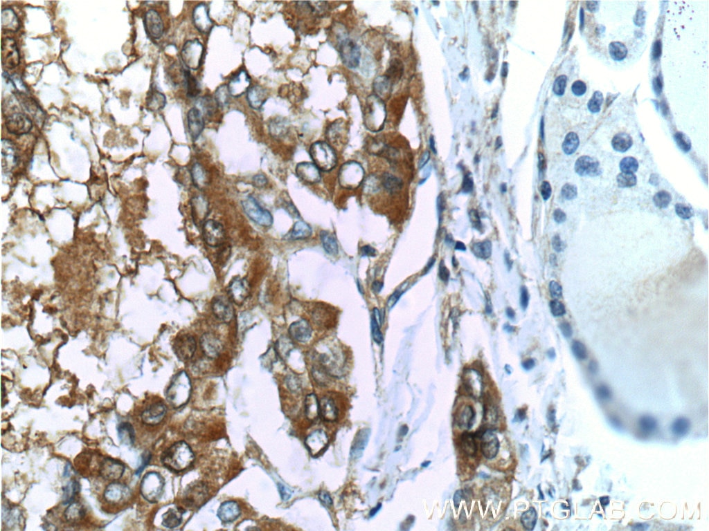
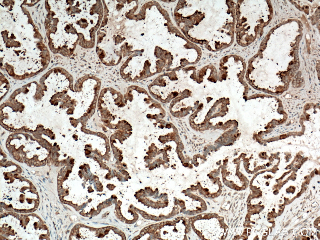
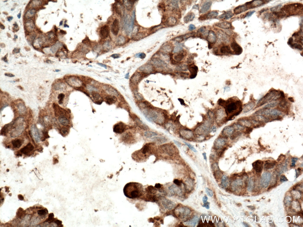
| Positive IHC detected in | human thyroid cancer tissue, human colon tissue, human stomach tissue, human thyroid cancer tissue, human stomach cancer tissue, human ovary tumor tissue Note: suggested antigen retrieval with TE buffer pH 9.0; (*) Alternatively, antigen retrieval may be performed with citrate buffer pH 6.0 |
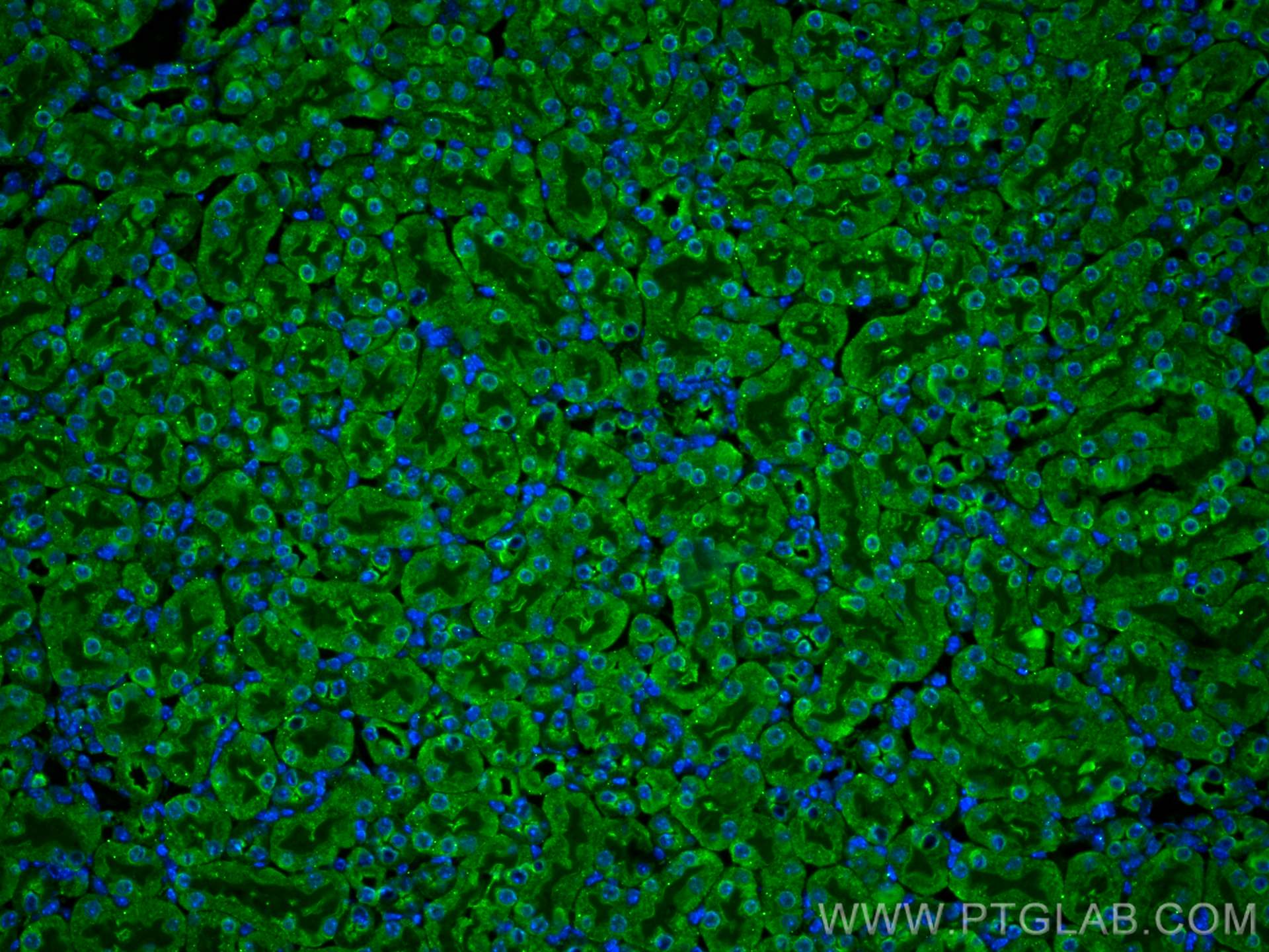
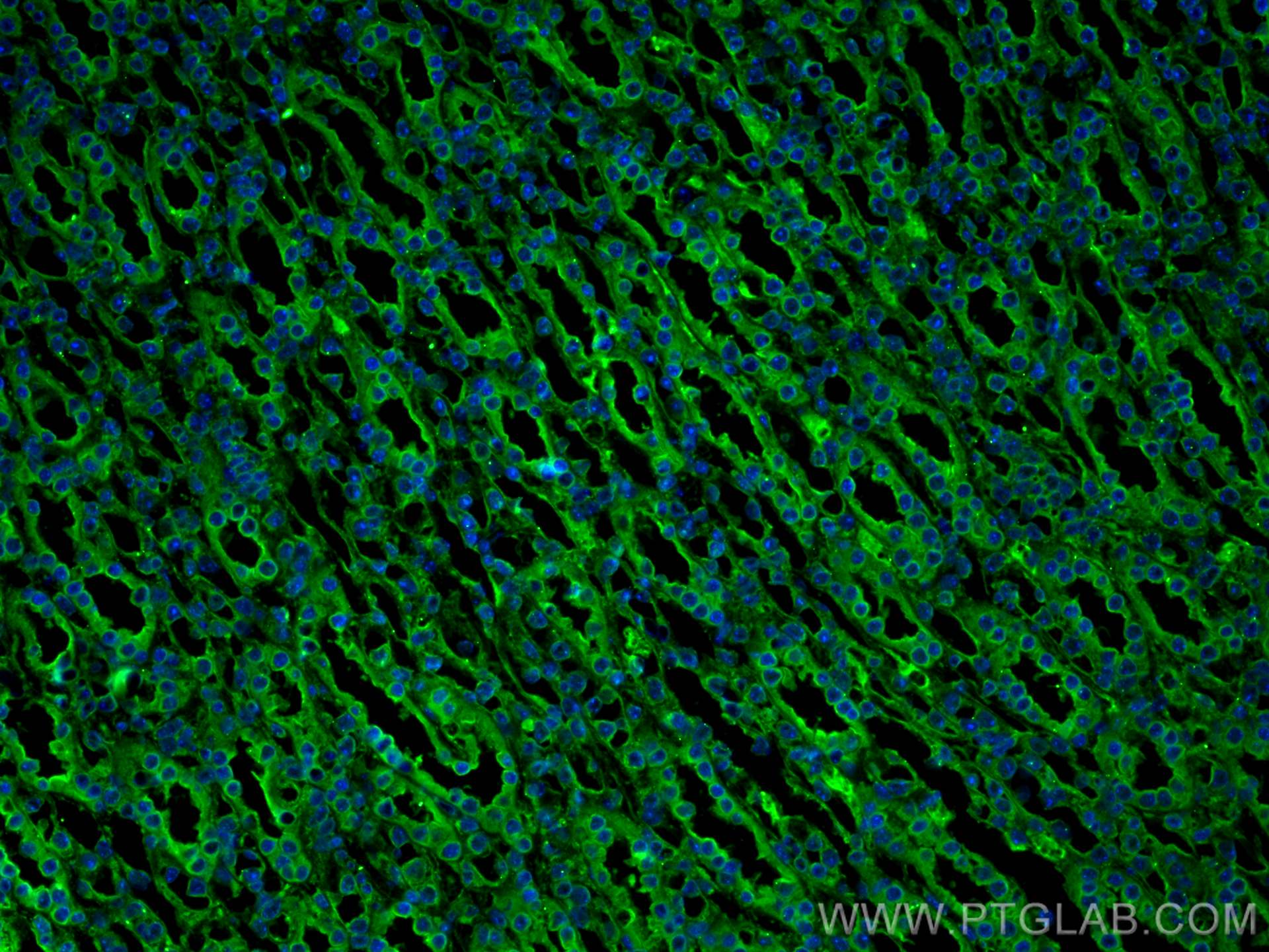
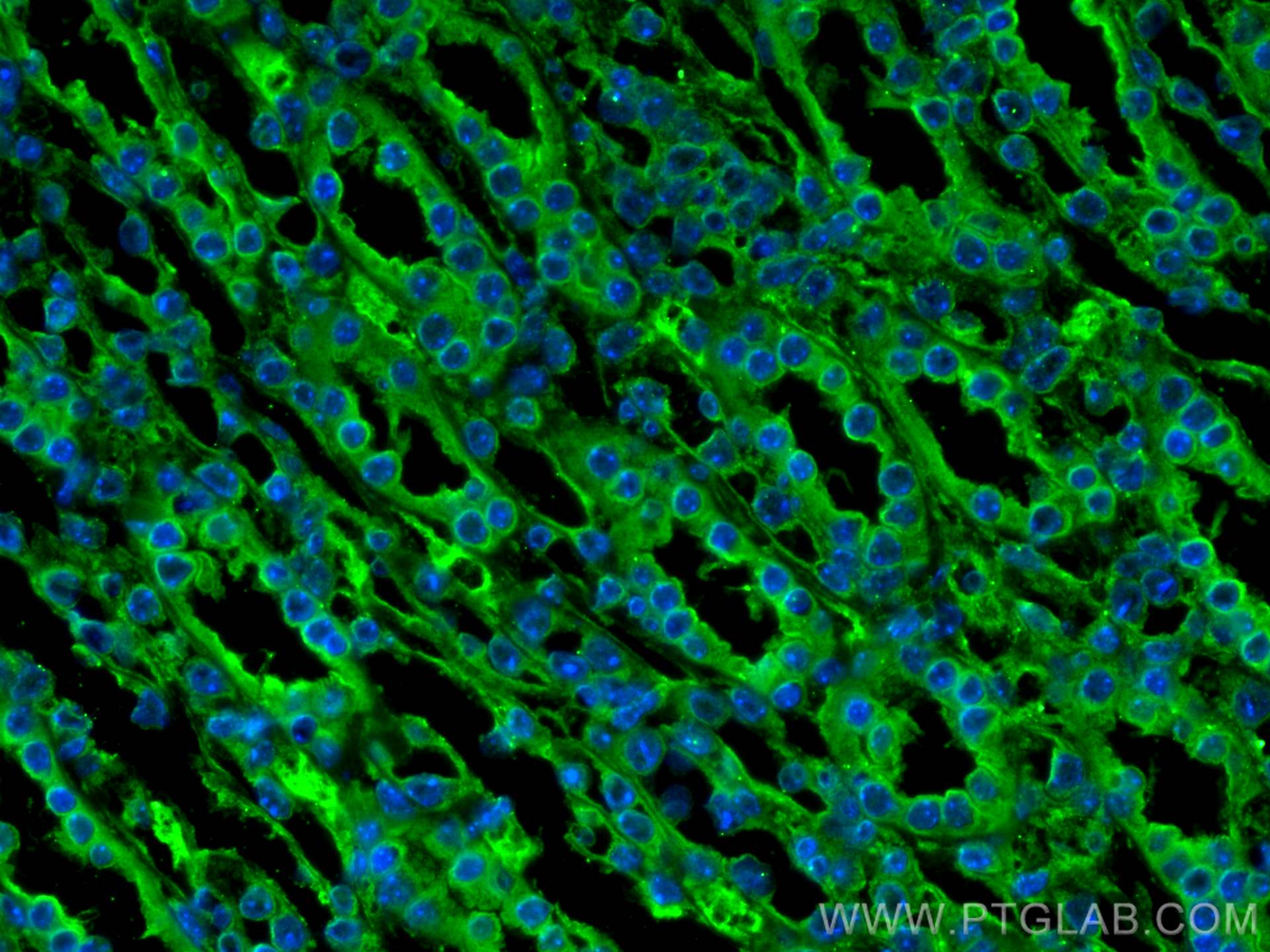
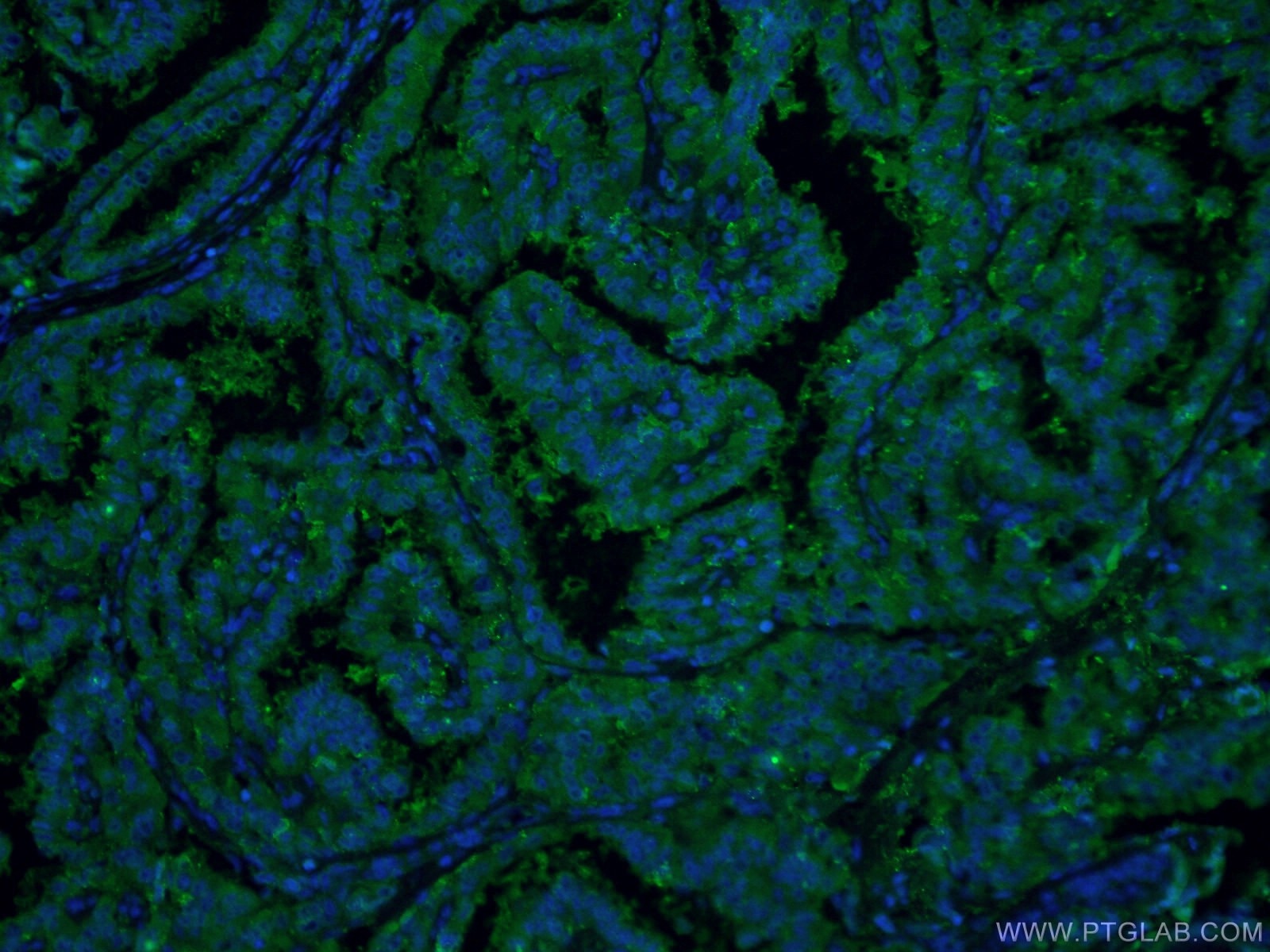
| Positive IF-P detected in | mouse kidney tissue, human thyroid cancer tissue |
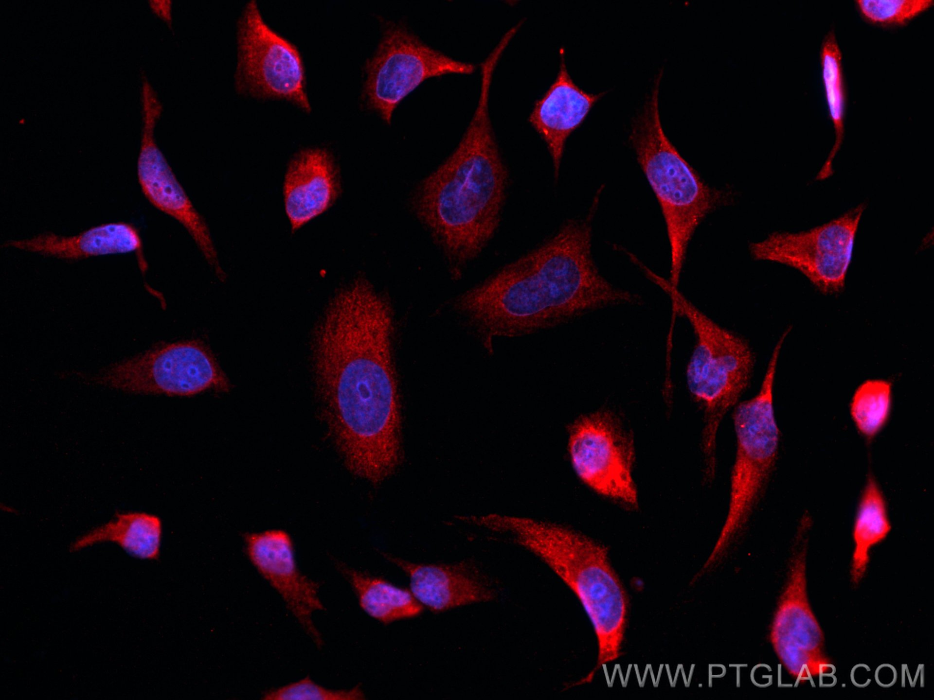
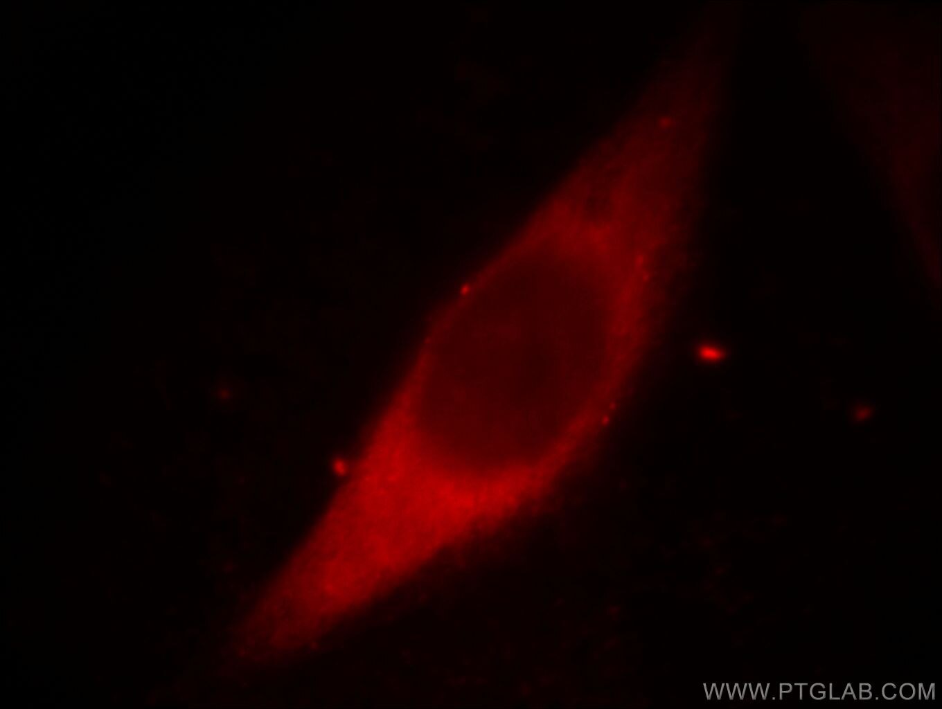
| Positive IF/ICC detected in | HeLa cells, mouse kidney tissue, MCF-7 cells, human thyroid cancer tissue |
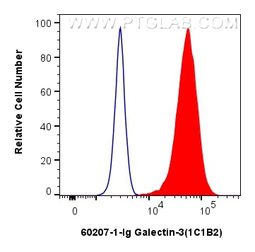
| Positive FC (Intra) detected in | HeLa cells |
Recommended dilution
| Application | Dilution |
|---|---|
| Western Blot (WB) | WB : 1:5000-1:50000 |
| Immunohistochemistry (IHC) | IHC : 1:1000-1:4000 |
| Immunofluorescence (IF)-P | IF-P : 1:200-1:800 |
| Immunofluorescence (IF)/ICC | IF/ICC : 1:400-1:1600 |
| Flow Cytometry (FC) (INTRA) | FC (INTRA) : 0.80 ug per 10^6 cells in a 100 µl suspension |
| It is recommended that this reagent should be titrated in each testing system to obtain optimal results. | |
| Sample-dependent, Check data in validation data gallery. | |
Product Information
60207-1-Ig targets Galectin-3 in WB, IHC, IF/ICC, IF-P, FC (Intra), IP, CoIP, ELISA applications and shows reactivity with human, mouse samples.
| Tested Reactivity | human, mouse |
| Cited Reactivity | human, mouse, rat |
| Host / Isotype | Mouse / IgG2b |
| Class | Monoclonal |
| Type | Antibody |
| Immunogen |
CatNo: Ag6753 Product name: Recombinant human Galectin 3 protein Source: e coli.-derived, PET28a Tag: 6*His Domain: 1-250 aa of BC001120 Sequence: MADNFSLHDALSGSGNPNPQGWPGAWGNQPAGAGGYPGASYPGAYPGQAPPGAYPGQAPPGAYPGAPGAYPGAPAPGVYPGPPSGPGAYPSSGQPSATGAYPATGPYGAPAGPLIVPYNLPLPGGVVPRMLITILGTVKPNANRIALDFQRGNDVAFHFNPRFNENNRRVIVCNTKLDNNWGREERQSVFPFESGKPFKIQVLVEPDHFKVAVNDAHLLQYNHRVKKLNEISKLGISGDIDLTSASYTMI Predict reactive species |
| Full Name | lectin, galactoside-binding, soluble, 3 |
| Calculated Molecular Weight | 26 kDa |
| Observed Molecular Weight | 31 kDa |
| GenBank Accession Number | BC001120 |
| Gene Symbol | Galectin-3 |
| Gene ID (NCBI) | 3958 |
| RRID | AB_10951109 |
| Conjugate | Unconjugated |
| Form | Liquid |
| Purification Method | Protein A purification |
| UNIPROT ID | P17931 |
| Storage Buffer | PBS with 0.02% sodium azide and 50% glycerol, pH 7.3. |
| Storage Conditions | Store at -20°C. Stable for one year after shipment. Aliquoting is unnecessary for -20oC storage. 20ul sizes contain 0.1% BSA. |
Background Information
Galectins are a family of animal lectins defined by shared characteristic amino-acid sequences and affinity for β-galactose-containing oligosac-charides (PMID: 8063692). Galectin-3, a member of the β-galactoside-binding proteins, contains one carbohydrate recognition domain (CRD) and a proline- and glycine-rich N-terminal domain through which is able to form oligomers (PMID: 14758078). Galectin-3 is widely expressed in many normal tissues and a variety of tumors. It is found intracellularly in nucleus and cytoplasm or secreted outside of cell, being present on the cell surface or in the extracellular space (PMID: 16478649). Galectin-3 is involved in various biological processes including cell growth, adhesion, differentiation, apoptosis, angiogenesis, immune response, neoplastic transformation and metastasis (PMID: 16478649; 14758078).
Protocols
| Product Specific Protocols | |
|---|---|
| FC protocol for Galectin-3 antibody 60207-1-Ig | Download protocol |
| IF protocol for Galectin-3 antibody 60207-1-Ig | Download protocol |
| IHC protocol for Galectin-3 antibody 60207-1-Ig | Download protocol |
| WB protocol for Galectin-3 antibody 60207-1-Ig | Download protocol |
| Standard Protocols | |
|---|---|
| Click here to view our Standard Protocols |
Publications
What published studies show
The Galectin-3 Monoclonal antibody (catalog number 60207-1-Ig) has 82 citations. These appear in journals including Cancer Cell, Cell, Nature Cell Biology, Nature Communications, Autophagy, Bioactive Materials, Hepatology, Cell Death & Differentiation, FASEB Journal, eLife, and Journal of Immunology, among others. Research fields span oncology (lung, breast, liver, thyroid, and nasopharyngeal cancers), lysosomal biology and autophagy, neuroinflammation and neurodegeneration, cardiovascular disease, atherosclerosis, fibrosis, and immunology. Applications include WB, IF, IHC, and CoIP across human, mouse, rat, and other species.
| Species | Application | Title |
|---|---|---|
Cancer Cell Overcoming tyrosine kinase inhibitor resistance in lung cancer brain metastasis with CTLA4 blockade | ||
Adv Mater Lysosome Targeted Nanoparticle Aggregation Reverses Immunosuppressive Tumor Microenvironment for Cancer Immunotherapy | ||
Bioact Mater Lysosomal destabilization: A missing link between pathological calcification and osteoarthritis | ||
Bioact Mater Ultrasonic-driven force-electric nanodomains orchestrate lysosome-to-mitochondria functional cascade to accelerate bone regeneration. | ||
Nat Cell Biol Cholesterol sensing by the SCAP-FAM134B complex regulates ER-phagy and STING innate immunity. |
Reviews
What customers say
Customers consistently rate this antibody 4–5 stars across Western Blot and Immunofluorescence applications. Users report reliable performance in human cells (HUVEC, NRVCMs, U2OS) and mouse kidney tissue. One reviewer confirmed specificity through siRNA knockdown validation in U2OS cells. Recommended dilutions range from 1:200 to 1:1000 depending on the application. Overall, reviewers describe the antibody as working well for both imaging and blotting, with no major issues noted.
The reviews below have been submitted by verified Proteintech customers who received an incentive for providing their feedback.
Boyan (Verified Customer) (06-19-2026) | good for both imaging and WB
|
Marco (Verified Customer) (07-09-2024) | works perfectly fine
|
Kenzo (Verified Customer) (08-29-2023) | I used this antibody in mouse kidney tissues and it worked well.
|
Muhammad (Verified Customer) (09-13-2022) | Work best in HUVECs
|
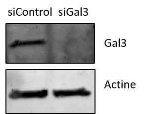
Noémie (Verified Customer) (10-20-2021) | I used this antibody for Western Blot against Gal3 in mamalian cells (U2OS), and did a validation by siRNA.
 |























