

Tested Applications
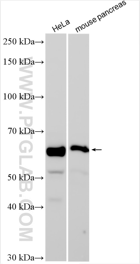
| Positive WB detected in | HeLa cells, mouse pancreas tissue |
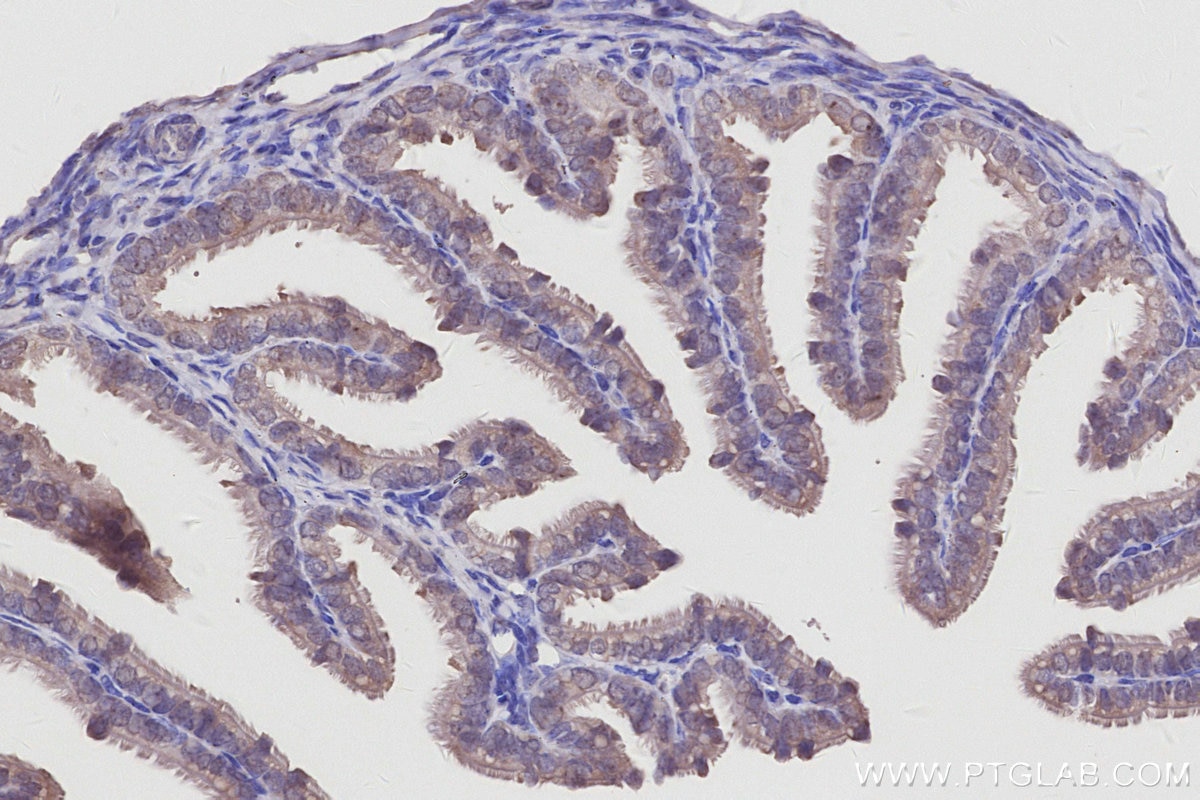
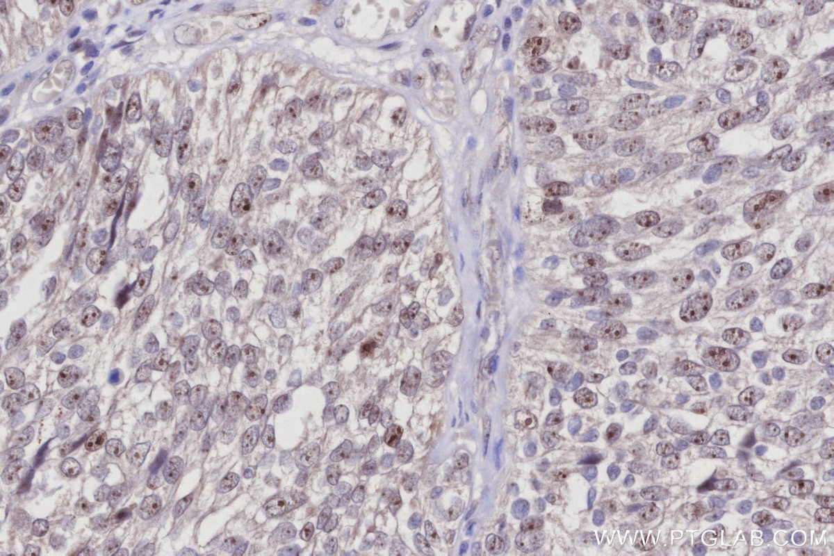
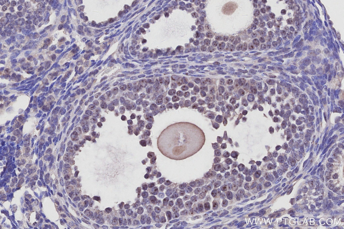
| Positive IHC detected in | human ovary cancer tissue, mouse ovary tissue Note: suggested antigen retrieval with TE buffer pH 9.0; (*) Alternatively, antigen retrieval may be performed with citrate buffer pH 6.0 |
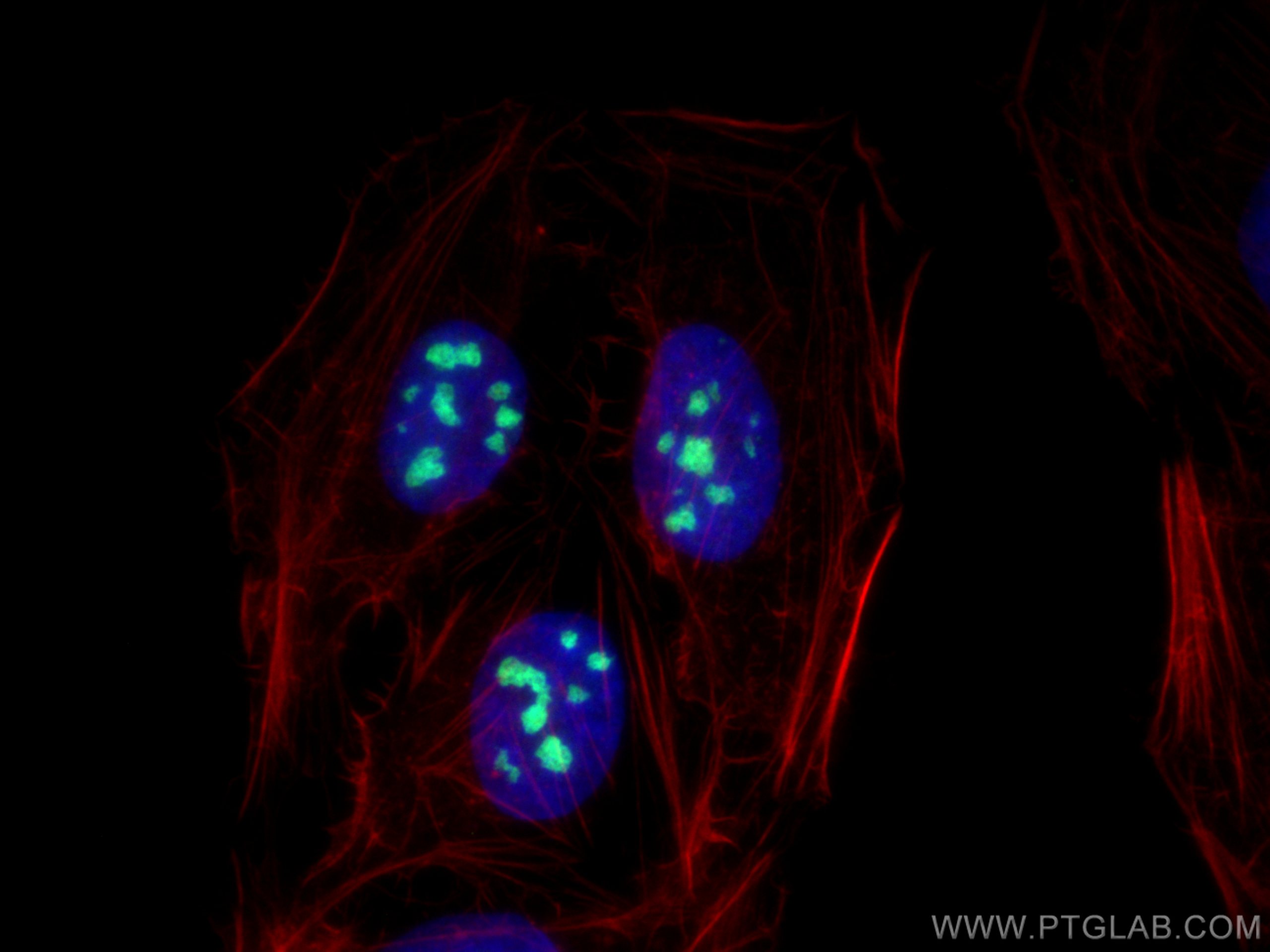
| Positive IF/ICC detected in | U2OS cells |
Recommended dilution
| Application | Dilution |
|---|---|
| Western Blot (WB) | WB : 1:2000-1:16000 |
| Immunohistochemistry (IHC) | IHC : 1:500-1:2000 |
| Immunofluorescence (IF)/ICC | IF/ICC : 1:50-1:500 |
| It is recommended that this reagent should be titrated in each testing system to obtain optimal results. | |
| Sample-dependent, Check data in validation data gallery. | |
Product Information
27353-1-AP targets GLTSCR2 in WB, IHC, IF/ICC, ELISA applications and shows reactivity with human, mouse samples.
| Tested Reactivity | human, mouse |
| Cited Reactivity | human |
| Host / Isotype | Rabbit / IgG |
| Class | Polyclonal |
| Type | Antibody |
| Immunogen |
CatNo: Ag26358 Product name: Recombinant human GLTSCR2 protein Source: e coli.-derived, PGEX-4T Tag: GST Domain: 141-440 aa of BC010095 Sequence: NAKKLRRKEQLWEKLAKQGELPREVRRAQARLLNPSATRAKPGPQDTVERPFYDLWASDNPLDRPLVGQDEFFLEQTKKKGVKRPARLHTKPSQAPAVEVAPAGASYNPSFEDHQTLLSAAHEVELQRQKEAEKLERQLALPATEQAATQESTFQELCEGLLEESDGEGEPGQGEGPEAGDAEVCPTPARLATTEKKTEQQRRREKAVHRLRVQQAALRAARLRHQELFRLRGIKAQVALRLAELARRRRRRQARREAEADKPRRLGRLKYQAPDIDVQLSSELTDSLRTLKPEGNILRD Predict reactive species |
| Full Name | glioma tumor suppressor candidate region gene 2 |
| Calculated Molecular Weight | 54 kDa |
| Observed Molecular Weight | 55-60 kDa |
| GenBank Accession Number | BC010095 |
| Gene Symbol | GLTSCR2 |
| Gene ID (NCBI) | 29997 |
| RRID | AB_2880852 |
| Conjugate | Unconjugated |
| Form | Liquid |
| Purification Method | Antigen affinity purification |
| UNIPROT ID | Q9NZM5 |
| Storage Buffer | PBS with 0.02% sodium azide and 50% glycerol, pH 7.3. |
| Storage Conditions | Store at -20°C. Stable for one year after shipment. Aliquoting is unnecessary for -20oC storage. 20ul sizes contain 0.1% BSA. |
Protocols
| Product Specific Protocols | |
|---|---|
| IF protocol for GLTSCR2 antibody 27353-1-AP | Download protocol |
| IHC protocol for GLTSCR2 antibody 27353-1-AP | Download protocol |
| WB protocol for GLTSCR2 antibody 27353-1-AP | Download protocol |
| Standard Protocols | |
|---|---|
| Click here to view our Standard Protocols |
Publications
What published studies show
The GLTSCR2 Polyclonal antibody (Cat# 27353-1-AP) has 5 total citations. These appear in Science Advances, Developmental Cell, Biochemical and Biophysical Research Communications (×2), and bioRxiv. The cited research spans ribosome biogenesis, autophagy regulation, centrosome proteomics, pre-rRNA processing, and nucleolar dynamics, all in human samples using Western blot.
| Species | Application | Title |
|---|---|---|
Sci Adv mTOR-NAA10-C7orf50 axis senses nutritional status to coordinate ribosome biogenesis and autophagy | ||
Dev Cell Proteomic profiling of centrosomes across multiple mammalian cell and tissue types by an affinity capture method | ||
Biochem Biophys Res Commun Pre-ribosomal WDR74 module coordinates the early and late pre-rRNA processing stages for the NVL2-mediated regulation of 60S ribosome biogenesis | ||
Biochem Biophys Res Commun MTR4 adaptor PICT1 functions in two distinct steps during pre-rRNA processing
| ||
Reviews
What customers say
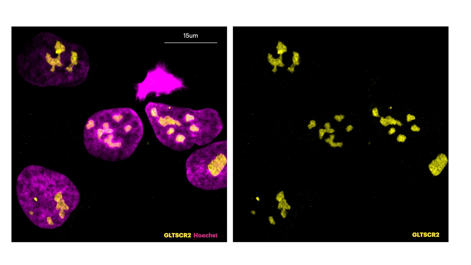
One reviewer rated this antibody 5/5 for immunofluorescence on U2OS cells at a 1:200 dilution. The protocol involved 4% PFA fixation, 0.3% Triton permeabilization, and 5% BSA blocking. The antibody was incubated for 1 hour at room temperature, followed by an Alexa-555 anti-rabbit secondary at 1:600. The resulting image showed clear nucleolar staining of GLTSCR2 (yellow) alongside Hoechst nuclear counterstain (magenta), demonstrating strong and specific signal.
The reviews below have been submitted by verified Proteintech customers who received an incentive for providing their feedback.
Elisa (Verified Customer) (03-15-2022) | U2OS cells were fixed in 4% PFA for 15' at Room temperature. Cells were washed once with PBS for. Membrane permeabilisation was then performed with 0.3% Triton in PBS for 5'. Cells were finally incubated with blocking buffer (5% BSA+ 0.1% Tween in PBS) for 30' at room temperature. Primary antibody was diluted in blocking buffer 1:200 and incubated for 1h at room temperature. Alexa-555-Anti-rabbit was used as secondary antibody (1:600 dilution) (1h at room temperature). In this picture, cells are stained for Hoechst (DNA marker, in magenta) and GLTSCR2 (nucleolar protein in yellow).
 |






