

Tested Applications
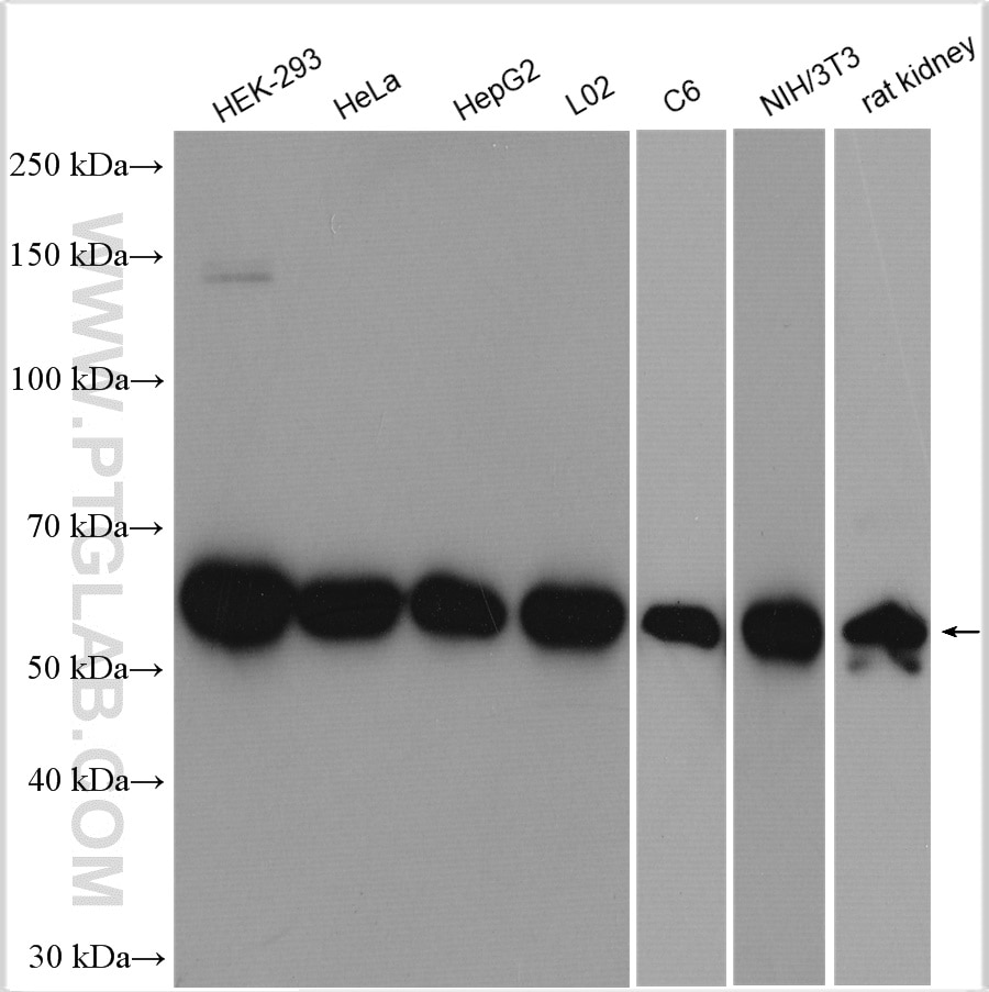
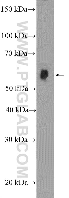
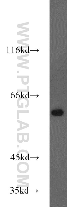
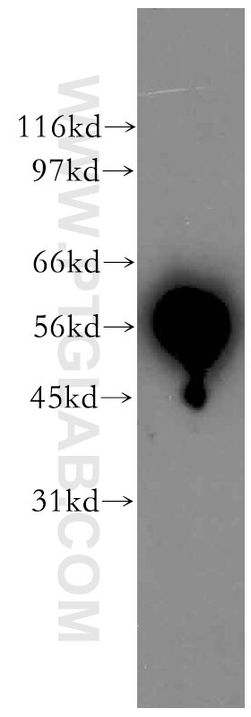
| Positive WB detected in | HEK-293 cells, human kidney tissue, MCF-7 cells, rat liver tissue, HeLa cells, HepG2 cells, L02 cells, C6 cells, NIH/3T3 cells, rat kidney tissue |
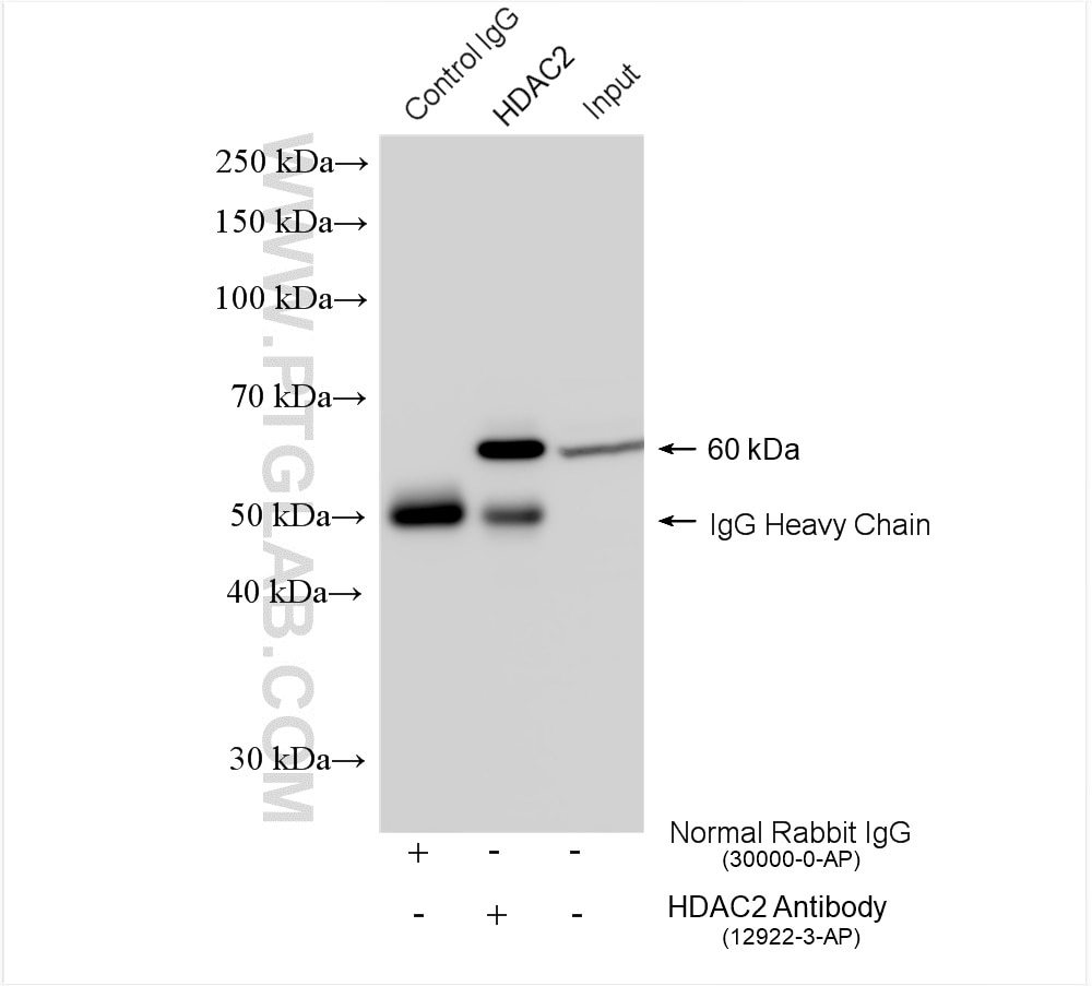
| Positive IP detected in | HEK-293 cells |
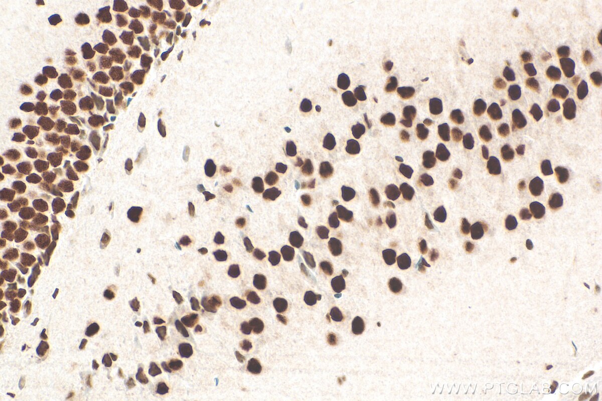
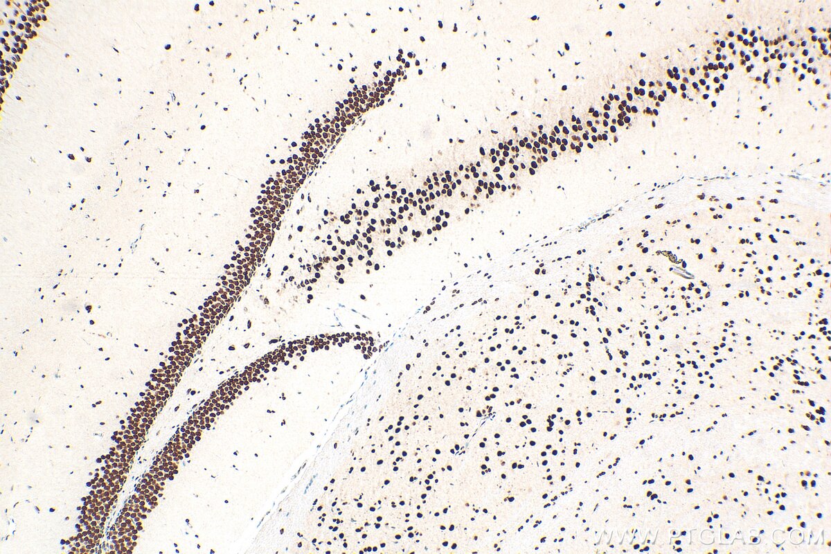
| Positive IHC detected in | mouse brain tissue Note: suggested antigen retrieval with TE buffer pH 9.0; (*) Alternatively, antigen retrieval may be performed with citrate buffer pH 6.0 |
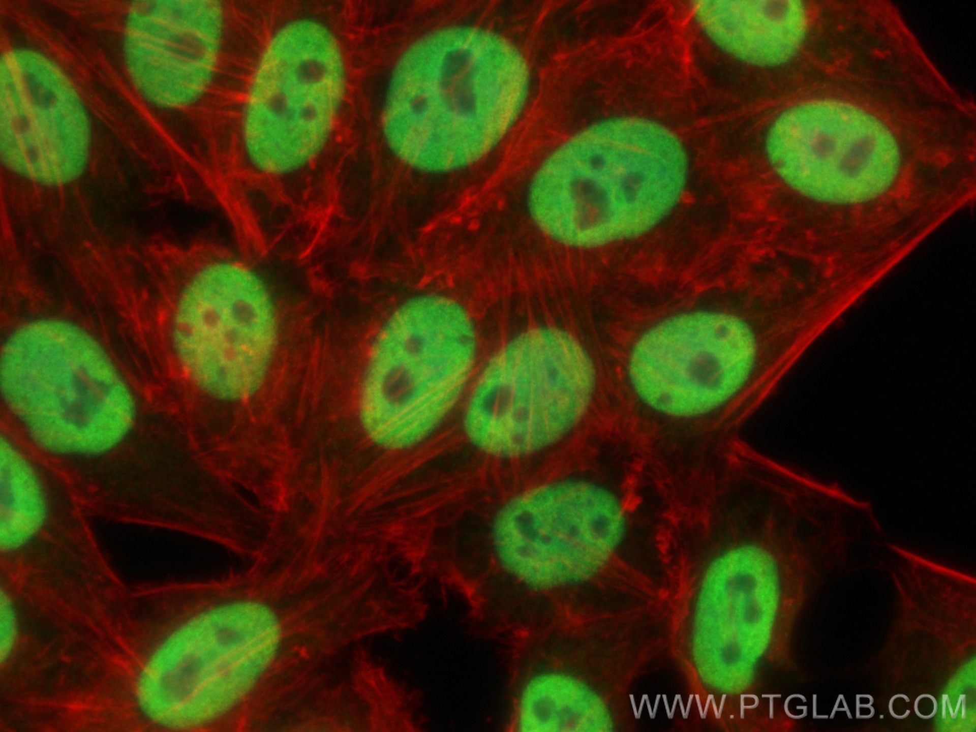
| Positive IF/ICC detected in | HepG2 cells |
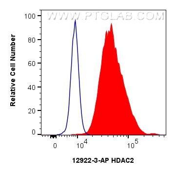
| Positive FC (Intra) detected in | HeLa cells |
Recommended dilution
| Application | Dilution |
|---|---|
| Western Blot (WB) | WB : 1:5000-1:50000 |
| Immunoprecipitation (IP) | IP : 0.5-4.0 ug for 1.0-3.0 mg of total protein lysate |
| Immunohistochemistry (IHC) | IHC : 1:2000-1:8000 |
| Immunofluorescence (IF)/ICC | IF/ICC : 1:200-1:800 |
| Flow Cytometry (FC) (INTRA) | FC (INTRA) : 0.25 ug per 10^6 cells in a 100 µl suspension |
| It is recommended that this reagent should be titrated in each testing system to obtain optimal results. | |
| Sample-dependent, Check data in validation data gallery. | |
Product Information
12922-3-AP targets HDAC2 in WB, IHC, IF/ICC, FC (Intra), IP, CoIP, ChIP, ELISA, CUT&RUN, CUT&Tag applications and shows reactivity with human, mouse, rat samples.
| Tested Reactivity | human, mouse, rat |
| Cited Reactivity | human, mouse, rat |
| Host / Isotype | Rabbit / IgG |
| Class | Polyclonal |
| Type | Antibody |
| Immunogen |
CatNo: Ag3607 Product name: Recombinant human HDAC2 protein Source: e coli.-derived, PGEX-4T Tag: GST Domain: 274-571 aa of BC031055 Sequence: TDRVMTVSFHKYGEYFPGTGDLRDIGAGKGKYYAVNFPMRDGIDDESYGQIFKPIISKVMEMYQPSAVVLQCGADSLSGDRLGCFNLTVKGHAKCVEVVKTFNLPLLMLGGGGYTIRNVARCWTHETAVALDCEIPNELPYNDYFEYFGPDFKLHISPSNMTNQNTPEYMEKIKQRLFENLRMLPHAPGVQMQAIPEDAVHEDSGDEDGEDPDKRISIRASDKRIACDEEFSDSEDEGEGGRRNVADHKKGAKKARIEEDKKETEDKKTDVKEEDKSKDNSGEKTDTKGTKSEQLSNP Predict reactive species |
| Full Name | histone deacetylase 2 |
| Calculated Molecular Weight | 458 aa, 52 kDa; 488 aa,55 kDa |
| Observed Molecular Weight | 55-60 kDa |
| GenBank Accession Number | BC031055 |
| Gene Symbol | HDAC2 |
| Gene ID (NCBI) | 3066 |
| RRID | AB_2118516 |
| Conjugate | Unconjugated |
| Form | Liquid |
| Purification Method | Antigen affinity purification |
| UNIPROT ID | Q92769 |
| Storage Buffer | PBS with 0.02% sodium azide and 50% glycerol, pH 7.3. |
| Storage Conditions | Store at -20°C. Stable for one year after shipment. Aliquoting is unnecessary for -20oC storage. 20ul sizes contain 0.1% BSA. |
Background Information
Histone deacetylases(HDAC) are a class of enzymes that remove the acetyl groups from the lysine residues leading to the formation of a condensed and transcriptionally silenced chromatin.Histone deacetylases act via the formation of large multiprotein complexes, and are responsible for the deacetylation of lysine residues at the N-terminal regions of core histones (H2A, H2B, H3 and H4). At least 4 classes of HDAC were identified. As a class I HDAC, HDAC2 was primarily found in the nucleus. HDAC2 forms transcriptional repressor complexes by associating with many different proteins, including YY1, a mammalian zinc-finger transcription factor. Thus, it plays an important role in transcriptional regulation, cell cycle progression and developmental events. This antibody is a rabbit polyclonal antibody raised against residues near the C terminus of human HDAC2.
Protocols
| Product Specific Protocols | |
|---|---|
| FC protocol for HDAC2 antibody 12922-3-AP | Download protocol |
| IF protocol for HDAC2 antibody 12922-3-AP | Download protocol |
| IHC protocol for HDAC2 antibody 12922-3-AP | Download protocol |
| IP protocol for HDAC2 antibody 12922-3-AP | Download protocol |
| WB protocol for HDAC2 antibody 12922-3-AP | Download protocol |
| Standard Protocols | |
|---|---|
| Click here to view our Standard Protocols |
Publications
What published studies show
HDAC2 Polyclonal antibody (Cat# 12922-3-AP) has accumulated 134 citations published in high-impact journals including Nature Communications, Cell Metabolism, Molecular Cell, Journal of Clinical Investigation, Cell Reports, PNAS, Oncogene, and Autophagy. The research fields span epigenetics, oncology, cardiology, neuroscience, immunology, metabolism, toxicology, and infectious disease. Primary applications include Western blot, immunoprecipitation, ChIP, and IHC, validated in human, mouse, and rat.
| Species | Application | Title |
|---|---|---|
Kidney Int Histone deacetylase 4 selectively contributes to podocyte injury in diabetic nephropathy. | ||
Autophagy Acetylation of STX17 (syntaxin 17) controls autophagosome maturation.
| ||
Nat Commun Targeting SMC3 deacetylation synergizes with XPO1 inhibition to reprogram the epigenetic landscape and suppress NPM1-mutated acute myeloid leukemia | ||
Nat Commun The methyltransferase METTL3 negatively regulates nonalcoholic steatohepatitis (NASH) progression. | ||
Nat Commun Exercise-induced histone lactylation in monocyte-derived macrophages restores cardiac immune homeostasis and function in sepsis-induced cardiomyopathy. |
Reviews
What customers say
Users consistently rate this HDAC2 antibody highly (average 4.8/5 across 5 reviews). It performs well in Western blot, immunohistochemistry, immunofluorescence, and immunoprecipitation. Reviewers highlight bright, clean single bands at the expected molecular weight, excellent confocal imaging, and strong tissue staining. Typical dilutions are 1:600–1:1000 for WB and 1:100 for IHC. One reviewer noted two closely spaced bands on WB, possibly reflecting different isoforms, but still rated the overall quality positively.
The reviews below have been submitted by verified Proteintech customers who received an incentive for providing their feedback.
Mounika (Verified Customer) (01-08-2026) | Bands are very bright, also best under confocal microscope
|
Sai Sindhura (Verified Customer) (01-08-2026) | HDAC2 works very nice
|
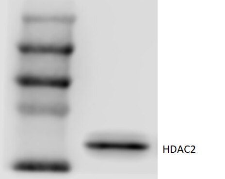
Zhongwen (Verified Customer) (09-25-2023) | There are two bands close to each other. Maybe they are different isoforms. But the quality is quite good.
 |
Iram (Verified Customer) (09-17-2020) | HDAC2 antibody is very good. Giving very clear bands and very good staining in tissue sections
|
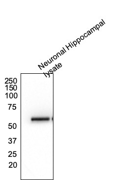
Dipen (Verified Customer) (09-04-2020) | Excellent antibody. One single clean band at the correct molecular weight.
 |










