
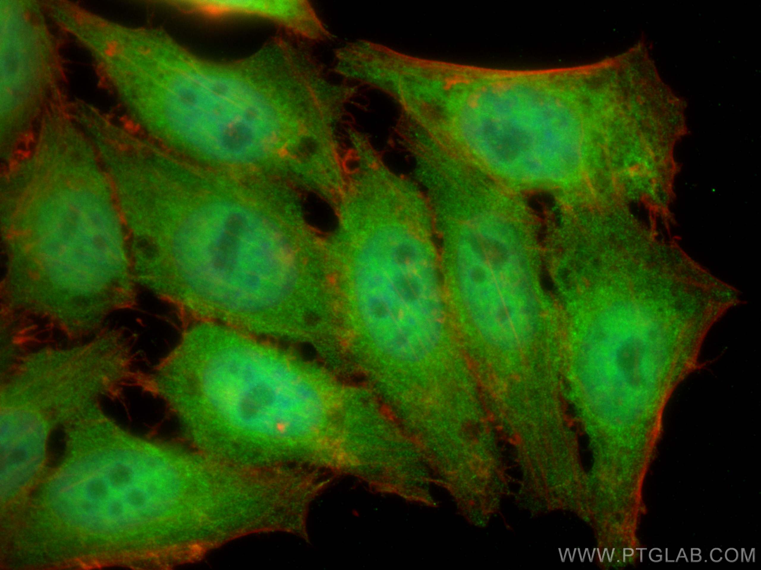

Tested Applications
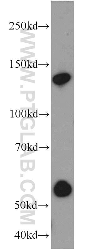
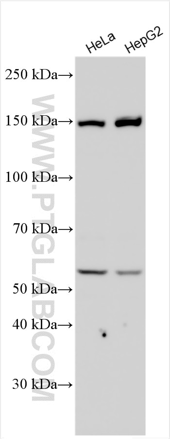
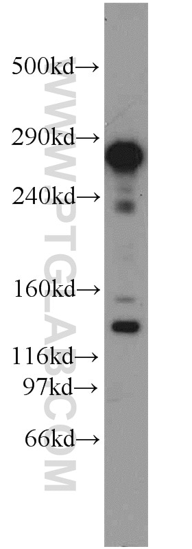
| Positive WB detected in | mouse liver tissue, HeLa cells, HepG2 cells |
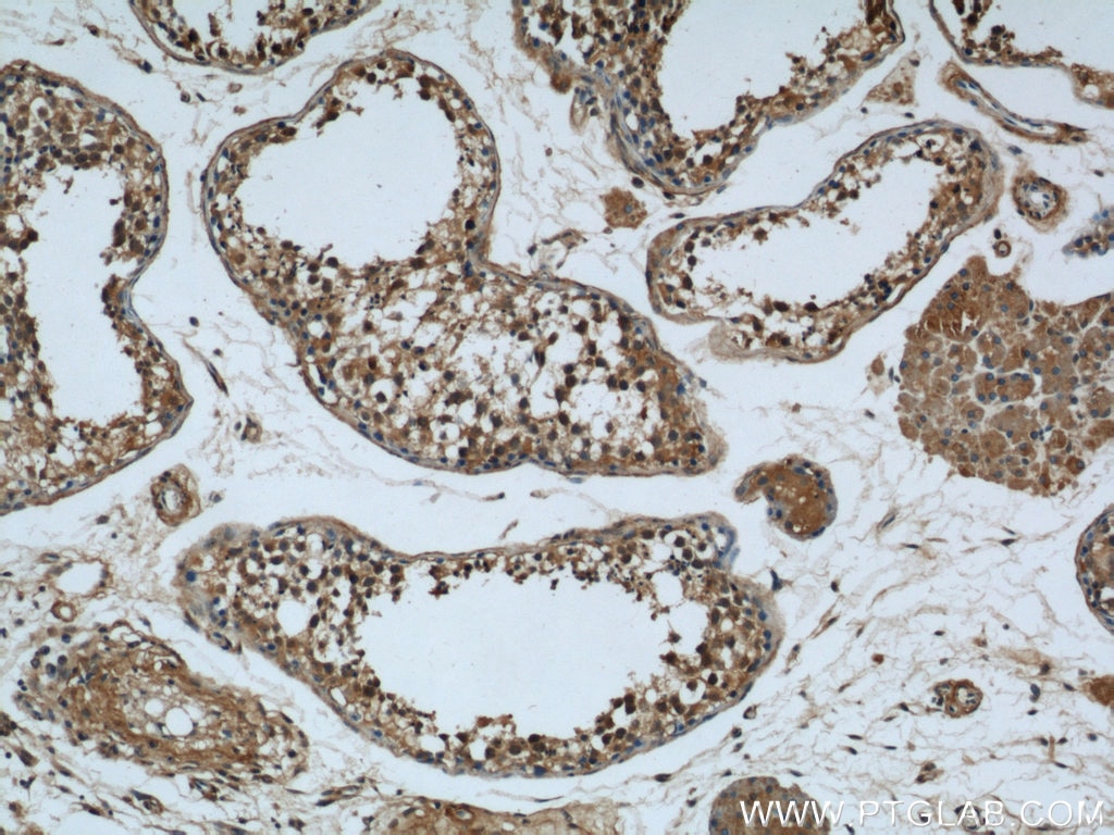
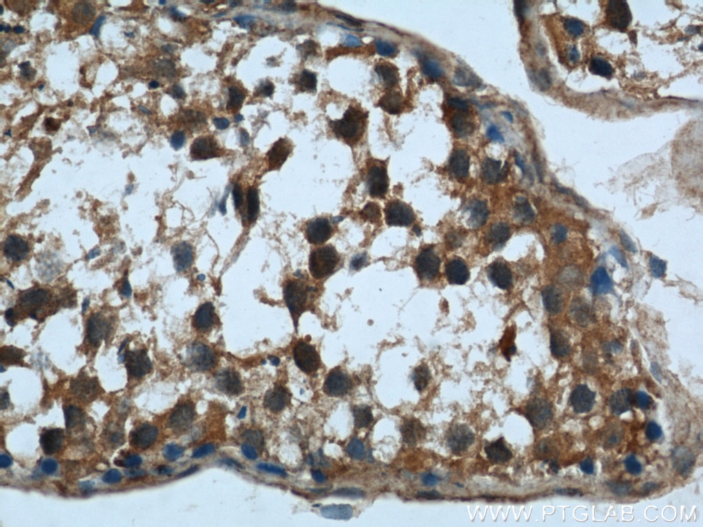
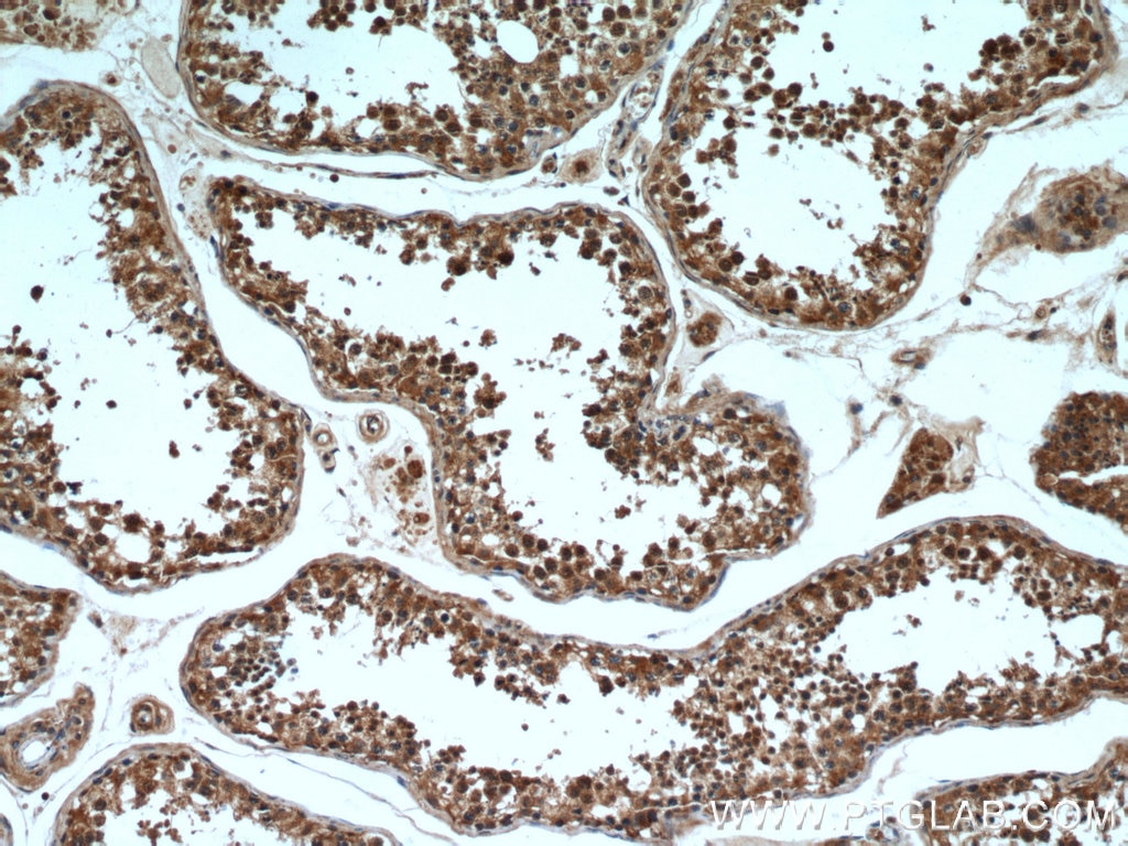
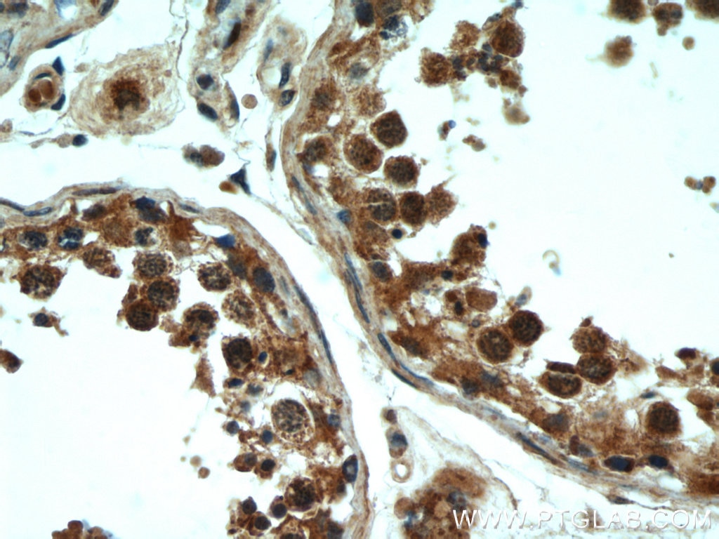
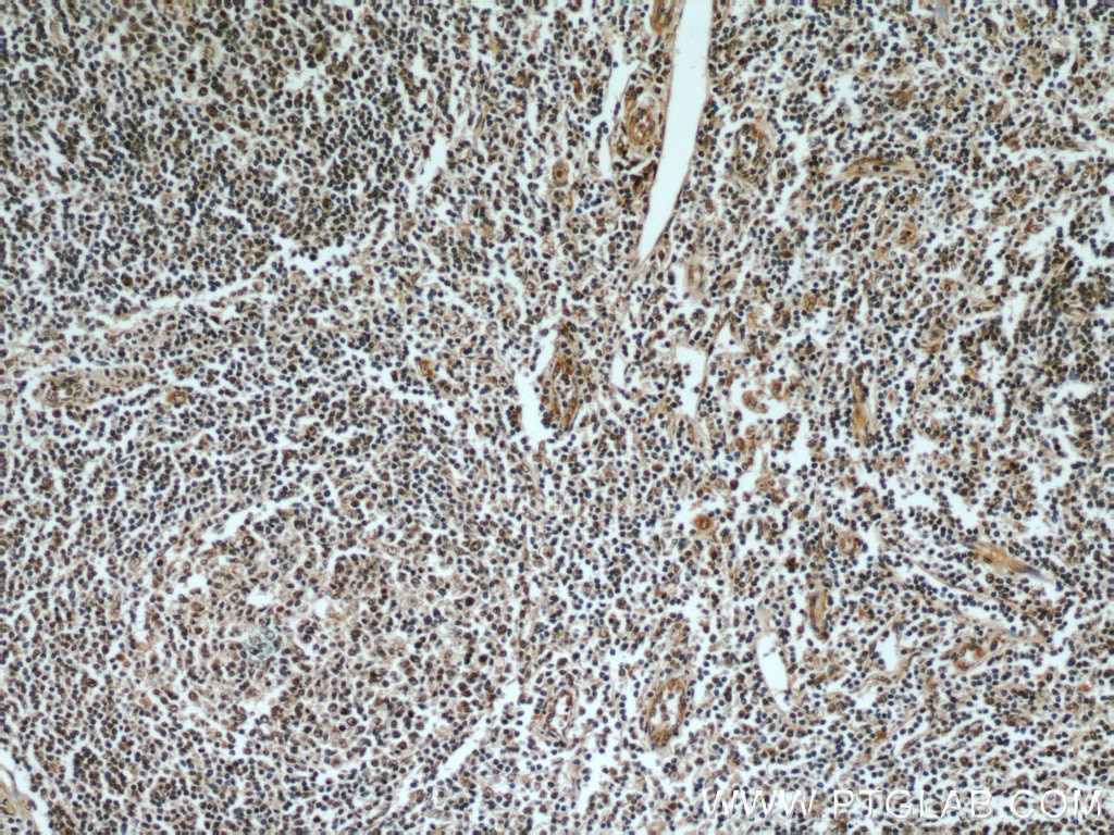
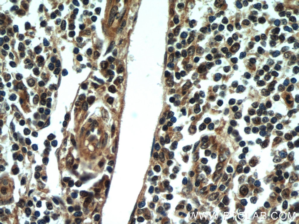
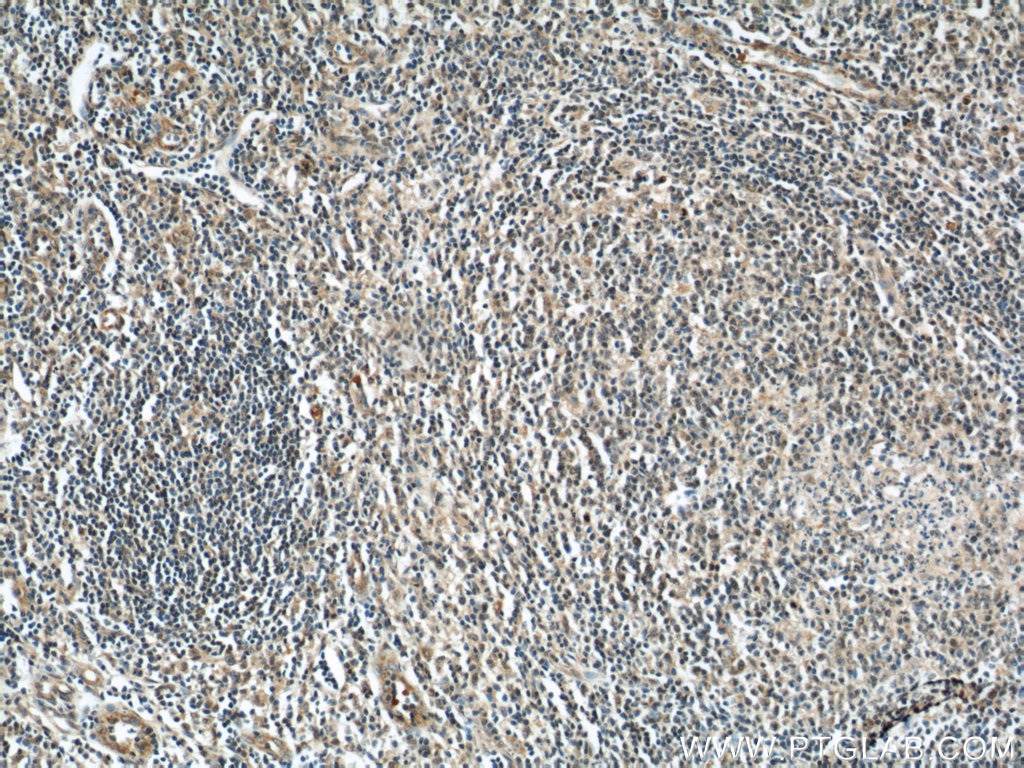
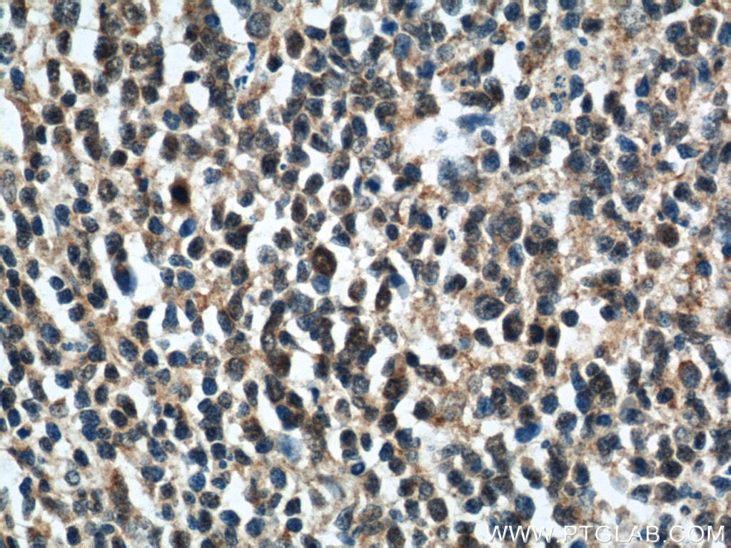
| Positive IHC detected in | human testis tissue, human tonsillitis tissue Note: suggested antigen retrieval with TE buffer pH 9.0; (*) Alternatively, antigen retrieval may be performed with citrate buffer pH 6.0 |
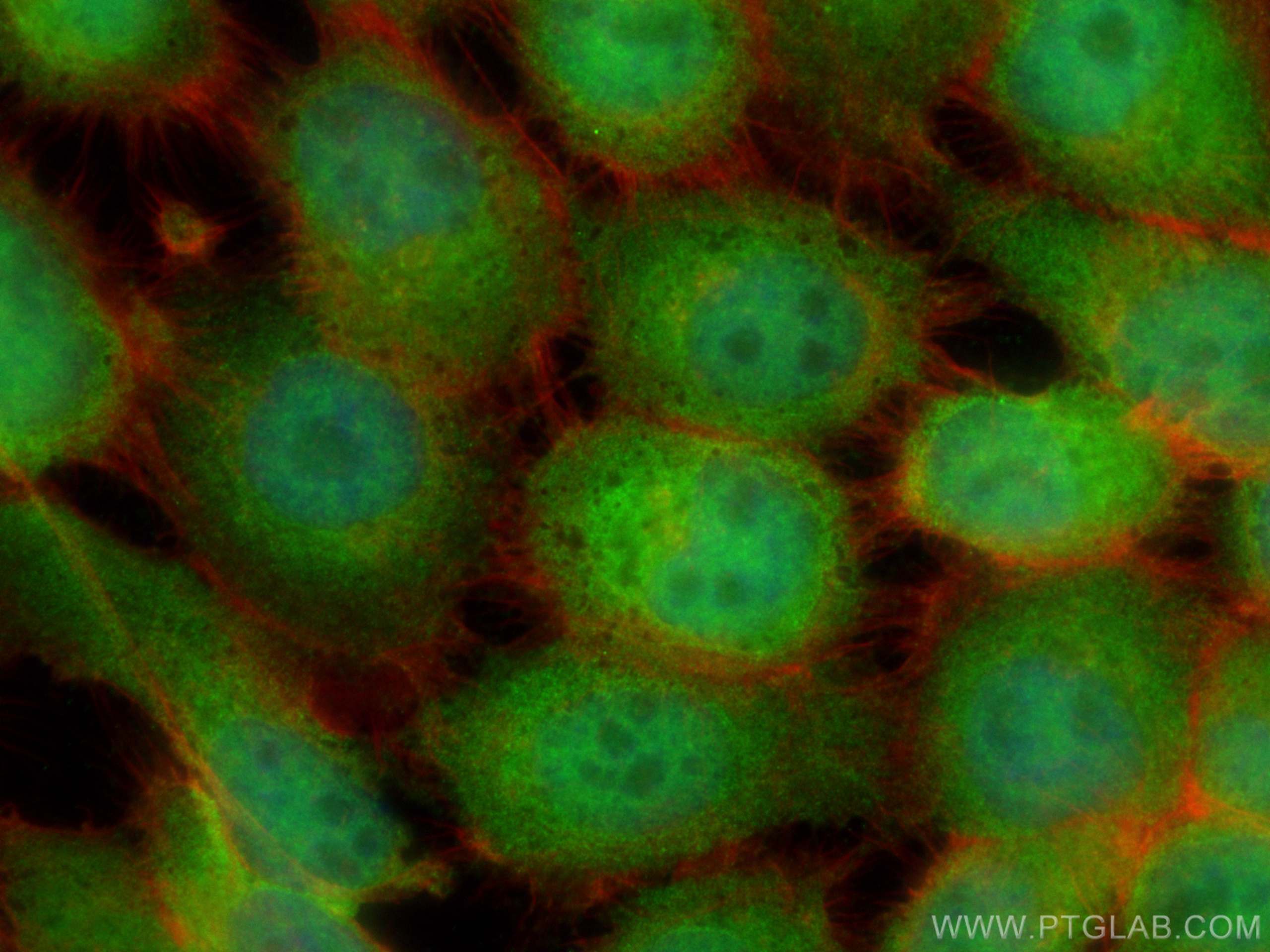
| Positive IF/ICC detected in | HepG2 cells, A431 cells |
Recommended dilution
| Application | Dilution |
|---|---|
| Western Blot (WB) | WB : 1:500-1:2000 |
| Immunohistochemistry (IHC) | IHC : 1:20-1:200 |
| Immunofluorescence (IF)/ICC | IF/ICC : 1:50-1:500 |
| It is recommended that this reagent should be titrated in each testing system to obtain optimal results. | |
| Sample-dependent, Check data in validation data gallery. | |
Product Information
21444-1-AP targets Rubicon in WB, IHC, IF/ICC, IP, CoIP, ELISA applications and shows reactivity with human, mouse, rat samples.
| Tested Reactivity | human, mouse, rat |
| Cited Reactivity | human, mouse, rat, escherichia coli |
| Host / Isotype | Rabbit / IgG |
| Class | Polyclonal |
| Type | Antibody |
| Immunogen |
CatNo: Ag13715 Product name: Recombinant human KIAA0226 protein Source: e coli.-derived, PET28a Tag: 6*His Domain: 61-290 aa of BC033615 Sequence: VAELWLQHSLQYHCLSAQLRPLLGDRQYIRKFYTDAAFLLSDAHVTAMLQCLEAVEQNNPRLLAQIDASMFARKHESPLLVTKSQSLTALPSSTYTPPNSYAQHSYFGSFSSLHQSVPNNGSERRSTSFPLSGPPRKPQESRGHVSPAEDQTIQAPPVSVSALARDSPLTPNEMSSSTLTSPIEASWVSSQNDSPGDASEGPEYLAIGNLDPRGRTASCQSHSSNAESSS Predict reactive species |
| Full Name | KIAA0226 |
| Calculated Molecular Weight | 972 aa, 109 kDa |
| Observed Molecular Weight | 130~150 kDa |
| GenBank Accession Number | BC033615 |
| Gene Symbol | Rubicon |
| Gene ID (NCBI) | 9711 |
| RRID | AB_11182941 |
| Conjugate | Unconjugated |
| Form | Liquid |
| Purification Method | Antigen affinity purification |
| UNIPROT ID | Q92622 |
| Storage Buffer | PBS with 0.02% sodium azide and 50% glycerol, pH 7.3. |
| Storage Conditions | Store at -20°C. Stable for one year after shipment. Aliquoting is unnecessary for -20oC storage. 20ul sizes contain 0.1% BSA. |
Background Information
Rubicon (Run domain Beclin-1 interacting and cysteine-rich containing protein) consists of an N-terminal RUN domain, a middle region that includes its PI3K-binding domain (PIKBD), a coiled-coil domain (CCD), a serine-rich region (S-R), a helix-coil-rich domain (H-C) and a C-terminal Rubicon homology domain (RH) (PMID: 36288306). Rubicon is ubiquitously expressed in most tissues and organs. Western analysis detected bands at 130~150 kDa due to post-translational modification (PMID: 37898101, 35235784).
Protocols
| Product Specific Protocols | |
|---|---|
| IF protocol for Rubicon antibody 21444-1-AP | Download protocol |
| IHC protocol for Rubicon antibody 21444-1-AP | Download protocol |
| WB protocol for Rubicon antibody 21444-1-AP | Download protocol |
| Standard Protocols | |
|---|---|
| Click here to view our Standard Protocols |
Publications
What published studies show
The anti-Rubicon (RUBCN) antibody, catalog number 21444-1-AP, has 28 citations. These appear in journals including Cell Mol Immunol, Autophagy, Nat Commun, ACS Nano, Redox Biol, Adv Sci, Cancer Lett, Int J Biol Sci, Cell Commun Signal, Acta Pharmacol Sin, Genes Dev, PLoS Biol, Cell Insight, J Lipid Res, Sci Rep, J Cereb Blood Flow Metab, J Neurochem, Lab Invest, Immunol Lett, J Immunol Res, Int J Clin Oncol, PLoS One, J Cardiovasc Transl Res, Access Microbiol, and Front Physiol. Research fields span autophagy, cancer, neurology, immunology, and metabolism.
| Species | Application | Title |
|---|---|---|
Autophagy RUBCN/rubicon and EGFR regulate lysosomal degradative processes in the retinal pigment epithelium (RPE) of the eye.
| ||
Autophagy The HGF-MET axis coordinates liver cancer metabolism and autophagy for chemotherapeutic resistance. | ||
Autophagy STYK1 promotes autophagy through enhancing the assembly of autophagy-specific class III phosphatidylinositol 3-kinase complex I. | ||
Nat Commun A mosquito salivary protein promotes flavivirus transmission by activation of autophagy. |
Reviews
What customers say
The single review for this antibody (used for immunofluorescence on iBMDMs at 1:200 dilution) gave a 3-star rating. The reviewer found the antibody performed adequately, though they experienced difficulty distinguishing true positive signal from cellular autofluorescence. They noted this issue stemmed from their cell type rather than the antibody itself, indicating the reagent functioned as expected for its intended application.
The reviews below have been submitted by verified Proteintech customers who received an incentive for providing their feedback.
Daniel (Verified Customer) (07-21-2021) | The antibody seemed to work okay, although I had issues differentiating between autofluorescence and a positive signal. However, that is not the fault of the antibody, but rather my cells.
|














