Tested Applications
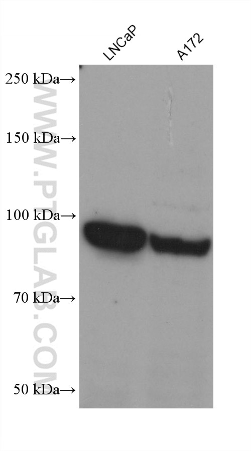
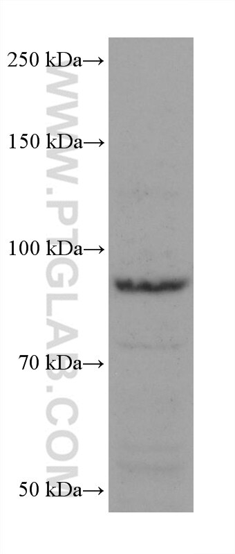
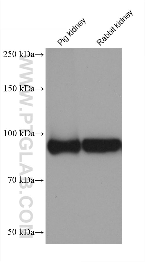
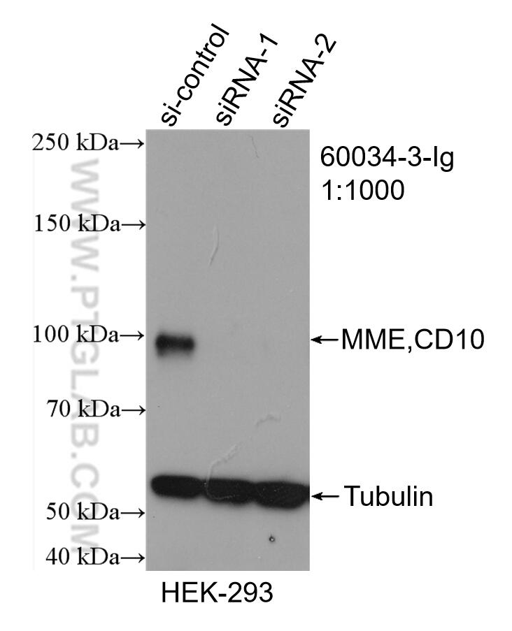
| Positive WB detected in | LNCaP cells, Daudi cells, HEK-293 cells, mouse kidney tissue, pig kidney tissue, Ramos cells, A172 cells, rabbit kidney tissue |
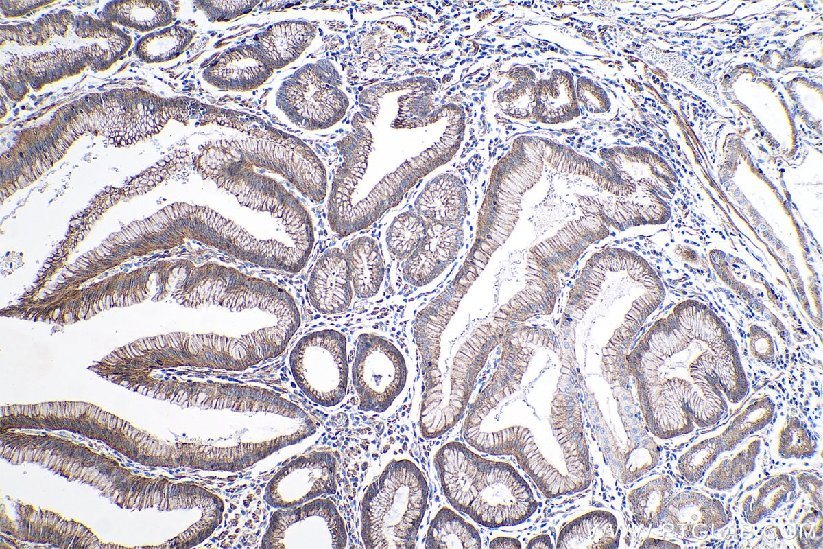
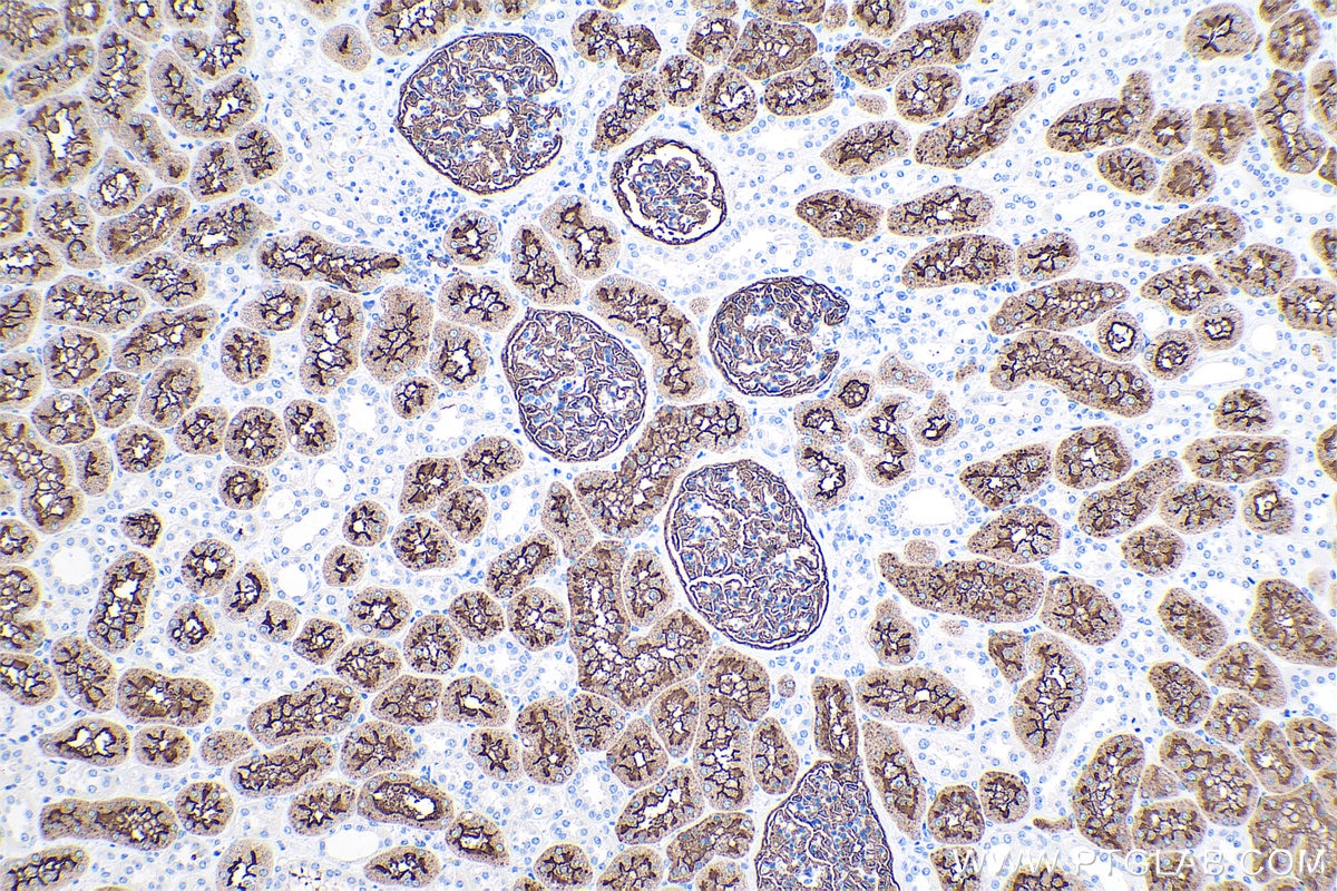
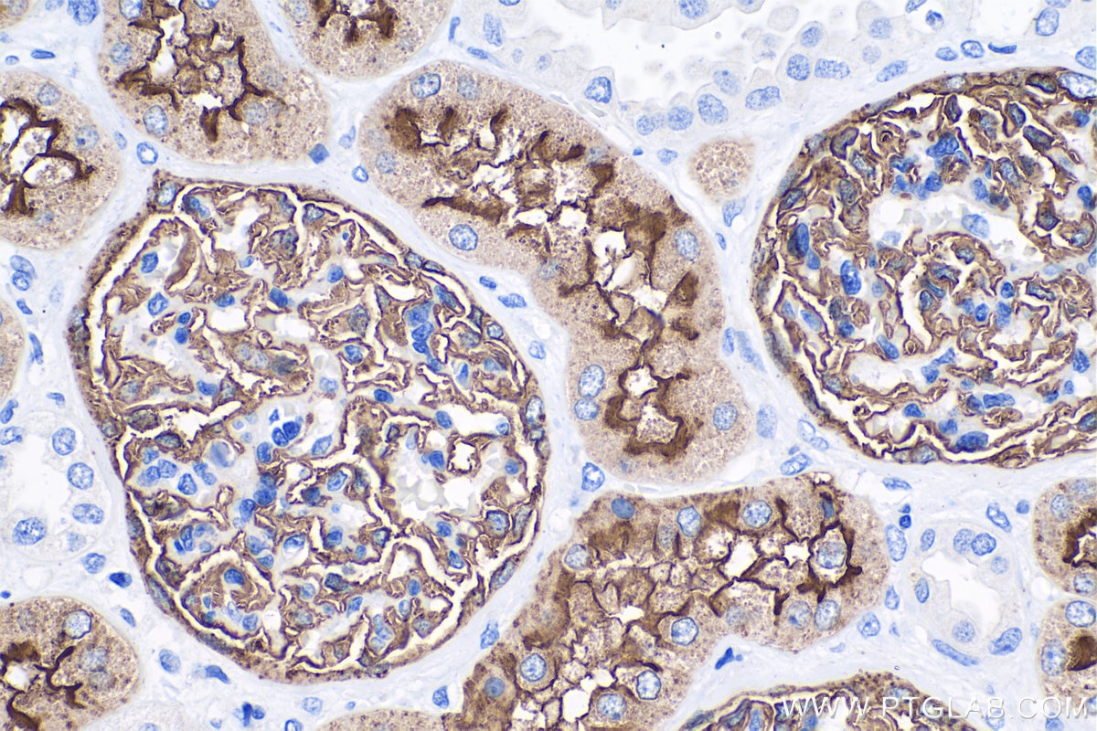
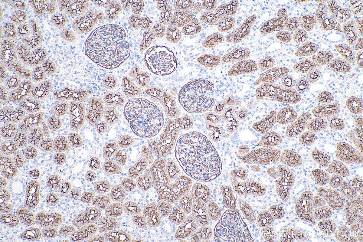
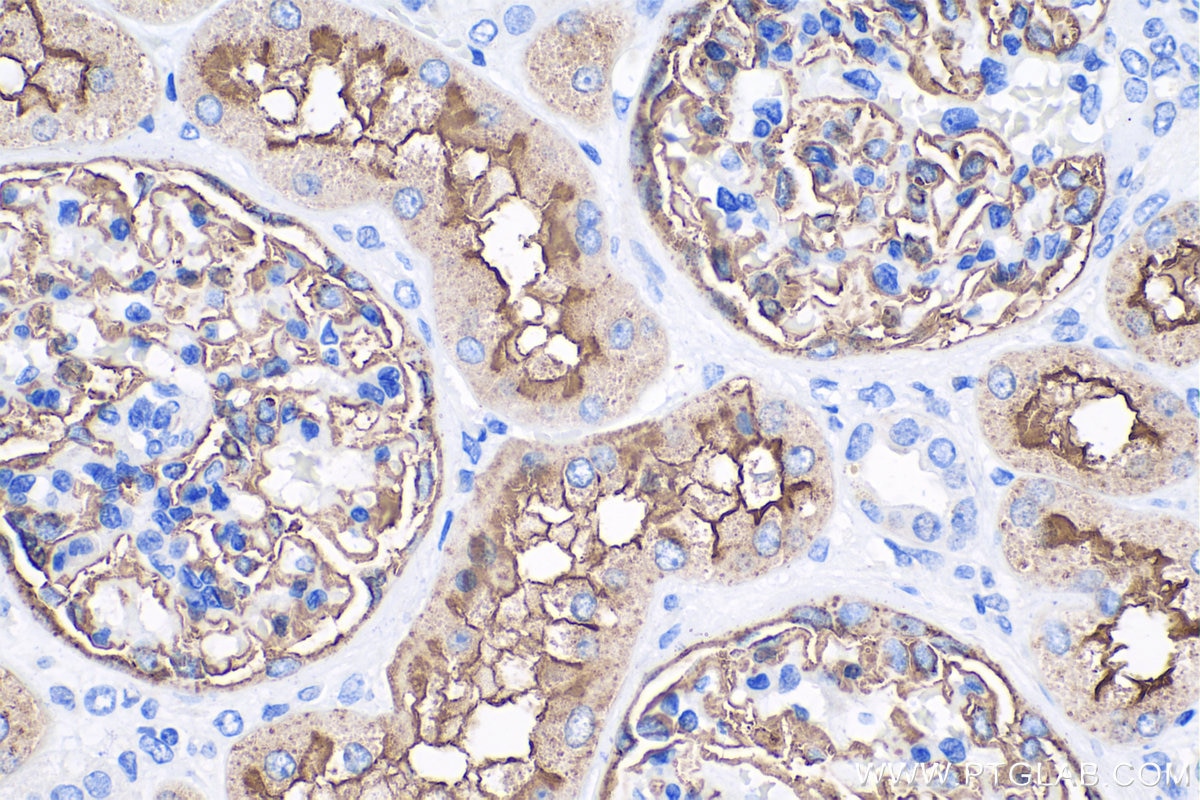
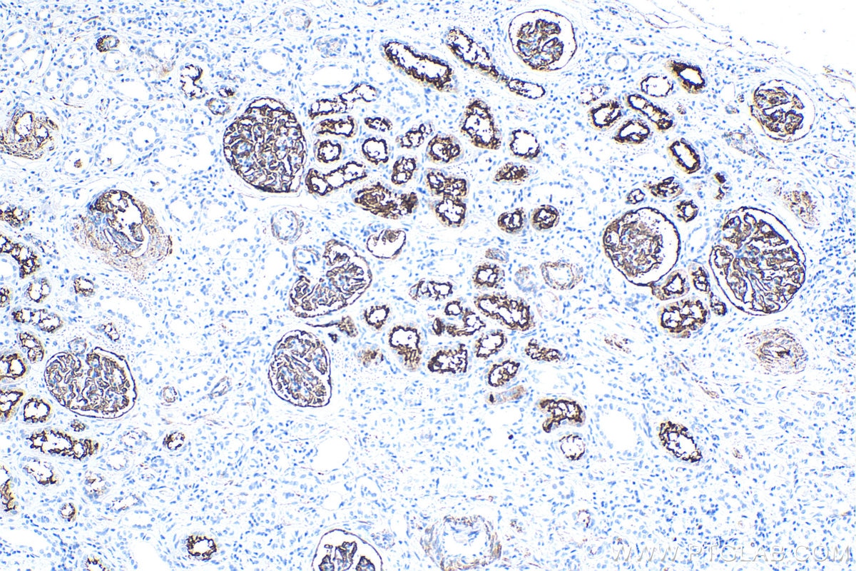
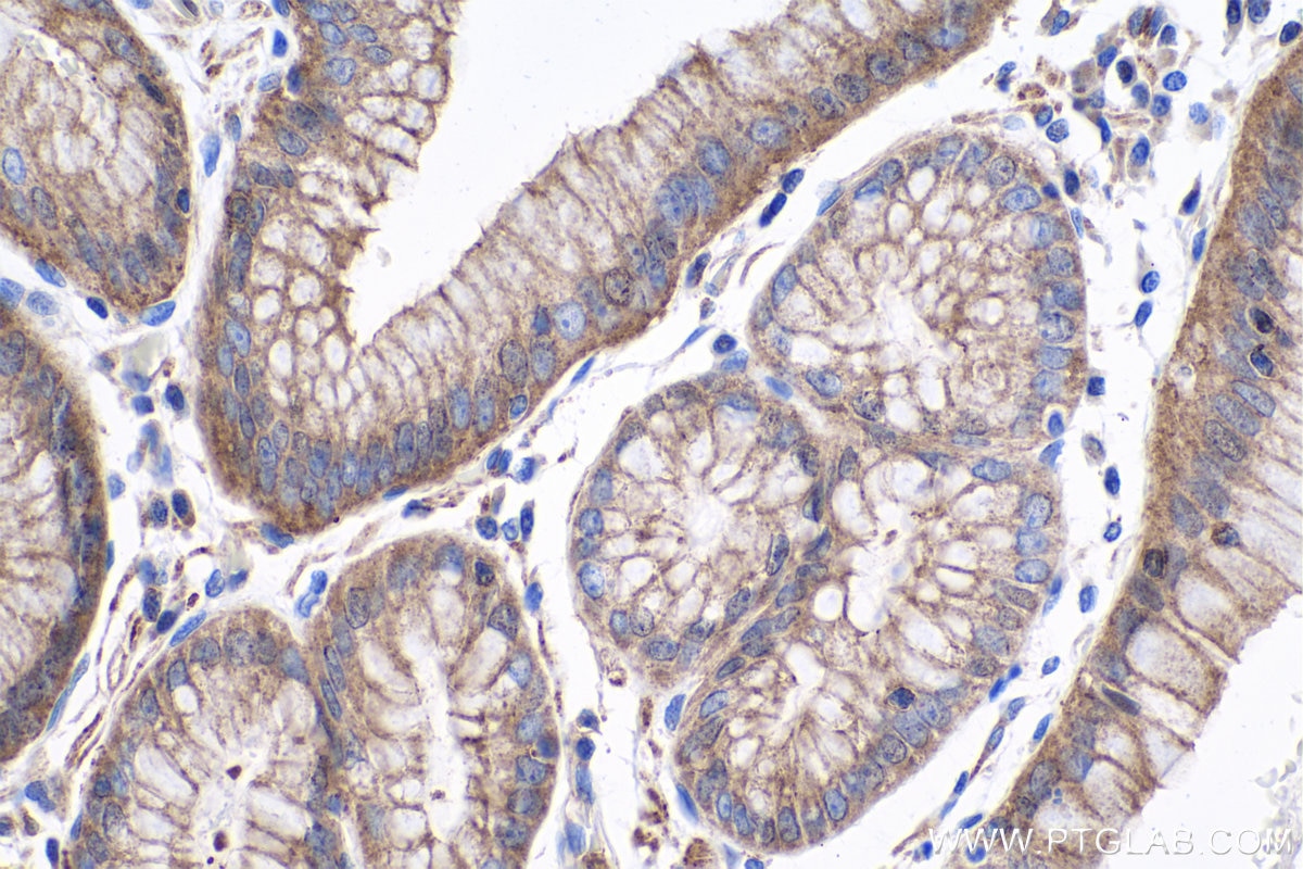
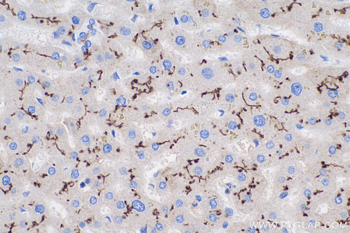
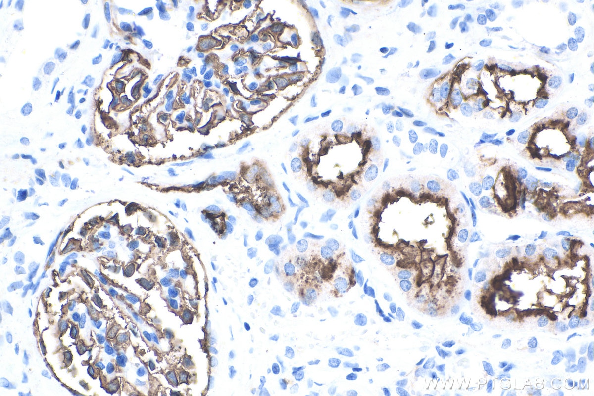
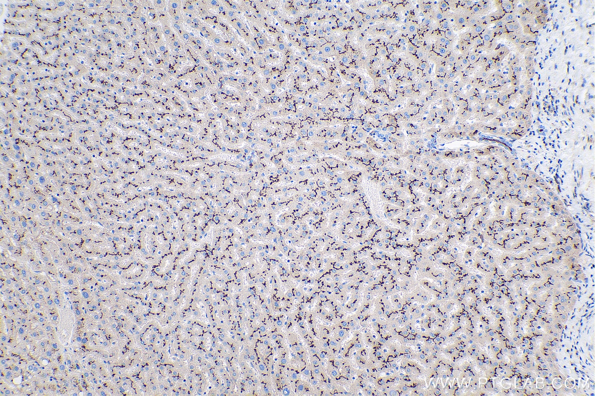
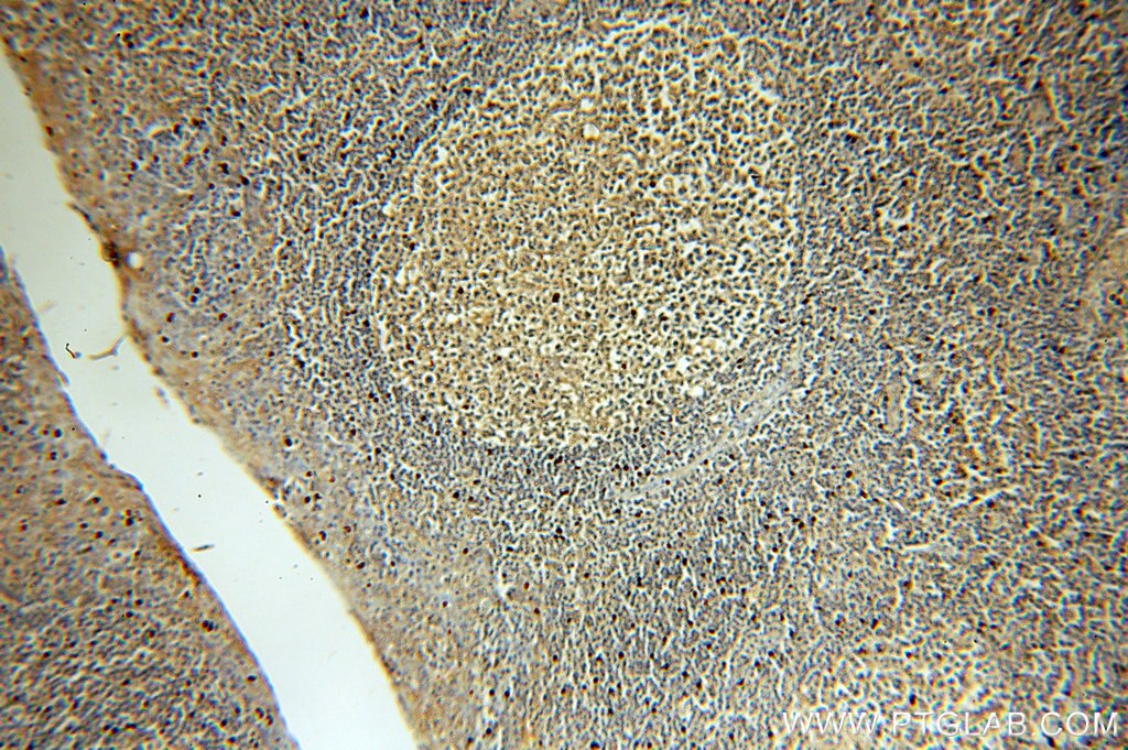
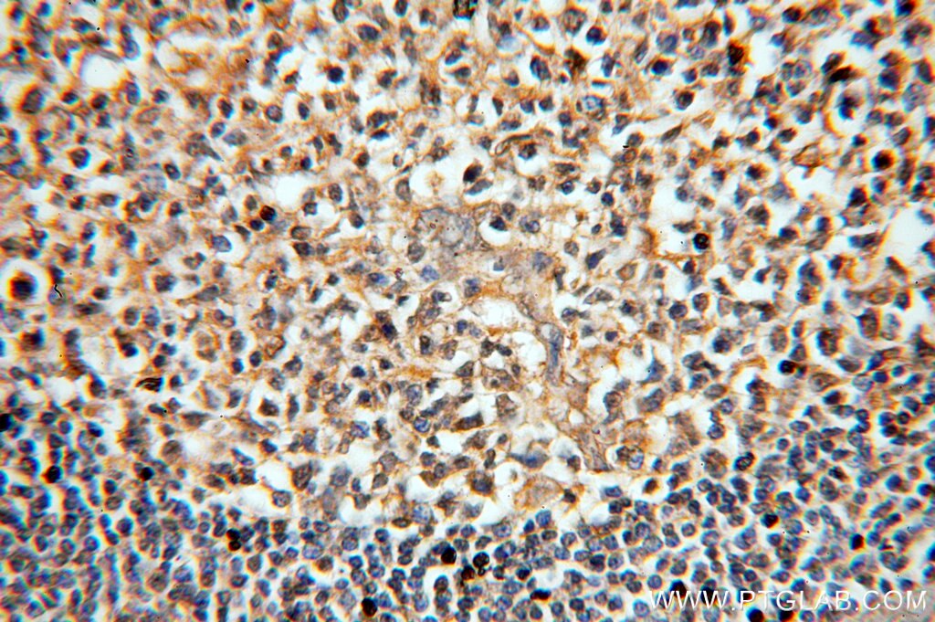
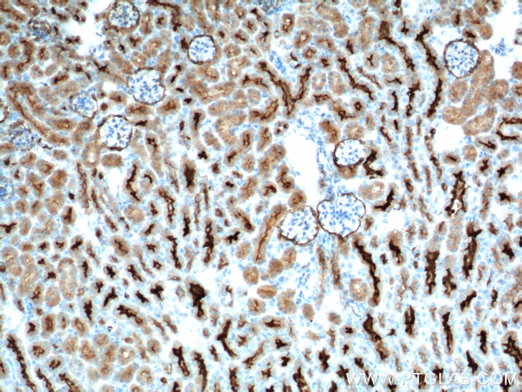
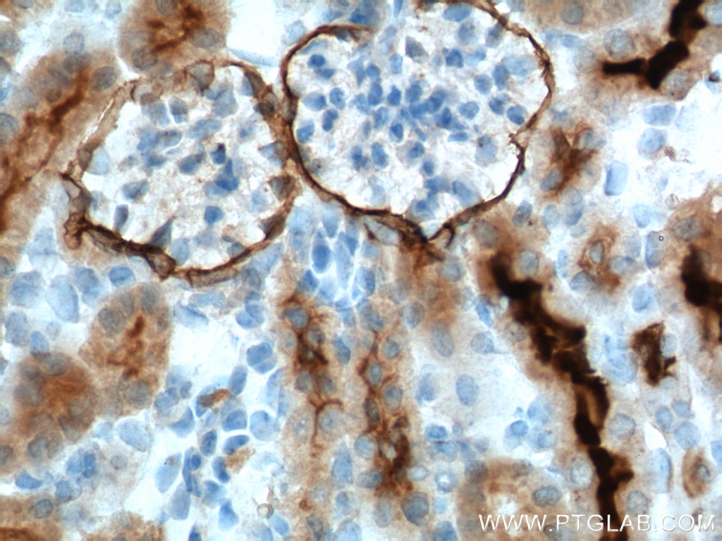
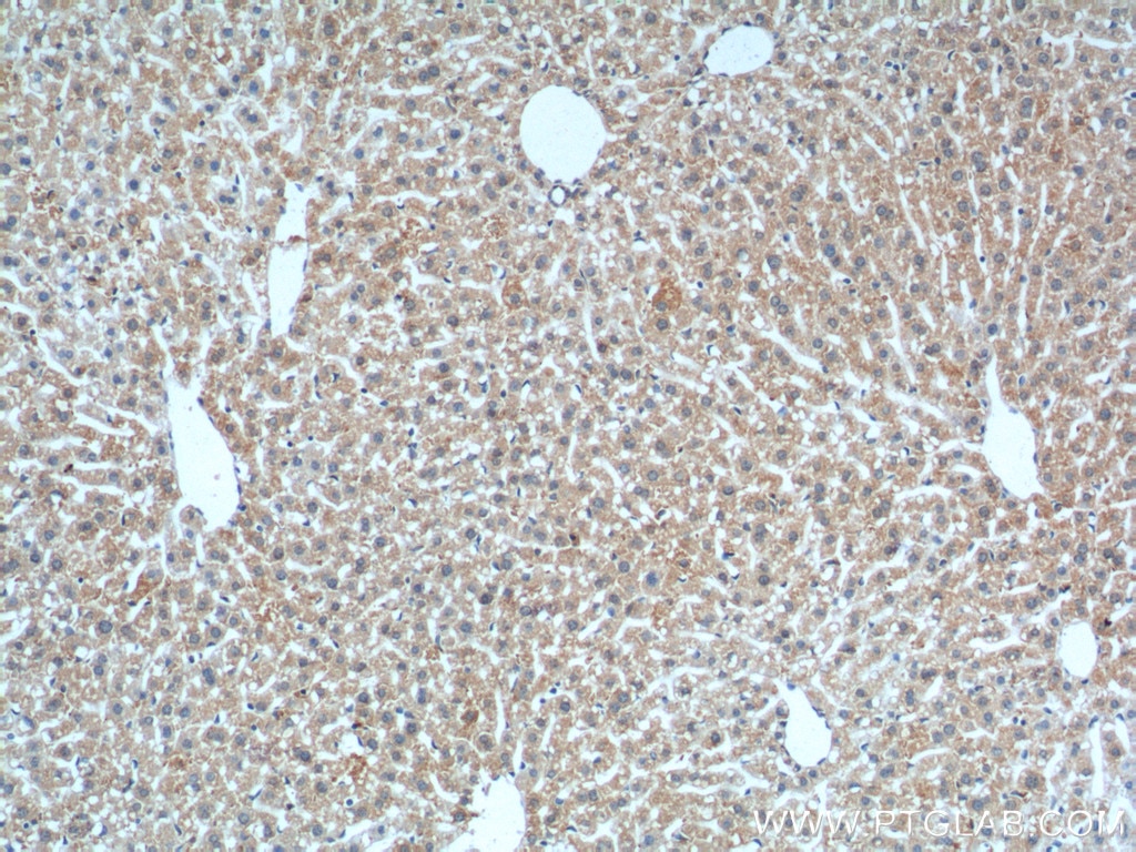
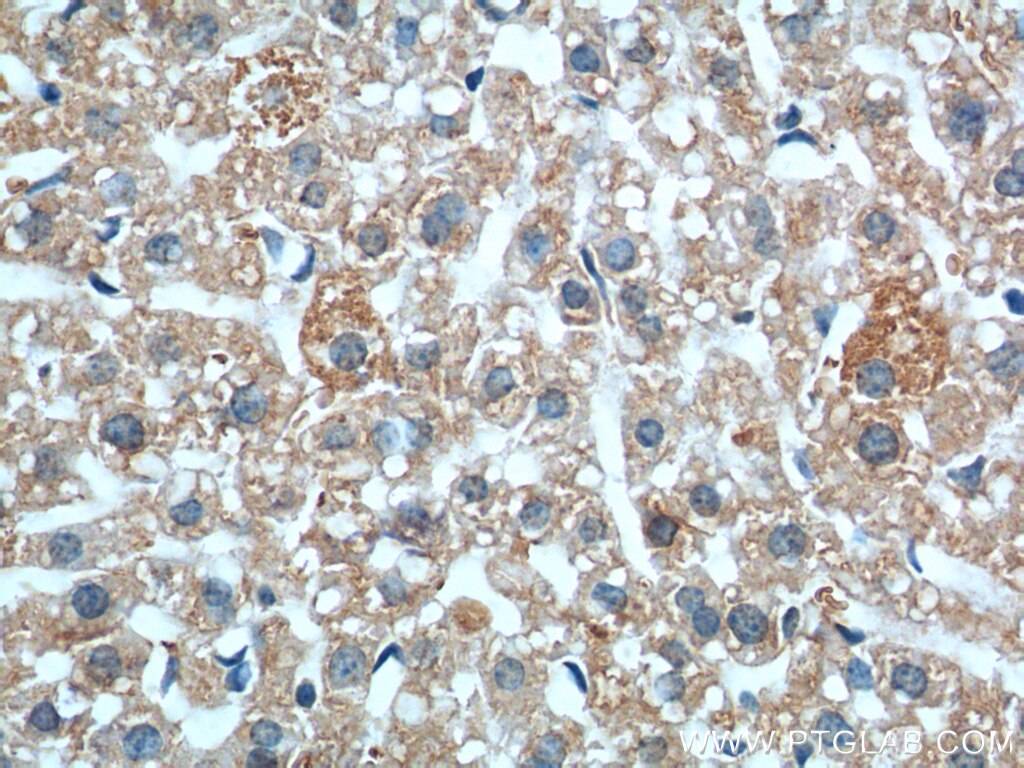
| Positive IHC detected in | human kidney tissue, human liver tissue, human renal cell carcinoma tissue, human stomach cancer tissue, human tonsil tissue, mouse kidney tissue, mouse liver tissue Note: suggested antigen retrieval with TE buffer pH 9.0; (*) Alternatively, antigen retrieval may be performed with citrate buffer pH 6.0 |
Recommended dilution
| Application | Dilution |
|---|---|
| Western Blot (WB) | WB : 1:5000-1:50000 |
| Immunohistochemistry (IHC) | IHC : 1:25000-1:100000 |
| It is recommended that this reagent should be titrated in each testing system to obtain optimal results. | |
| Sample-dependent, Check data in validation data gallery. | |
Product Information
60034-3-Ig targets Neprilysin/CD10 in WB, IHC, ELISA applications and shows reactivity with human, mouse, pig, rabbit samples.
| Tested Reactivity | human, mouse, pig, rabbit |
| Host / Isotype | Mouse / IgG1 |
| Class | Monoclonal |
| Type | Antibody |
| Immunogen |
CatNo: Ag0427 Product name: Recombinant human MME,CD10 protein Source: e coli.-derived, PGEX-4T Tag: GST Domain: 52-252 aa of BC101658 Sequence: YDDGICKSSDCIKSAARLIQNMDATTEPCTDFFKYACGGWLKRNVIPETSSRYGNFDILRDELEVVLKDVLQEPKTEDIVAVQKAKALYRSCINESAIDSRGGEPLLKLLPDIYGWPVATENWEQKYGASWTAEKAIAQLNSKYGKKVLINLFVGTDDKNSVNHVIHIDQPRLGLPSRDYYECTGIYKEACTAYVDFMISV Predict reactive species |
| Full Name | membrane metallo-endopeptidase |
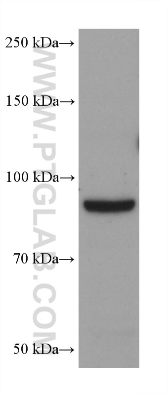
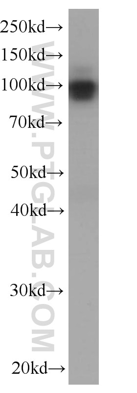
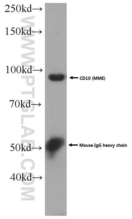
| Calculated Molecular Weight | 750 aa, 86 kDa |
| Observed Molecular Weight | 100 kDa |
| GenBank Accession Number | BC101658 |
| Gene Symbol | CD10 |
| Gene ID (NCBI) | 4311 |
| RRID | AB_10641991 |
| Conjugate | Unconjugated |
| Form | Liquid |
| Purification Method | Protein G purification |
| UNIPROT ID | P08473 |
| Storage Buffer | PBS with 0.02% sodium azide and 50% glycerol, pH 7.3. |
| Storage Conditions | Store at -20°C. Stable for one year after shipment. Aliquoting is unnecessary for -20oC storage. 20ul sizes contain 0.1% BSA. |
Background Information
CD10, also known as neprilysin, membrane metallo-endopeptidase (MME), neutral endopeptidase (NEP), or common acute lymphoblastic leukemia antigen (CALLA), is a 100-kDa type II transmembrane glycoprotein belonging to peptidase M13 family (PMID: 7760013; 8102558). Among hematopoietic cells, CD10 is expressed on granulocytes, B cell precursors, mature germinal center B cells, a subset of immature thymocytes (PMID: 13679451). CD10 is also expressed on a variety of nonhematopoietic cell types, including bronchial epithelial cells, cultured fibroblasts, bone marrow stromal cells, renal proximal tubular epithelial cells, breast myoepithelium, biliary canaliculi (PMID: 8102558). CD10 is a cell surface peptidase that cleaves peptide bonds on the amino side of hydrophobic amino acids and inactivates a variety of physiologically active peptides. Loss or decreases in CD10 expression have been reported in a variety of malignancies (PMID: 16054017).
Protocols
| Product Specific Protocols | |
|---|---|
| IHC protocol for Neprilysin/CD10 antibody 60034-3-Ig | Download protocol |
| WB protocol for Neprilysin/CD10 antibody 60034-3-Ig | Download protocol |
| Standard Protocols | |
|---|---|
| Click here to view our Standard Protocols |