


Tested Applications
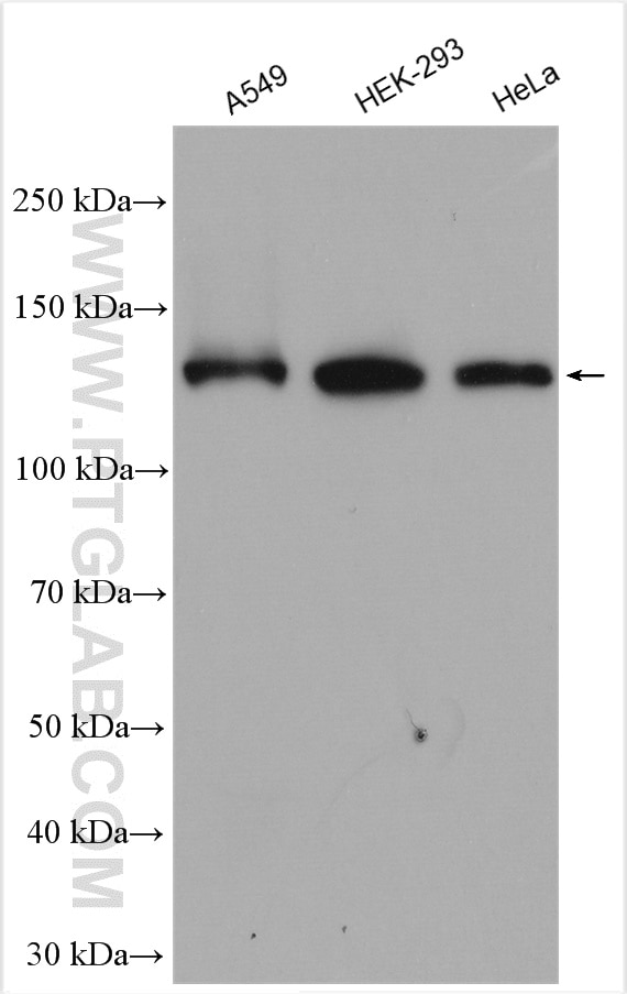
| Positive WB detected in | A549 cells, HEK-293 cells, HeLa cells |
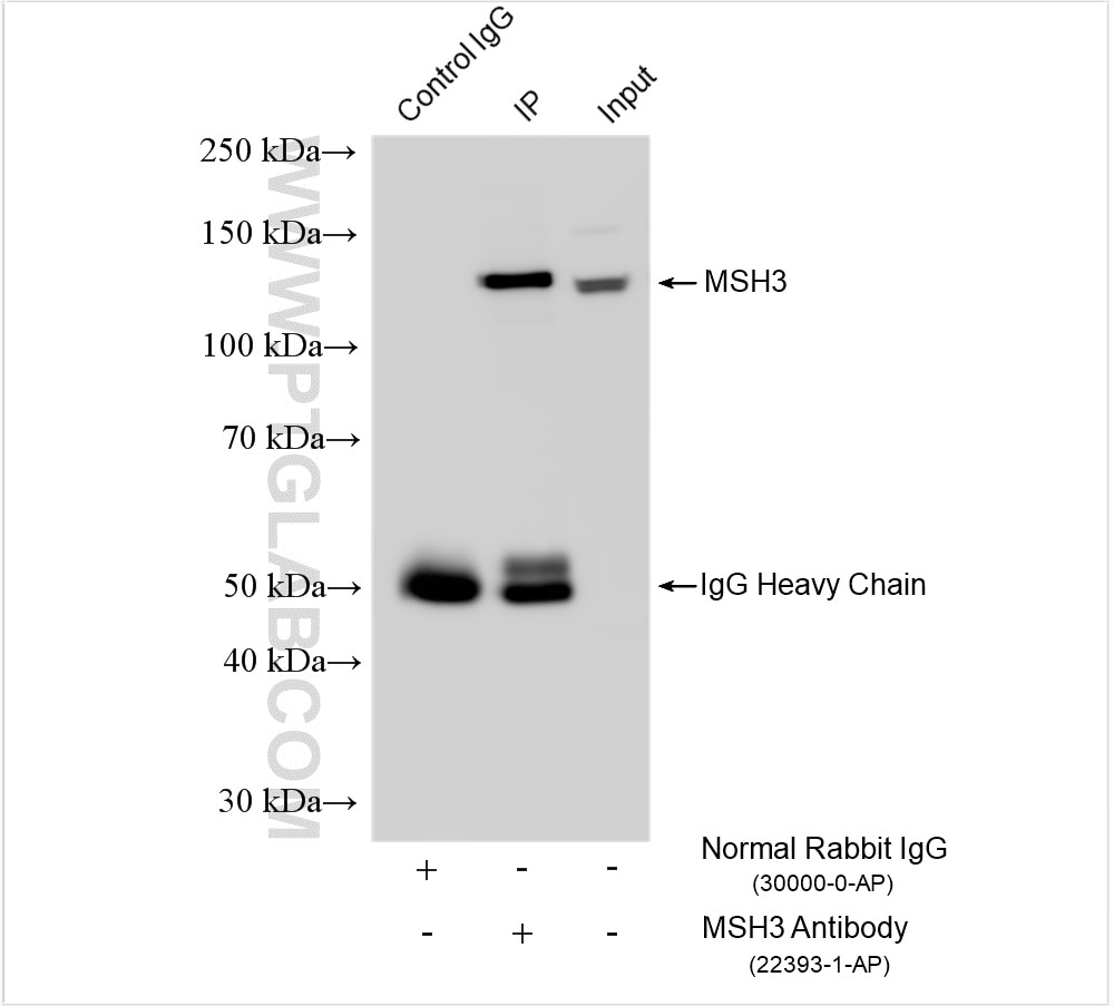
| Positive IP detected in | HeLa cells |
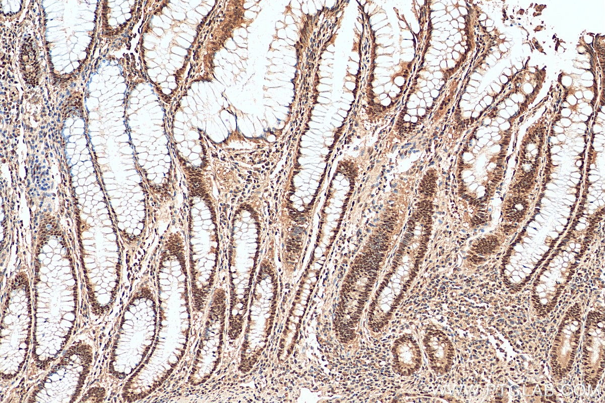
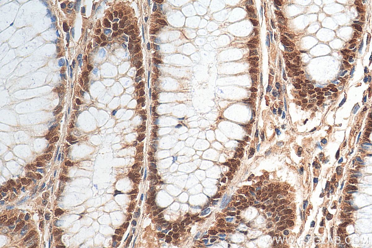
| Positive IHC detected in | human colon cancer tissue Note: suggested antigen retrieval with TE buffer pH 9.0; (*) Alternatively, antigen retrieval may be performed with citrate buffer pH 6.0 |
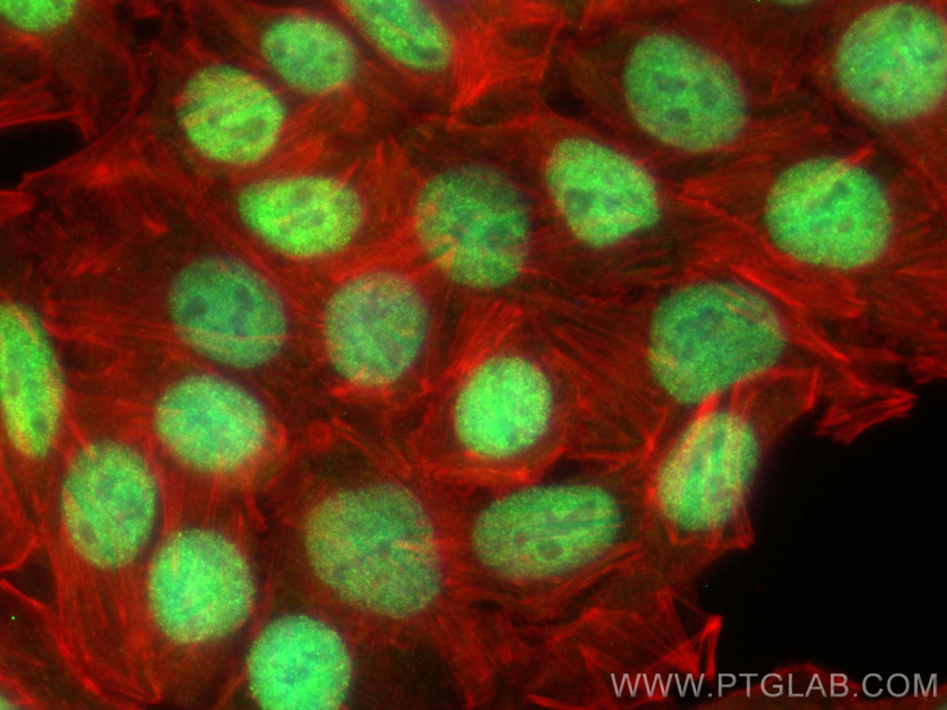
| Positive IF/ICC detected in | HT-29 cells, U2OS cells |
Recommended dilution
| Application | Dilution |
|---|---|
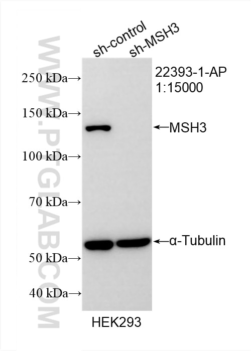
| Western Blot (WB) | WB : 1:5000-1:50000 |
| Immunoprecipitation (IP) | IP : 0.5-4.0 ug for 1.0-3.0 mg of total protein lysate |
| Immunohistochemistry (IHC) | IHC : 1:50-1:500 |
| Immunofluorescence (IF)/ICC | IF/ICC : 1:50-1:500 |
| It is recommended that this reagent should be titrated in each testing system to obtain optimal results. | |
| Sample-dependent, Check data in validation data gallery. | |
Product Information
22393-1-AP targets MSH3 in WB, IHC, IF/ICC, IP, ELISA applications and shows reactivity with human samples.
| Tested Reactivity | human |
| Cited Reactivity | human, mouse |
| Host / Isotype | Rabbit / IgG |
| Class | Polyclonal |
| Type | Antibody |
| Immunogen |
CatNo: Ag18060 Product name: Recombinant human MSH3 protein Source: e coli.-derived, PGEX-4T Tag: GST Domain: 1-350 aa of BC130434 Sequence: MSRRKPASGGLAASSSAPARQAVLSRFFQSTGSLKSTSSSTGAADQVDPGAAAAAAAAAATAPPAPPAPAFPPQLPPHIATEIDRRKKRPLENDGPVKKKVKKVQQKEGGSDLGMSGNSEPKKCLRTRNVSKSLEKLKEFCCDSALPQSRVQTESLQERFAVLPKCTDFDDISLLHAKNAVSSEDSKRQINQKDTTLFDLSQFGSSNTSHENLQKTASKSANKRSKSIYTPLELQYIEMKQQHKDAVLCVECGYKYRFFGEDAEIAARELNIYCHLDHNFMTASIPTHRLFVHVRRLVAKGYKVGVVKQTETAALKAIGDNRSSLFSRKLTALYTKSTLIGEDVNPLIKL Predict reactive species |
| Full Name | mutS homolog 3 (E. coli) |
| Calculated Molecular Weight | 1137 aa, 127 kDa |
| Observed Molecular Weight | 127 kDa |
| GenBank Accession Number | BC130434 |
| Gene Symbol | MSH3 |
| Gene ID (NCBI) | 4437 |
| RRID | AB_11232405 |
| Conjugate | Unconjugated |
| Form | Liquid |
| Purification Method | Antigen affinity purification |
| UNIPROT ID | P20585 |
| Storage Buffer | PBS with 0.02% sodium azide and 50% glycerol, pH 7.3. |
| Storage Conditions | Store at -20°C. Stable for one year after shipment. Aliquoting is unnecessary for -20oC storage. 20ul sizes contain 0.1% BSA. |
Background Information
MSH3 is a DNA mismatch repair (MMR) gene that heterodimerizes with MSH2 to form MutS beta which binds to DNA mismatches thereby initiating DNA repair. It undergoes frequent somatic mutation in colorectal cancers (CRCs) with MMR deficiency. MSH3 mismatch repair protein regulates sensitivity to cytotoxic drugs and a histone deacetylase inhibitor in human colon carcinoma cells (PMID: 23724141, PMID:32671096).
Protocols
| Product Specific Protocols | |
|---|---|
| IF protocol for MSH3 antibody 22393-1-AP | Download protocol |
| IHC protocol for MSH3 antibody 22393-1-AP | Download protocol |
| IP protocol for MSH3 antibody 22393-1-AP | Download protocol |
| WB protocol for MSH3 antibody 22393-1-AP | Download protocol |
| Standard Protocols | |
|---|---|
| Click here to view our Standard Protocols |
Publications
What published studies show
Able is busy answering your previous question. Please try again later.
| Species | Application | Title |
|---|---|---|
Mol Cancer CircLIFR synergizes with MSH2 to attenuate chemoresistance via MutSα/ATM-p73 axis in bladder cancer. | ||
Nucleic Acids Res RNA/DNA-binding protein TDP43 regulates DNA mismatch repair genes with implications for genome stability. | ||
Nucleic Acids Res Nuclear PI3P produced by the Beclin-1/Vps34 complex regulates DNA mismatch repair. | ||
Sci Adv Deficiency in mammalian STN1 promotes colon cancer development via inhibiting DNA repair | ||
J Immunother Cancer Protein arginine methyltransferase 6 enhances immune checkpoint blockade efficacy via the STING pathway in MMR-proficient colorectal cancer |
Reviews
What customers say
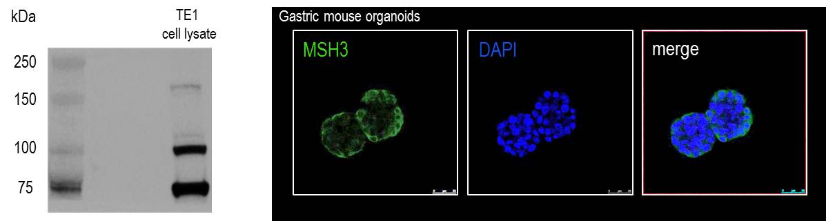
Reviews are mixed. One user reported excellent Western Blot results with a clean band matching the product page and no background, using a 1:1000 dilution in PBST 0.2% on human embryonic stem cells. Another user experienced issues: Western Blot showed multiple bands but none at the expected molecular weight, and immunofluorescence produced cytoplasmic staining rather than the expected nuclear localization of MSH3, tested on mouse organoids and an oesophageal cancer cell line. Overall, performance appears to vary by sample type and application.
The reviews below have been submitted by verified Proteintech customers who received an incentive for providing their feedback.
Chunky (Verified Customer) (03-20-2026) | The band looks as good as shown in the product page. No annoying background bands. Went with 1:1000 with PBST0.2%.
|
Daniela (Verified Customer) (03-17-2022) | In western blot we observed multiple bands, but none at the expected molecular weight. In immunofluorescene, staining was citoplasmatic, while MSH3 is expected to be in the nucleus.
 |








