
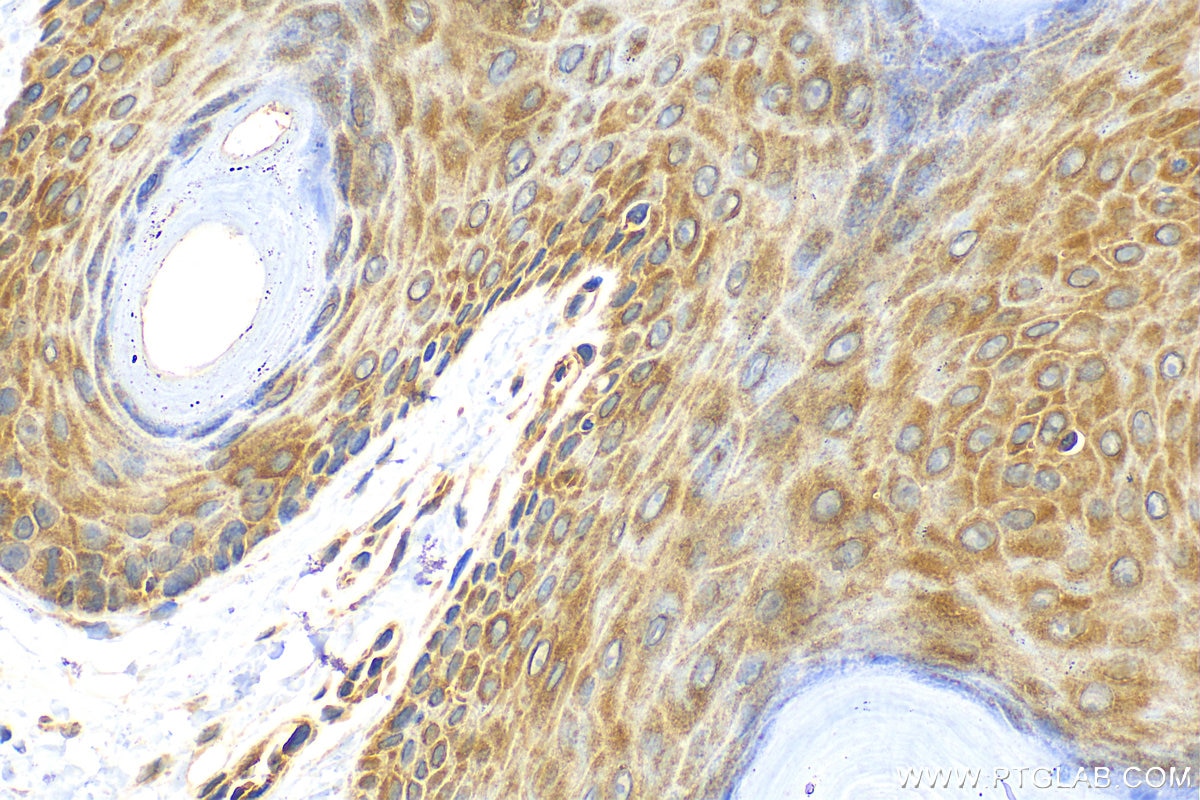
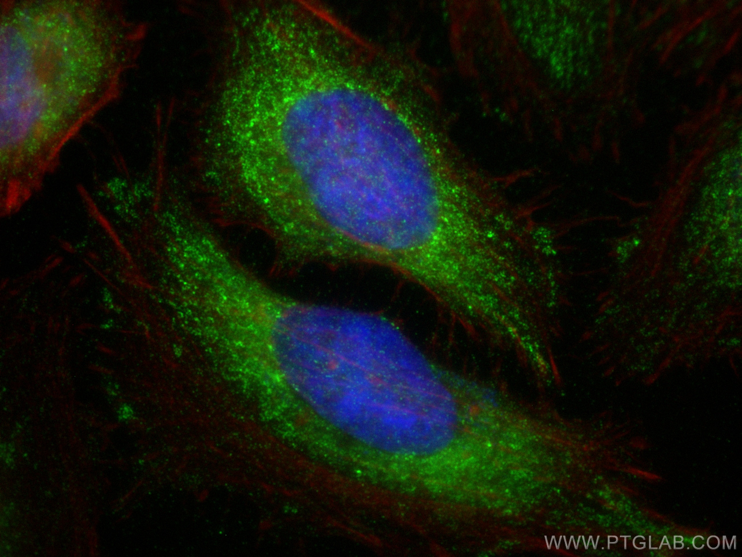

Tested Applications
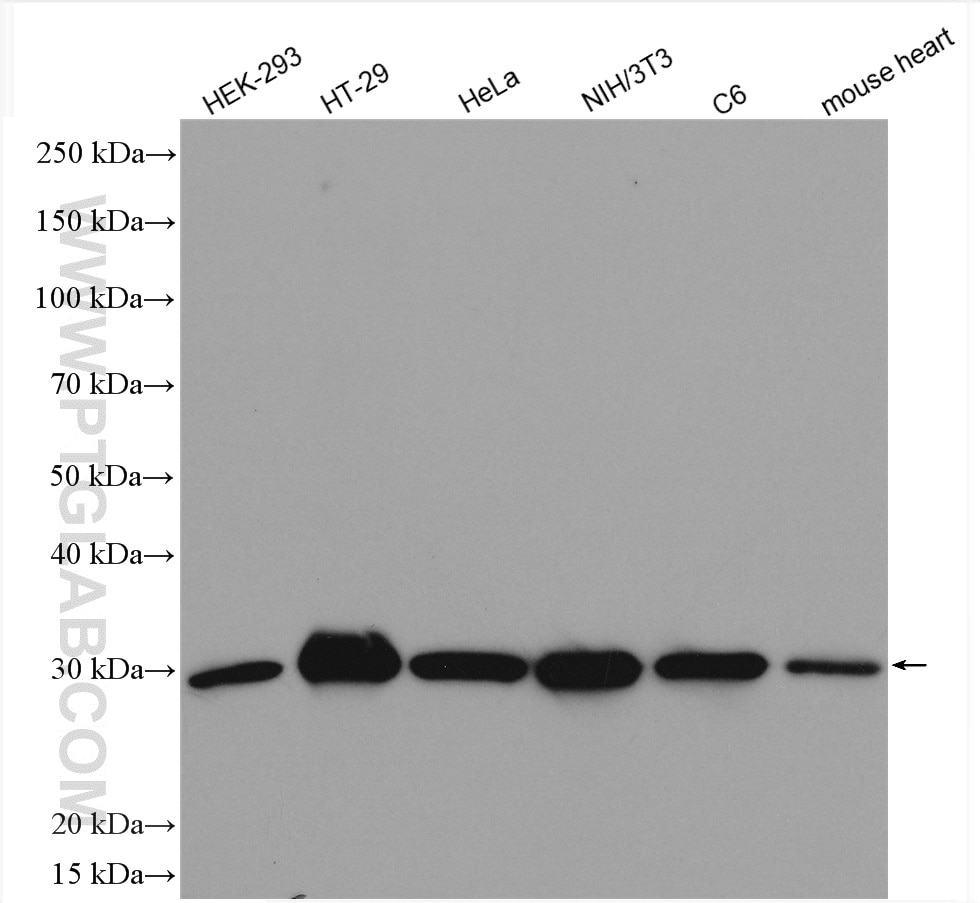
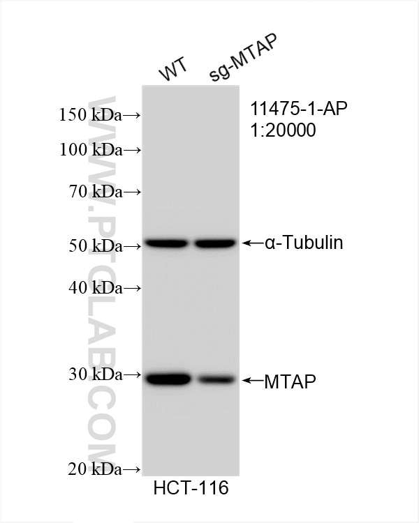
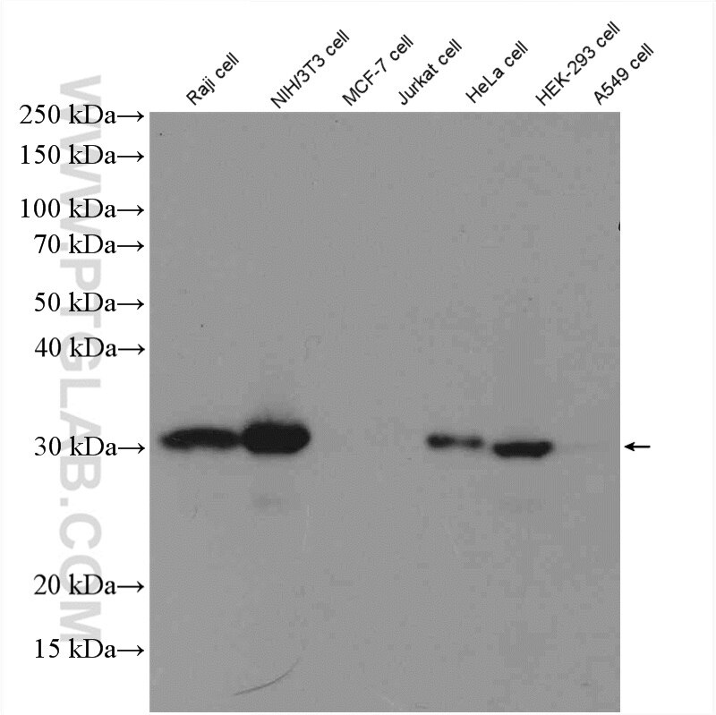
| Positive WB detected in | HEK-293 cells, HCT 116 cells, Raji cells, HT-29 cells, HeLa cells, NIH/3T3 cells, C6 cells, mouse heart tissue |
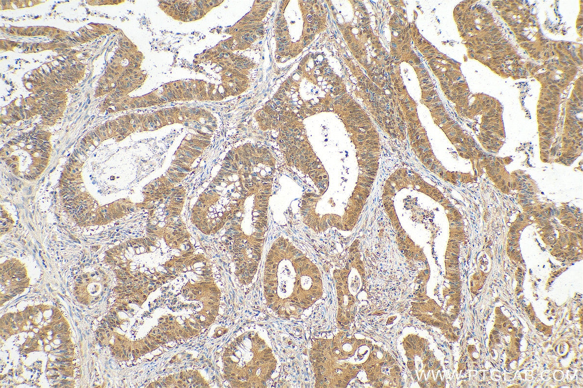
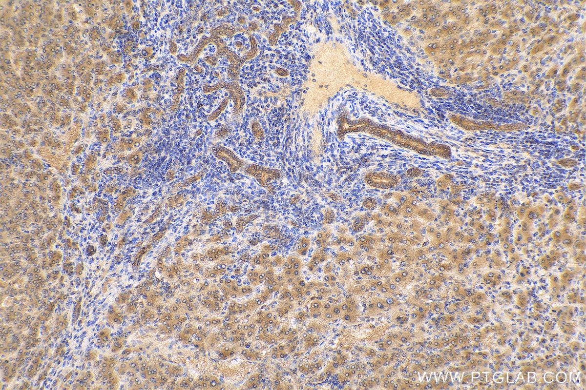
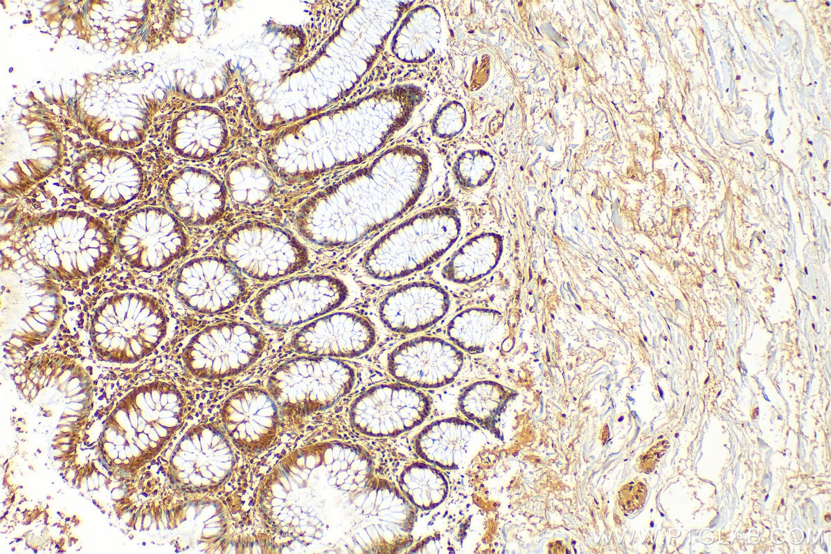
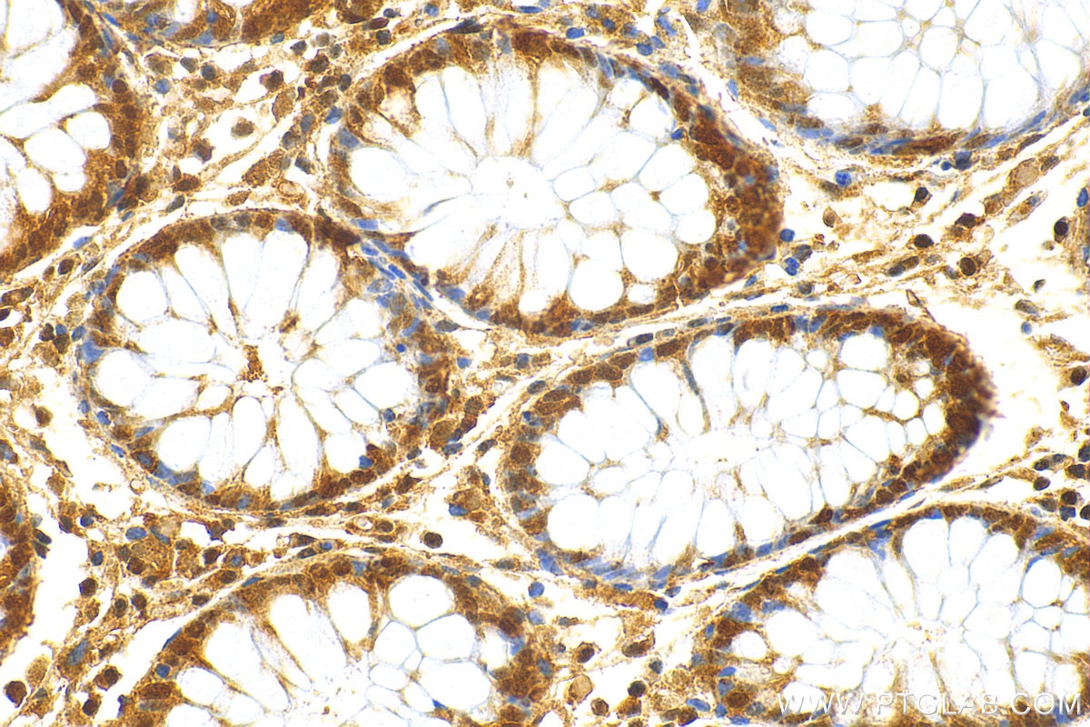
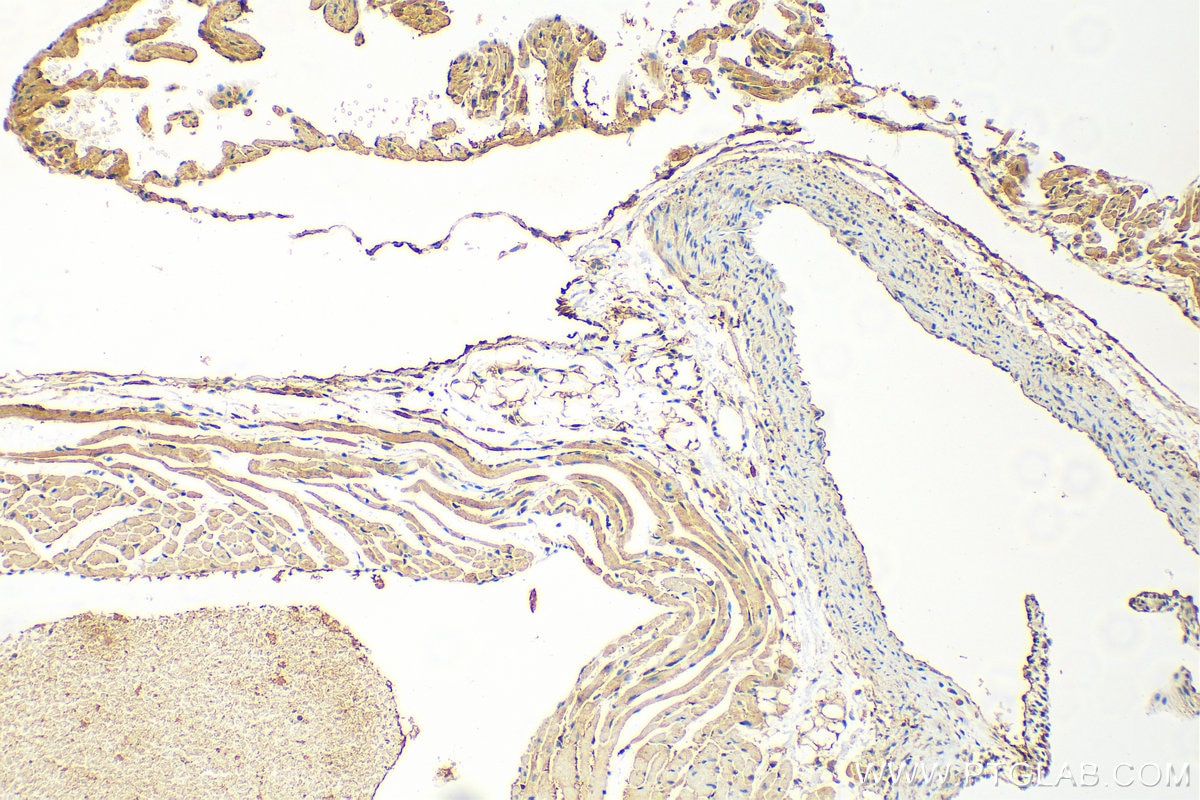
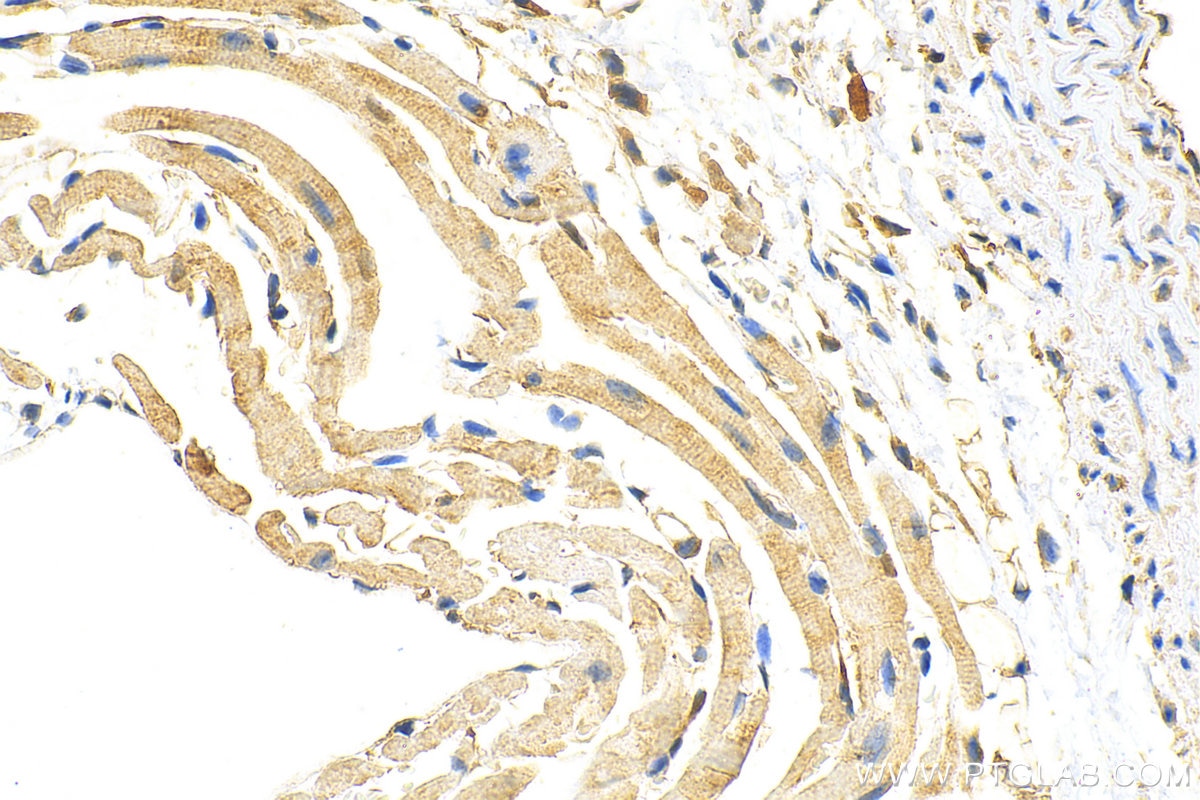
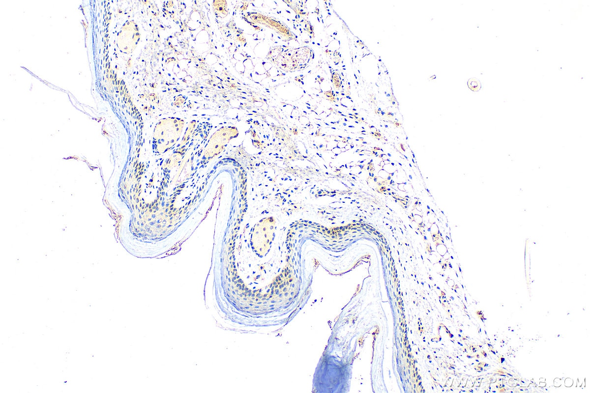
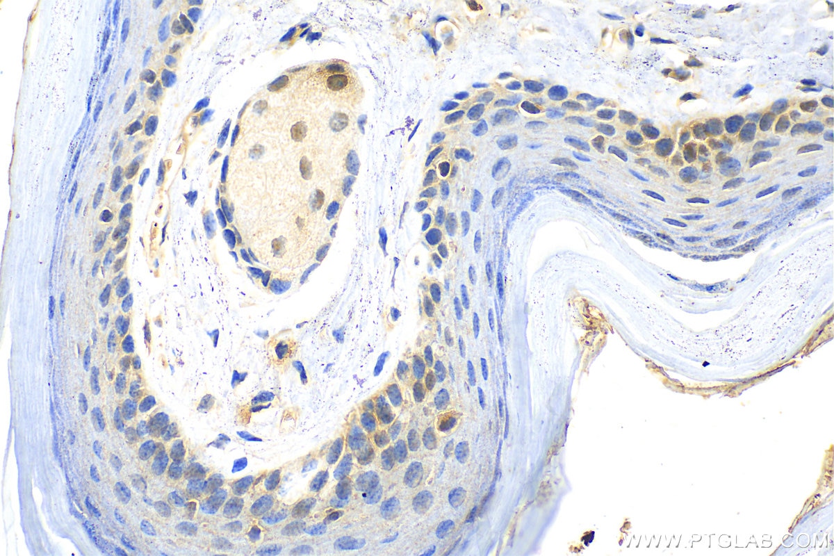
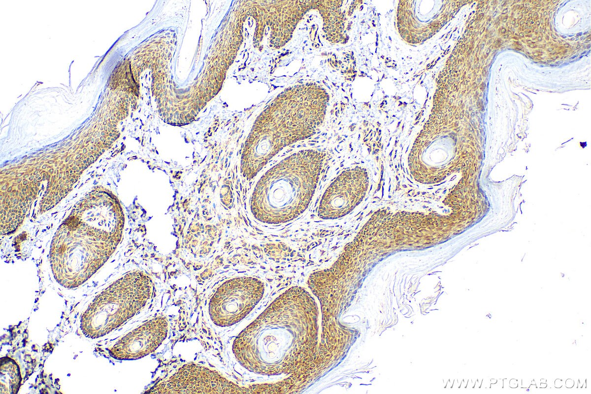
| Positive IHC detected in | human colon cancer tissue, human colon tissue, human liver cancer tissue, mouse heart tissue, mouse skin tissue, rat skin tissue Note: suggested antigen retrieval with TE buffer pH 9.0; (*) Alternatively, antigen retrieval may be performed with citrate buffer pH 6.0 |
| Positive IF/ICC detected in | HeLa cells |
Recommended dilution
| Application | Dilution |
|---|---|
| Western Blot (WB) | WB : 1:2000-1:12000 |
| Immunohistochemistry (IHC) | IHC : 1:300-1:1200 |
| Immunofluorescence (IF)/ICC | IF/ICC : 1:50-1:500 |
| It is recommended that this reagent should be titrated in each testing system to obtain optimal results. | |
| Sample-dependent, Check data in validation data gallery. | |
Product Information
11475-1-AP targets MTAP in WB, IHC, IF/ICC, ELISA applications and shows reactivity with human, mouse, rat samples.
| Tested Reactivity | human, mouse, rat |
| Cited Reactivity | human, mouse, rat |
| Host / Isotype | Rabbit / IgG |
| Class | Polyclonal |
| Type | Antibody |
| Immunogen |
CatNo: Ag2051 Product name: Recombinant human MTAP protein Source: e coli.-derived, PGEX-4T Tag: GST Domain: 1-154 aa of BC018625 Sequence: MASGTTTTAVKIGIIGGTGLDDPEILEGRTEKYVDTPFGKPSDALILGKIKNVDCILLARHGRQHTIMPSKVNYQANIWALKEEGCTHVIVTTACGSLREEIQPGDIVIIDQFIDSHVRRACFPFTFHHDCFQRPPQKPSRSHCVSCATCRTMS Predict reactive species |
| Full Name | methylthioadenosine phosphorylase |
| Calculated Molecular Weight | 283 aa, 31 kDa |
| Observed Molecular Weight | 31 kDa |
| GenBank Accession Number | BC018625 |
| Gene Symbol | MTAP |
| Gene ID (NCBI) | 4507 |
| RRID | AB_2147094 |
| Conjugate | Unconjugated |
| Form | Liquid |
| Purification Method | Antigen affinity purification |
| UNIPROT ID | Q13126 |
| Storage Buffer | PBS with 0.02% sodium azide and 50% glycerol, pH 7.3. |
| Storage Conditions | Store at -20°C. Stable for one year after shipment. Aliquoting is unnecessary for -20oC storage. 20ul sizes contain 0.1% BSA. |
Background Information
MTAP is a 5-deoxy-5-methylthioadenosine (MTA) phosphorylase, converting MTA to salvageable intermediates including adenine and 5-methylthioribose-1-phosphate. MTAP is abundant in normal cells, but it is frequently found to be deleted in a variety of cancers. Its deficiency is common in cancer cell lines as well as in primary leukemia, lung cancer, melanoma, bladder cancer, gliomas and breast cancer.
Protocols
| Product Specific Protocols | |
|---|---|
| IF protocol for MTAP antibody 11475-1-AP | Download protocol |
| IHC protocol for MTAP antibody 11475-1-AP | Download protocol |
| WB protocol for MTAP antibody 11475-1-AP | Download protocol |
| Standard Protocols | |
|---|---|
| Click here to view our Standard Protocols |
Publications
What published studies show
The MTAP antibody (Cat# 11475-1-AP) has accumulated 32 citations published in journals including Nature Metabolism, Nature Communications, Cell Discovery, Journal of Clinical Investigation, Clinical Cancer Research, European Urology Oncology, Oncotarget, Journal of Pathology, and Molecular Cancer Therapeutics. Associated research fields include cancer biology across multiple tumor types (prostate, melanoma, glioma, gastric, urothelial), polyamine metabolism, MTAP/CDKN2A-B deletion as prognostic markers, and novel therapeutic target discovery.
| Species | Application | Title |
|---|---|---|
Nat Metab Polyamine pathway activity promotes cysteine essentiality in cancer cells.
| ||
Nat Commun MTAP deficiency creates an exploitable target for antifolate therapy in 9p21-loss cancers. | ||
Nat Commun Pharmacological polyamine catabolism upregulation with methionine salvage pathway inhibition as an effective prostate cancer therapy. | ||
J Clin Invest SHARPIN-mediated regulation of protein arginine methyltransferase 5 controls melanoma growth. | ||
Clin Cancer Res Homozygous deletion of MTAP gene as a poor prognosticator in gastrointestinal stromal tumors. |
Reviews
What customers say
Both reviewers gave this antibody a perfect 5-star rating for Western Blot. One user confirmed it successfully marked a knockout sample in HEK 293 cells at a 1:1000 dilution. The other observed a band at the expected size with clean background and strong signal in mouse liver lysate at 1:2000 dilution. Overall, users report reliable specificity, strong signal, and clean results across different sample types and dilutions.
The reviews below have been submitted by verified Proteintech customers who received an incentive for providing their feedback.
Satyendra (Verified Customer) (07-16-2021) | I have marked the knockout as KO
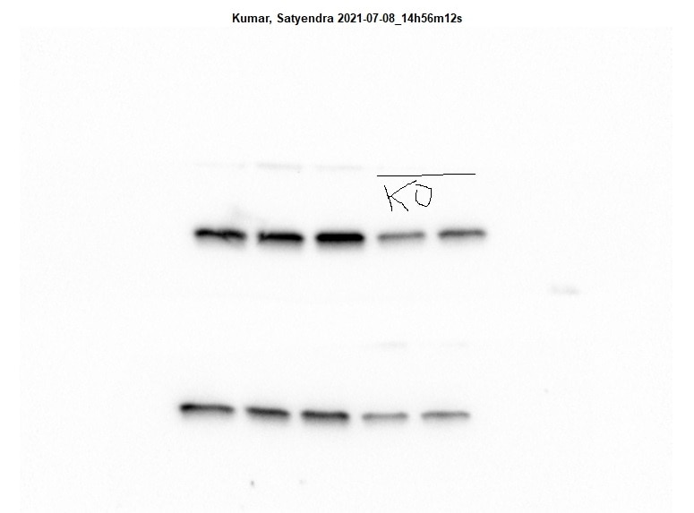 |
Hua (Verified Customer) (10-21-2020) | Observed band at the right size with a clean background; strong signal.
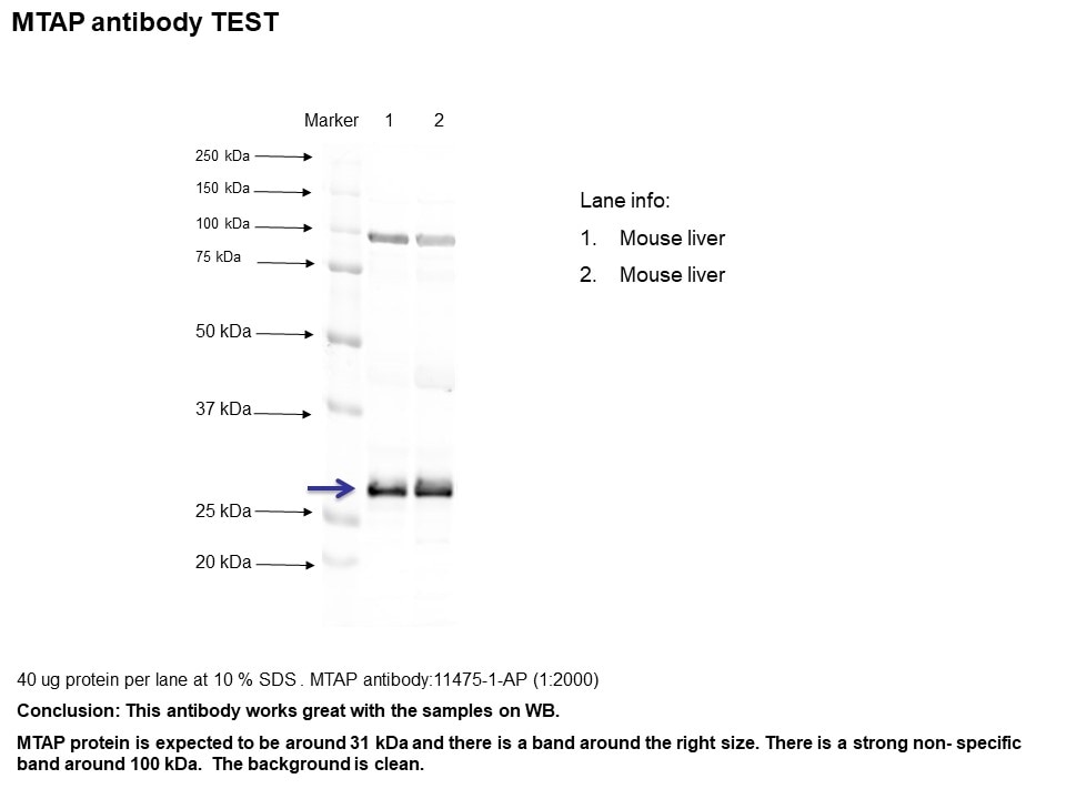 |















