

Tested Applications
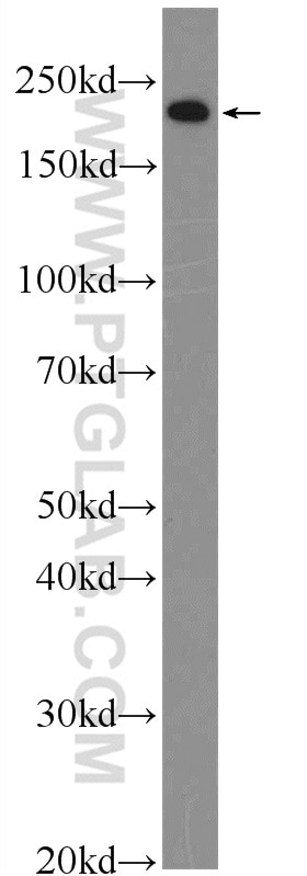
| Positive WB detected in | mouse embryo tissue |
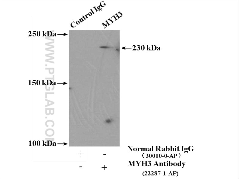
| Positive IP detected in | mouse skeletal muscle tissue |
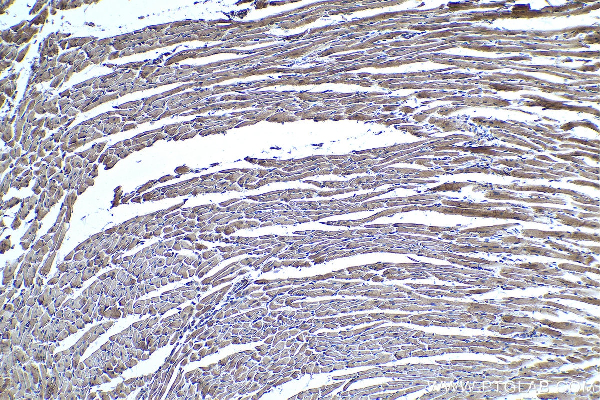
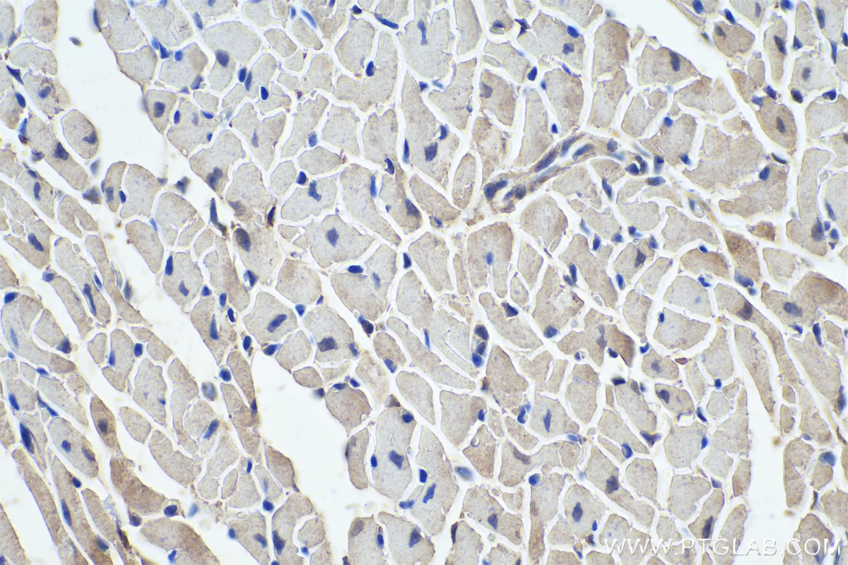
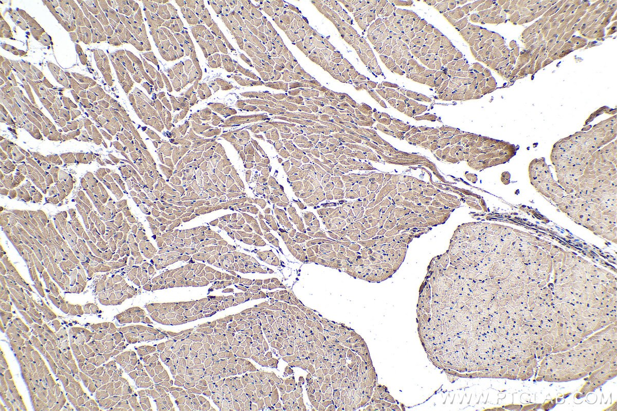
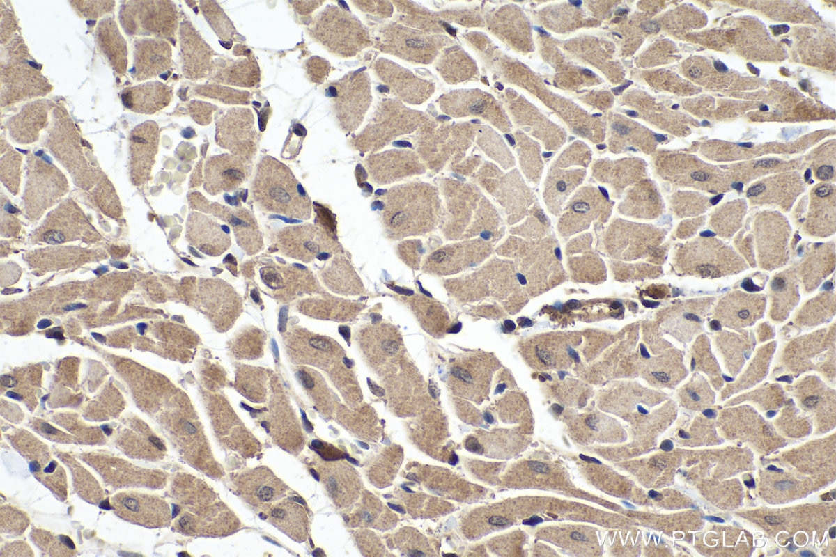
| Positive IHC detected in | rat heart tissue, mouse heart tissue Note: suggested antigen retrieval with TE buffer pH 9.0; (*) Alternatively, antigen retrieval may be performed with citrate buffer pH 6.0 |
Recommended dilution
| Application | Dilution |
|---|---|
| Western Blot (WB) | WB : 1:500-1:2000 |
| Immunoprecipitation (IP) | IP : 0.5-4.0 ug for 1.0-3.0 mg of total protein lysate |
| Immunohistochemistry (IHC) | IHC : 1:250-1:1000 |
| It is recommended that this reagent should be titrated in each testing system to obtain optimal results. | |
| Sample-dependent, Check data in validation data gallery. | |
Product Information
22287-1-AP targets MYH3 in WB, IHC, IF, IP, ELISA applications and shows reactivity with human, mouse, rat samples.
| Tested Reactivity | human, mouse, rat |
| Cited Reactivity | human, mouse, rat, pig, bovine |
| Host / Isotype | Rabbit / IgG |
| Class | Polyclonal |
| Type | Antibody |
| Immunogen |
Peptide Predict reactive species |
| Full Name | myosin, heavy chain 3, skeletal muscle, embryonic |
| Calculated Molecular Weight | 234 kDa |
| Observed Molecular Weight | 220-230 kDa |
| GenBank Accession Number | NM_002470 |
| Gene Symbol | MYH3 |
| Gene ID (NCBI) | 4621 |
| RRID | AB_2879060 |
| Conjugate | Unconjugated |
| Form | Liquid |
| Purification Method | Antigen affinity purification |
| UNIPROT ID | P11055 |
| Storage Buffer | PBS with 0.02% sodium azide and 50% glycerol, pH 7.3. |
| Storage Conditions | Store at -20°C. Stable for one year after shipment. Aliquoting is unnecessary for -20oC storage. 20ul sizes contain 0.1% BSA. |
Protocols
| Product Specific Protocols | |
|---|---|
| IHC protocol for MYH3 antibody 22287-1-AP | Download protocol |
| IP protocol for MYH3 antibody 22287-1-AP | Download protocol |
| WB protocol for MYH3 antibody 22287-1-AP | Download protocol |
| Standard Protocols | |
|---|---|
| Click here to view our Standard Protocols |
Publications
What published studies show
The MYH3 Polyclonal antibody (Cat# 22287-1-AP) has 24 citations spanning journals including Nature, Nature Communications, JCI, Cell Reports, Biomaterials, FASEB J, and BMC Genomics. Research fields encompass skeletal muscle regeneration, myogenesis, muscle satellite cell differentiation, cardiac biology, muscular dystrophy, muscle atrophy, and tissue engineering. Applications cited include WB, IF, and IHC across mouse, rat, human, bovine, and pig species.
| Species | Application | Title |
|---|---|---|
Nat Commun Hedgehog signaling via its ligand DHH acts as cell fate determinant during skeletal muscle regeneration | ||
J Clin Invest Activin A secretion by muscle-repairing macrophages induces heterotopic ossification in mice. | ||
Biomaterials Bioengineered human pre-vascularized microtissues reconstruct vascular niche and regenerative microenvironment to enhance functional skeletal muscle regeneration. | ||
Adv Healthc Mater A Peritoneum-Inspired Biomimetic Asymmetric Patch for Functional Repair and Anti-Adhesion in Abdominal Wall Defects. | ||
J Transl Med Microbiota-associated metabolite pantothenic acid enhances skeletal muscle contusion repair via epigenetic regulation of macrophage M2 polarization. |
Reviews
What customers say
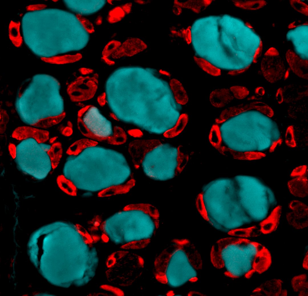
The single available review rates the antibody 4 out of 5 stars. It was used for immunofluorescence on Tibialis Anterior muscle cross sections at a 1:300 dilution, with antigen retrieval performed. The user successfully detected MYH3 in red, highlighting newly formed muscle fibers, alongside IgG in cyan marking dying fibers. Overall, the reviewer reported reliable performance for skeletal muscle/myoblast applications.
The reviews below have been submitted by verified Proteintech customers who received an incentive for providing their feedback.
Alessandra (Verified Customer) (03-23-2022) | Antigen retrieval was performed on Tibialis Anterior muscle cross sections. MYH3 in red (newly formed fibers); IgG in cyan (dying muscle fibers)
 |







