Tested Applications
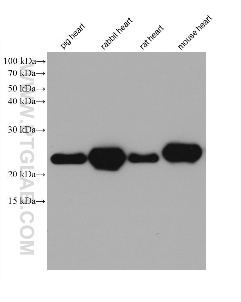
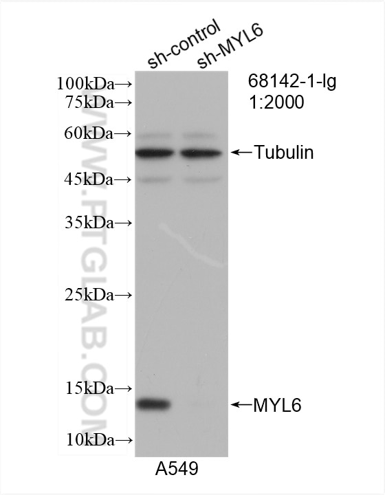
| Positive WB detected in | pig heart tissue, A549 cells, rabbit heart tissue, rat heart tissue, mouse heart tissue |
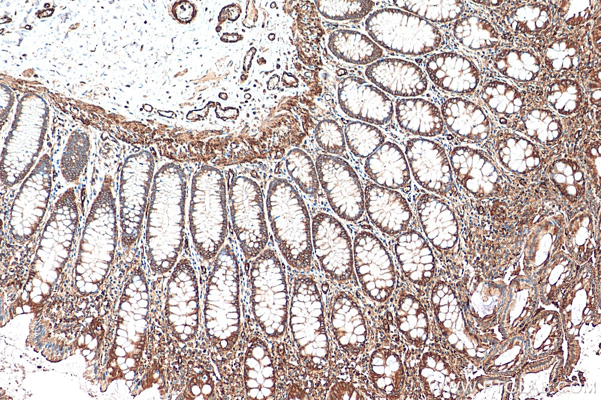
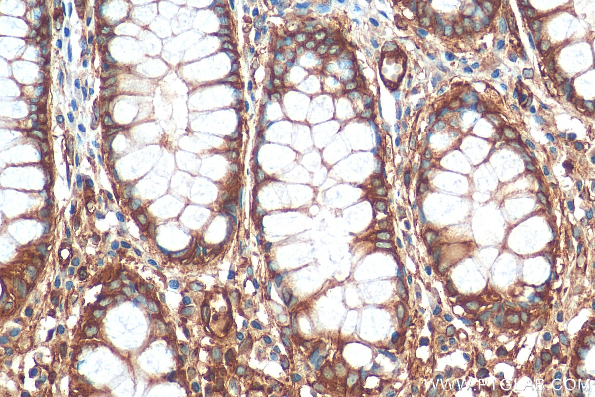
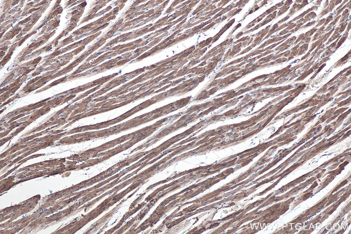
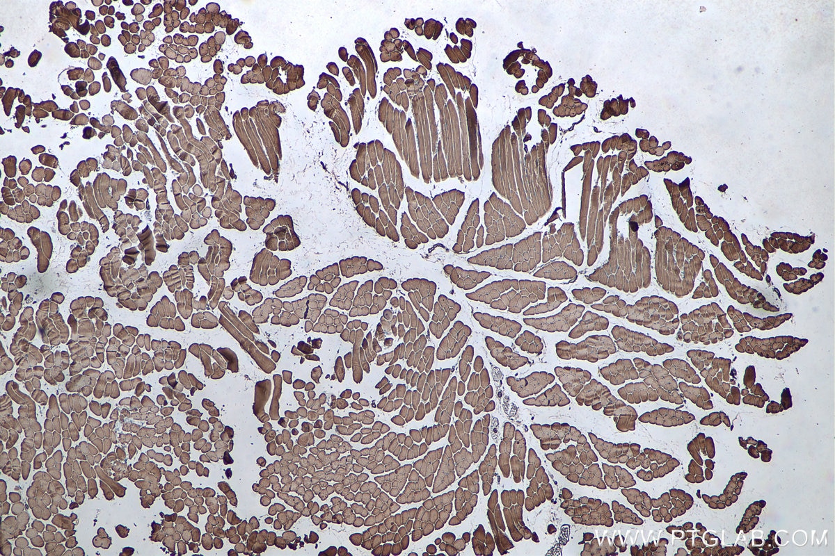
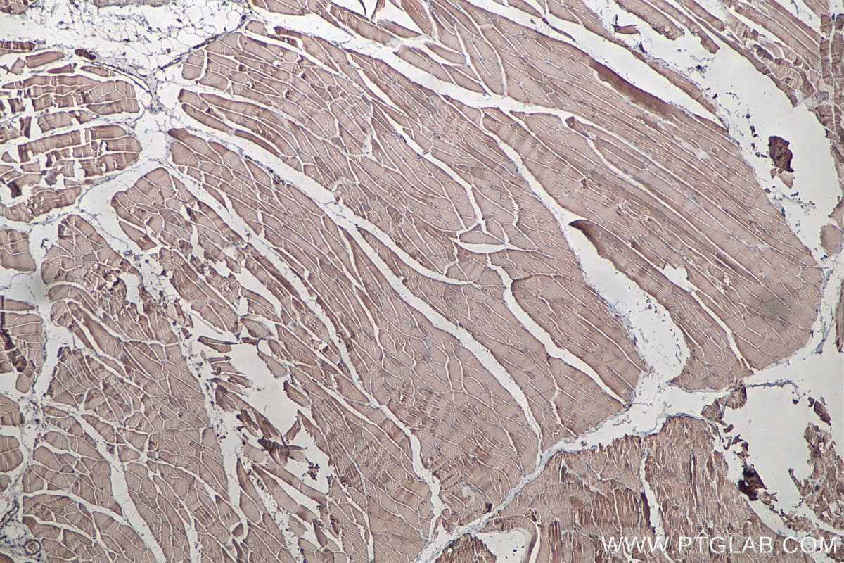
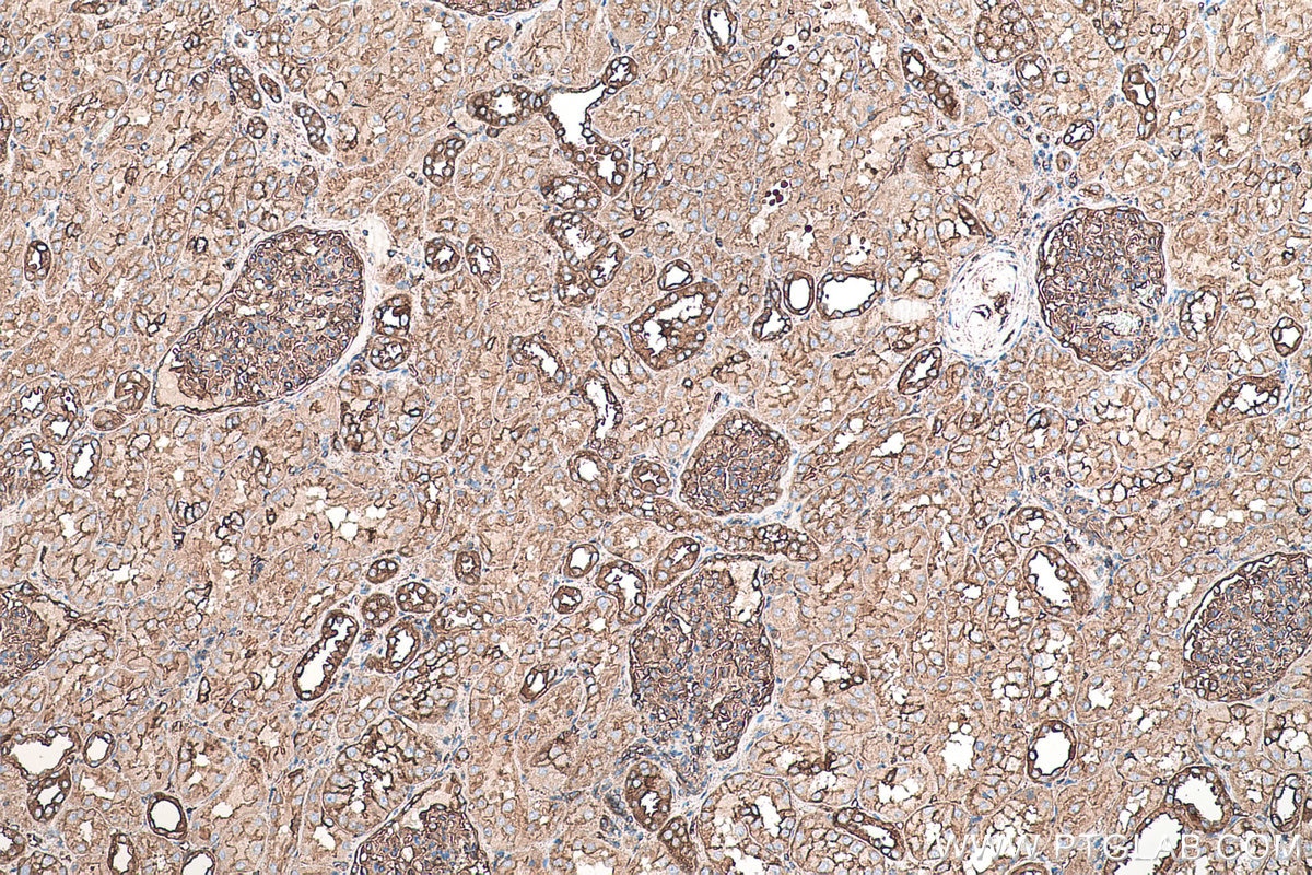
| Positive IHC detected in | human colon tissue, rat heart tissue, mouse skeletal muscle tissue, rat skeletal muscle tissue, human kidney tissue, mouse heart tissue Note: suggested antigen retrieval with TE buffer pH 9.0; (*) Alternatively, antigen retrieval may be performed with citrate buffer pH 6.0 |
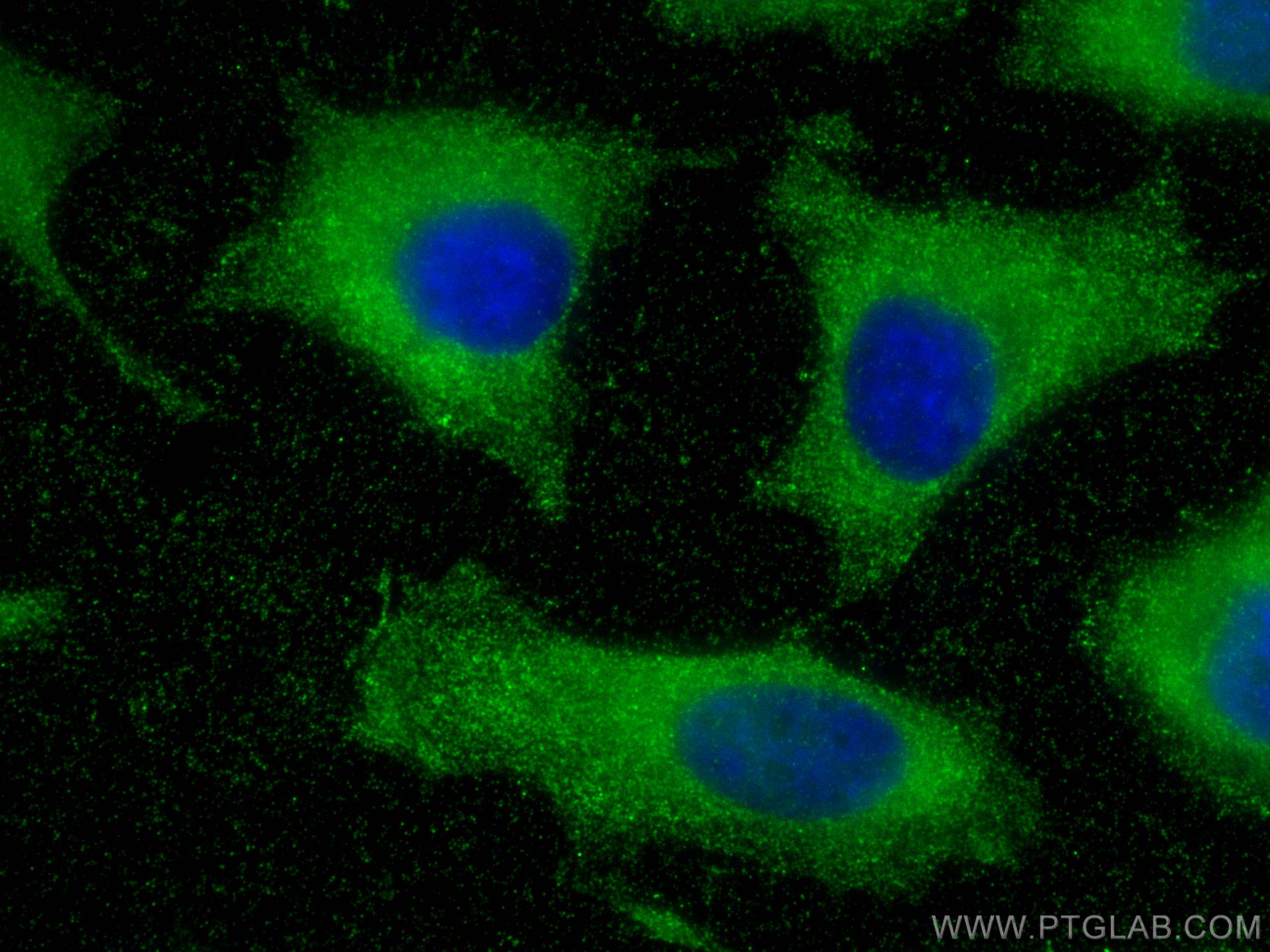
| Positive IF/ICC detected in | HeLa cells |
Recommended dilution
| Application | Dilution |
|---|---|
| Western Blot (WB) | WB : 1:5000-1:50000 |
| Immunohistochemistry (IHC) | IHC : 1:1000-1:4000 |
| Immunofluorescence (IF)/ICC | IF/ICC : 1:500-1:2000 |
| It is recommended that this reagent should be titrated in each testing system to obtain optimal results. | |
| Sample-dependent, Check data in validation data gallery. | |
Published Applications
| WB | See 2 publications below |
| IHC | See 4 publications below |
Product Information
68142-1-Ig targets MYL6 in WB, IHC, IF/ICC, ELISA applications and shows reactivity with Human, mouse, rat, pig, rabbit samples.
| Tested Reactivity | Human, mouse, rat, pig, rabbit |
| Cited Reactivity | human, mouse |
| Host / Isotype | Mouse / IgG1 |
| Class | Monoclonal |
| Type | Antibody |
| Immunogen |
CatNo: Ag13091 Product name: Recombinant human MYL6 protein Source: e coli.-derived, PET28a Tag: 6*His Domain: 1-151 aa of BC006781 Sequence: MCDFTEDQTAEFKEAFQLFDRTGDGKILYSQCGDVMRALGQNPTNAEVLKVLGNPKSDEMNVKVLDFEHFLPMLQTVAKNKDQGTYEDYVEGLRVFDKEGNGTVMGAEIRHVLVTLGEKMTEEEVEMLVAGHEDSNGCINYEAFVRHILSG Predict reactive species |
| Full Name | myosin, light chain 6, alkali, smooth muscle and non-muscle |
| Calculated Molecular Weight | 17 kDa |
| Observed Molecular Weight | 17-25 kDa |
| GenBank Accession Number | BC006781 |
| Gene Symbol | MYL6 |
| Gene ID (NCBI) | 4637 |
| RRID | AB_2935258 |
| Conjugate | Unconjugated |
| Form | Liquid |
| Purification Method | Protein G purification |
| UNIPROT ID | P60660 |
| Storage Buffer | PBS with 0.02% sodium azide and 50% glycerol, pH 7.3. |
| Storage Conditions | Store at -20°C. Stable for one year after shipment. Aliquoting is unnecessary for -20oC storage. 20ul sizes contain 0.1% BSA. |
Background Information
Myosin is a hexameric ATPase cellular motor protein. It is composed of two heavy chains, two nonphosphorylatable alkali light chains, and two phosphorylatable regulatory light chains. MYL6 (Myosin light polypeptide 6), encodes a myosin alkali light chain that is expressed in smooth muscle and non-muscle tissues. Expression of MYL6 is high in fibroblasts and myoblasts (PMID:8188229, PubMed:2304459, PMID:2722814).
Protocols
| Product Specific Protocols | |
|---|---|
| IF protocol for MYL6 antibody 68142-1-Ig | Download protocol |
| IHC protocol for MYL6 antibody 68142-1-Ig | Download protocol |
| WB protocol for MYL6 antibody 68142-1-Ig | Download protocol |
| Standard Protocols | |
|---|---|
| Click here to view our Standard Protocols |
Publications
| Species | Application | Title |
|---|---|---|
Transl Oncol Disulfidptosis-related gene expression reflects the prognosis of drug-resistant cancer patients and inhibition of MYH9 reverses sorafenib resistance | ||
Int J Biol Macromol Water-insoluble dietary fiber from walnut relieves constipation through Limosilactobacillus reuteri-mediated serotonergic synapse and neuroactive ligand-receptor pathways | ||
J Cell Mol Med A Novel Disulfidptosis-Related Diagnostic Gene Signature and Differential Expression Validation in Ischaemic Cardiomyopathy | ||
J Cardiothorac Surg Single-cell sequencing combined with transcriptome sequencing to reveal the molecular mechanisms related to integrated stress responses in atherosclerosis. |