

Tested Applications
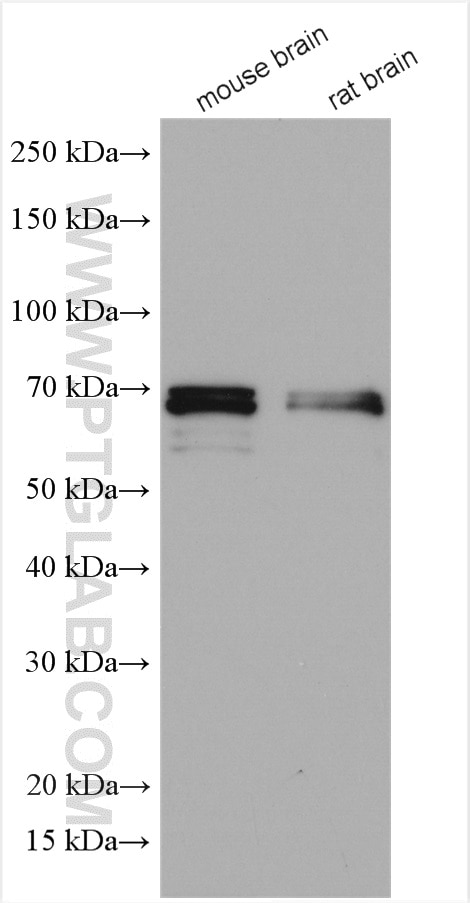
| Positive WB detected in | mouse brain tissue, rat brain tissue |
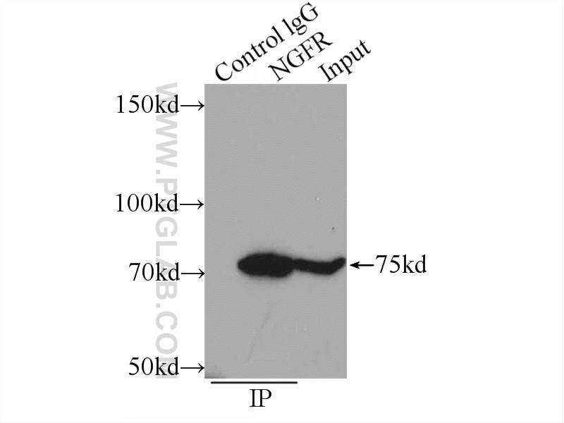
| Positive IP detected in | mouse brain tissue |
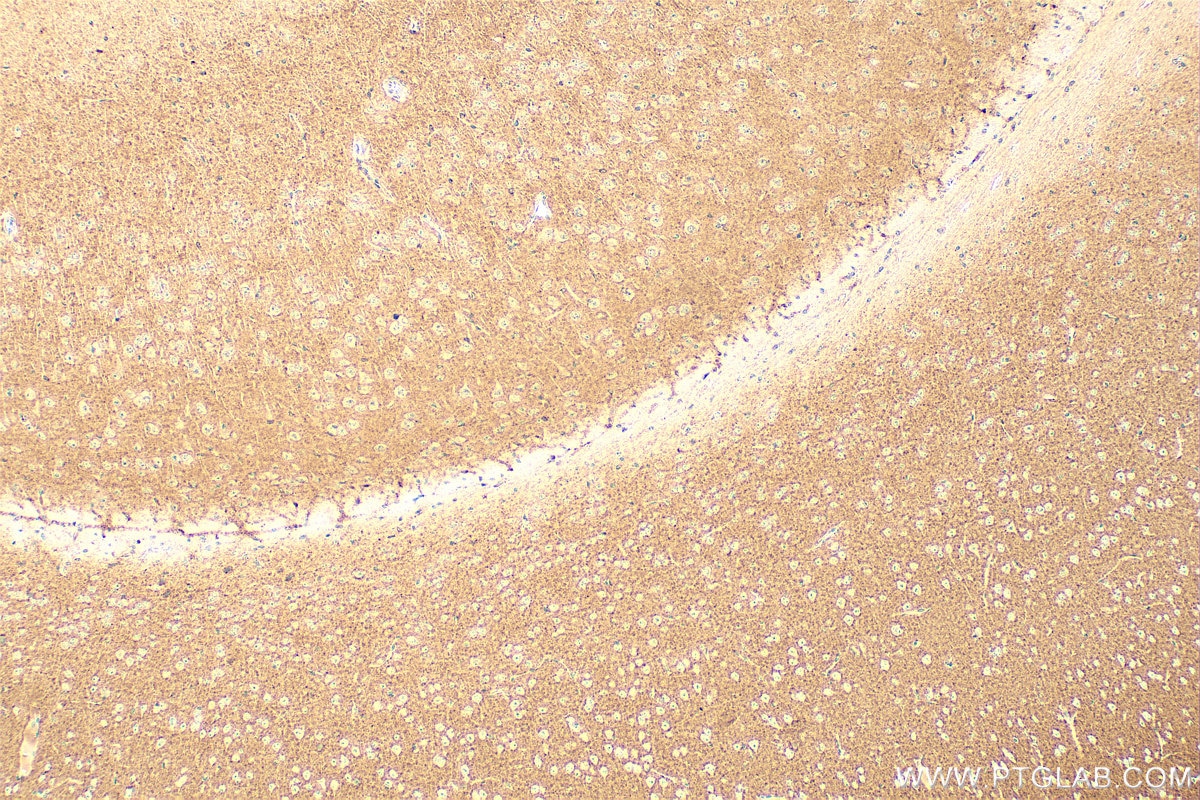
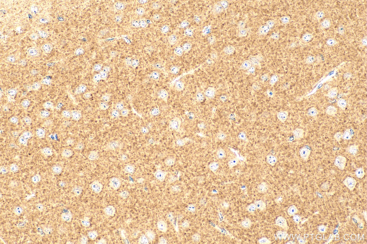
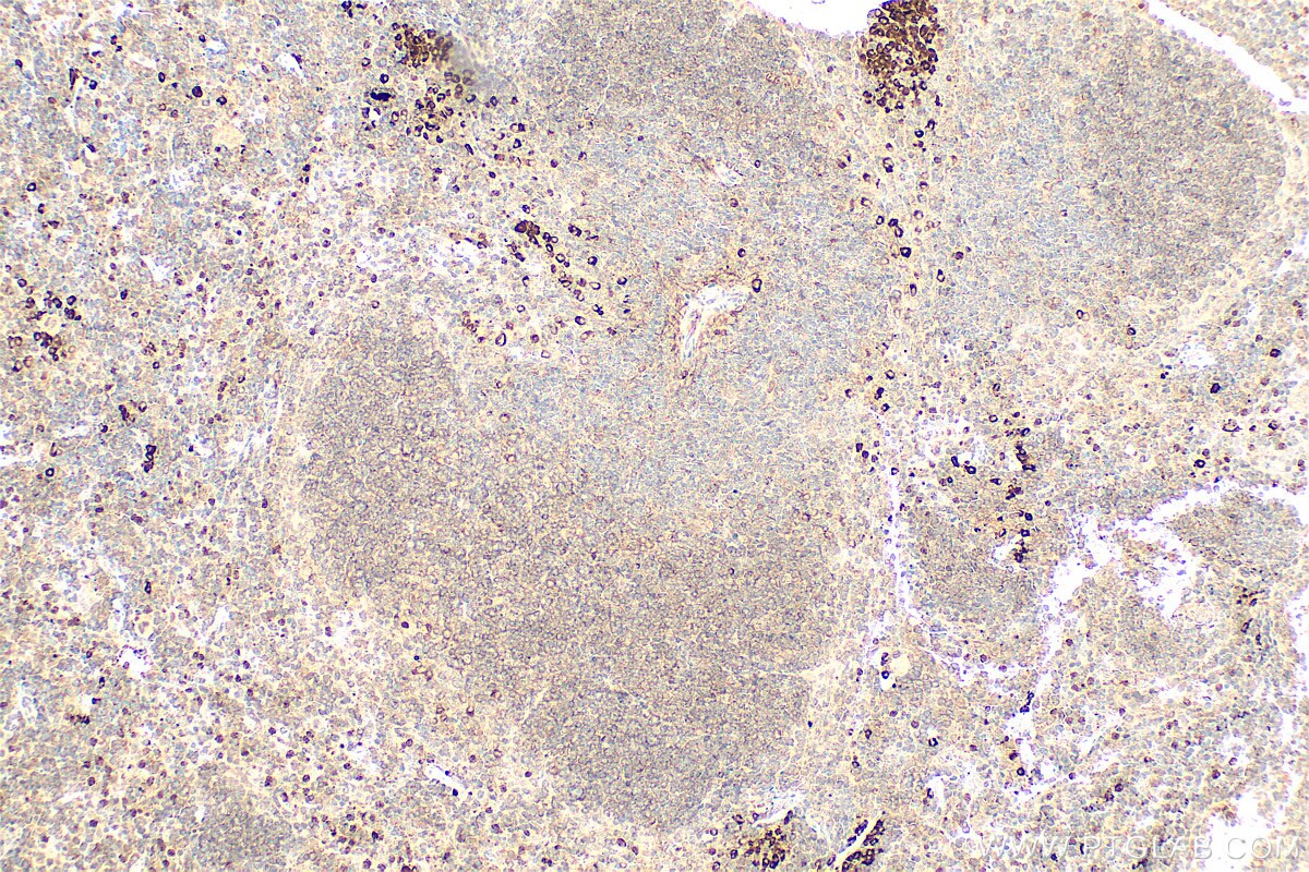
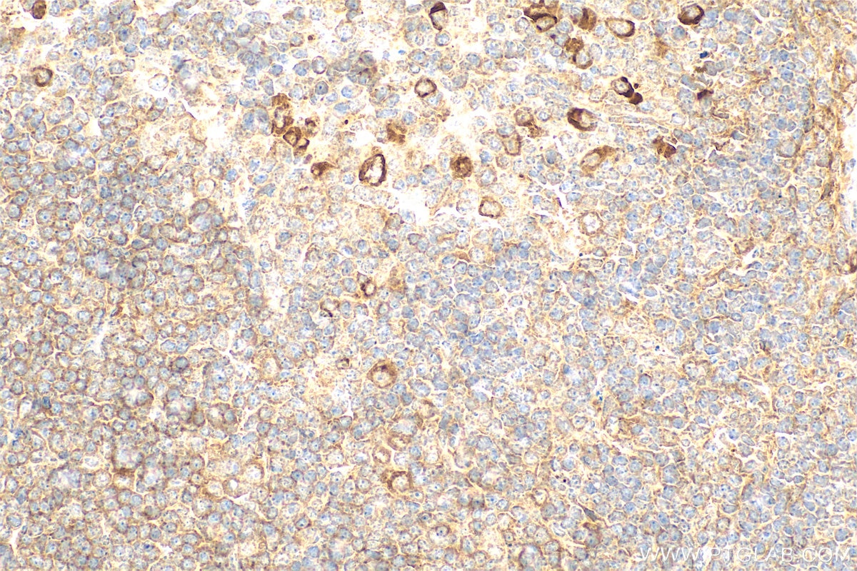
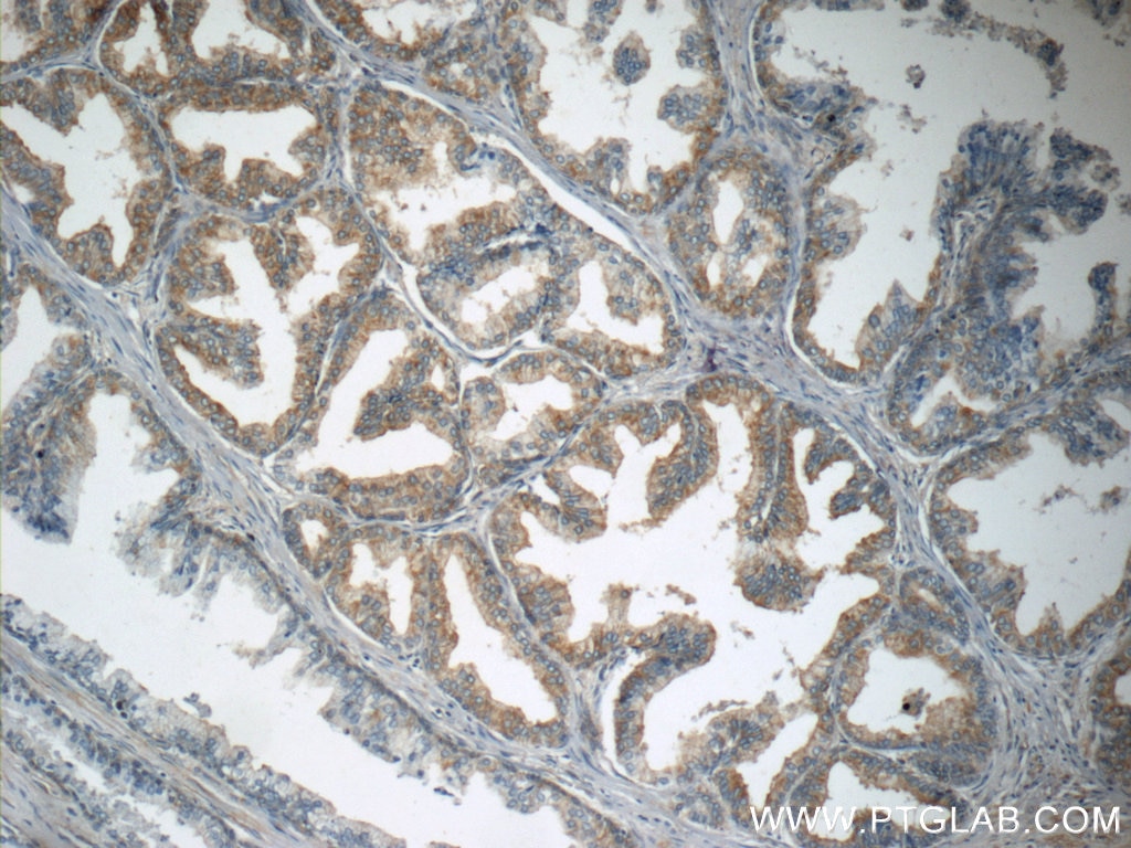
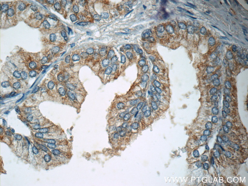
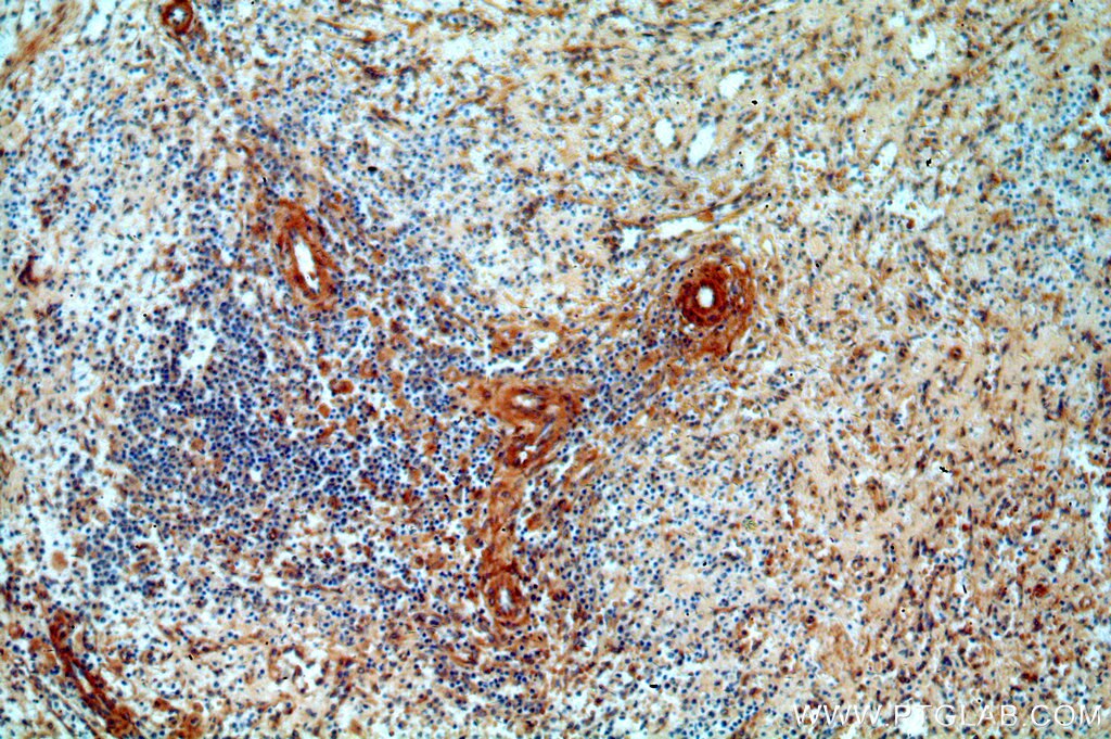
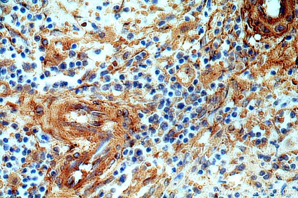
| Positive IHC detected in | mouse brain tissue, human prostate hyperplasia tissue, human spleen tissue, mouse spleen tissue Note: suggested antigen retrieval with TE buffer pH 9.0; (*) Alternatively, antigen retrieval may be performed with citrate buffer pH 6.0 |
Recommended dilution
| Application | Dilution |
|---|---|
| Western Blot (WB) | WB : 1:1000-1:4000 |
| Immunoprecipitation (IP) | IP : 0.5-4.0 ug for 1.0-3.0 mg of total protein lysate |
| Immunohistochemistry (IHC) | IHC : 1:200-1:800 |
| It is recommended that this reagent should be titrated in each testing system to obtain optimal results. | |
| Sample-dependent, Check data in validation data gallery. | |
Product Information
55014-1-AP targets NGFR/TNFRSF16 in WB, IHC, IP, CoIP, ELISA applications and shows reactivity with human, mouse, rat samples.
| Tested Reactivity | human, mouse, rat |
| Cited Reactivity | human, mouse, rat |
| Host / Isotype | Rabbit / IgG |
| Class | Polyclonal |
| Type | Antibody |
| Immunogen |
Peptide Predict reactive species |
| Full Name | nerve growth factor receptor (TNFR superfamily, member 16) |
| Calculated Molecular Weight | 45 kDa |
| Observed Molecular Weight | 70-75 kDa |
| GenBank Accession Number | NM_002507 |
| Gene Symbol | p75NTR |
| Gene ID (NCBI) | 4804 |
| RRID | AB_10858484 |
| Conjugate | Unconjugated |
| Form | Liquid |
| Purification Method | Antigen affinity purification |
| UNIPROT ID | P08138 |
| Storage Buffer | PBS with 0.02% sodium azide and 50% glycerol, pH 7.3. |
| Storage Conditions | Store at -20°C. Stable for one year after shipment. Aliquoting is unnecessary for -20oC storage. 20ul sizes contain 0.1% BSA. |
Background Information
NGFR, also named as TNFRSF16, Gp80-LNGFR, p75 ICD and CD271, is a low affinity receptor which can bind to NGF, BDNF, NT-3, and NT-4. It mediates cell survival as well as cell death of neural cells. The antibody is specific to NGFR.
Protocols
| Product Specific Protocols | |
|---|---|
| IHC protocol for NGFR/TNFRSF16 antibody 55014-1-AP | Download protocol |
| IP protocol for NGFR/TNFRSF16 antibody 55014-1-AP | Download protocol |
| WB protocol for NGFR/TNFRSF16 antibody 55014-1-AP | Download protocol |
| Standard Protocols | |
|---|---|
| Click here to view our Standard Protocols |
Publications
What published studies show
Anti-p75NTR/NGFR antibody (Cat# 55014-1-AP) has 44 total citations published in journals including Nature Communications, Journal of Experimental Medicine, Experimental Hematology & Oncology, Advanced Science, International Journal of Molecular Sciences, and Frontiers in Pharmacology. Associated research fields encompass neuroscience and peripheral nerve regeneration, oncology (hepatocellular, pancreatic, glioma, and esophageal cancers), hepatic and renal fibrosis, stem cell biology, pain mechanisms, cardiotoxicity, and ophthalmology. Applications are primarily Western blot and immunohistochemistry across human, mouse, rat, and other species.
| Species | Application | Title |
|---|---|---|
Signal Transduct Target Ther Activation of Nerve Growth Factor signaling limits the response to lenvatinib in hepatocellular carcinoma | ||
Nat Commun Tumor-derived GDF15 induces CCN3⁺ Schwann cells to promote cancer pain in pancreatic cancer. | ||
Exp Hematol Oncol MMP14 from BM-MSCs facilitates progression and Ara-C resistance in acute myeloid leukemia via the JAK/STAT pathway | ||
Adv Sci (Weinh) Bioactive Silk Sericin/Bioceramic Nerve Guidance Conduit for Effective Repair of Long-Gap Transected Peripheral Nerve Injury through Regulating Schwann Cells | ||
Burns Trauma Single cell deciphering of pruritic keloids: the interaction between fibroblasts and Schwann cells through the Midkine signaling. |
Reviews
What customers say
Users report mixed results depending on application. For Western blot, the antibody performs well, with two reviewers confirming clear detection of endogenous p75NTR at the expected molecular weight in cell lysates and brain tissue at 1:1000 dilution. One user noted three discernible bands but identified the correct one by size. For immunohistochemistry and immunofluorescence on spinal cord tissue, one reviewer found suboptimal staining, attributing it to the protein's ubiquitous expression causing a poor signal-to-noise ratio at 1:500 dilution. Overall service was rated positively.
The reviews below have been submitted by verified Proteintech customers who received an incentive for providing their feedback.
George (Verified Customer) (11-25-2025) | Very good service but have not been able to get good results from tissue staining. However this may be because of high ubqiuitous expression leading to a poor signal/noise ratio.
|
jelan (Verified Customer) (10-17-2025) | We use the p75NTR Rabbit PolyAb for recognizing endogenous levels of total p75NTR protein in westernblots, for cells that we have lysed according to our design experiment.
|
Renato (Verified Customer) (02-27-2019) | Produces 3 perfectly discerning bands, but one of which clearly identifiable as p75NTR as it shows at the expected molecular weight. Used as diluent 5% BSA.
|











