


Tested Applications
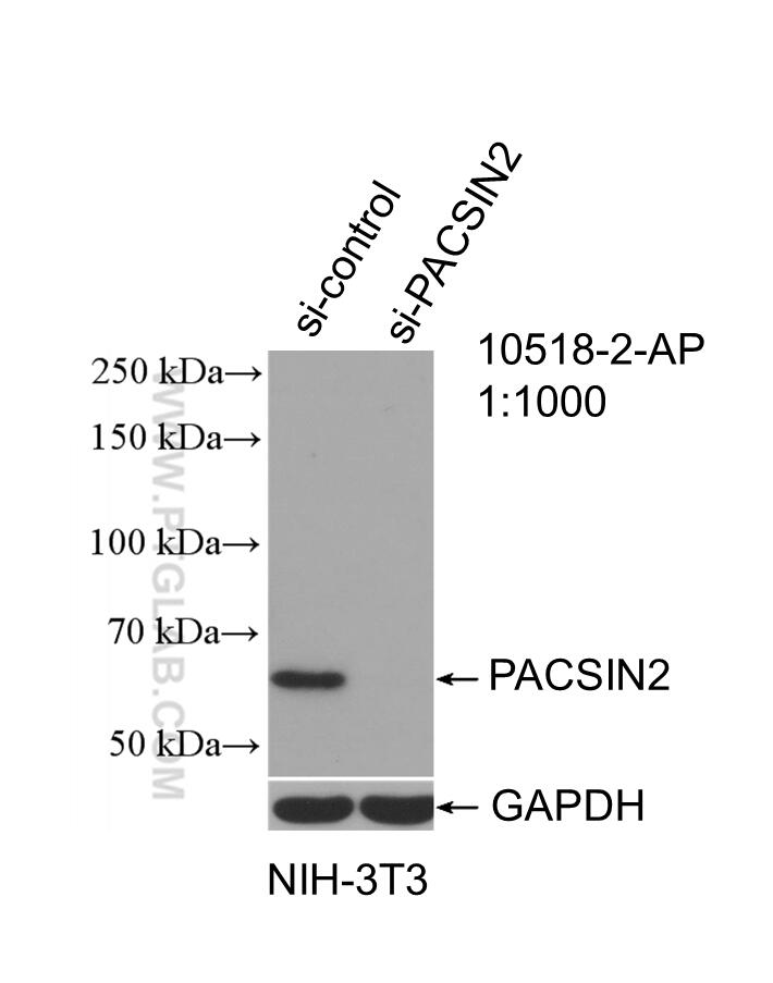
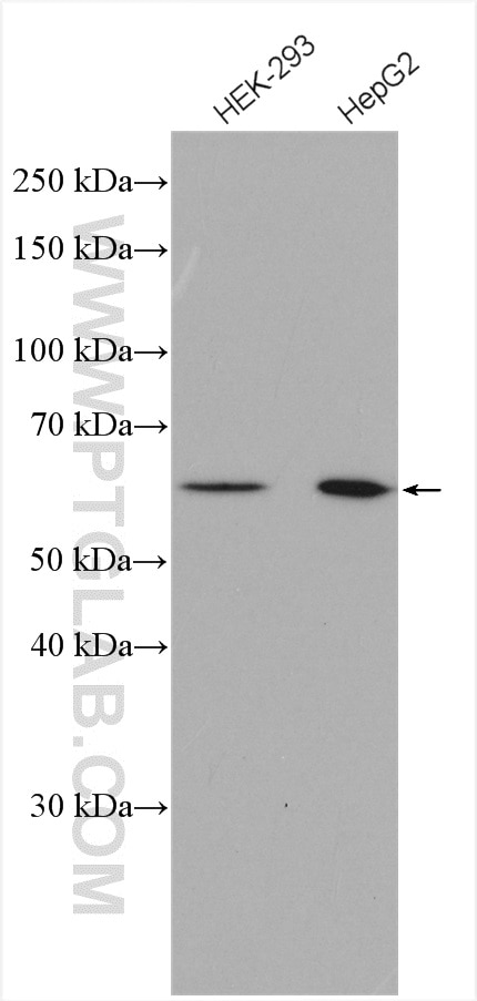
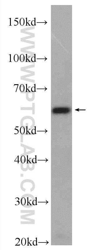
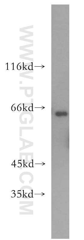
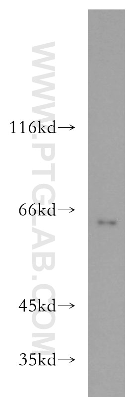
| Positive WB detected in | HEK-293 cells, mouse heart tissue, mouse lung tissue, NIH/3T3 cells, HepG2 cells |
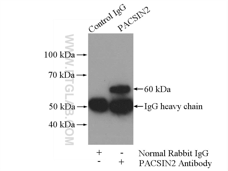
| Positive IP detected in | NIH/3T3 cells |
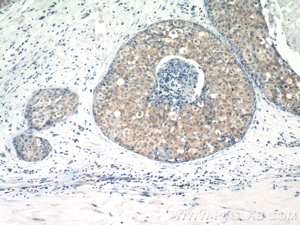
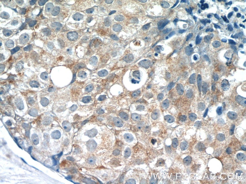
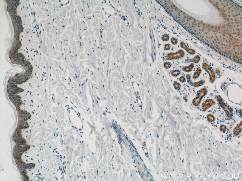
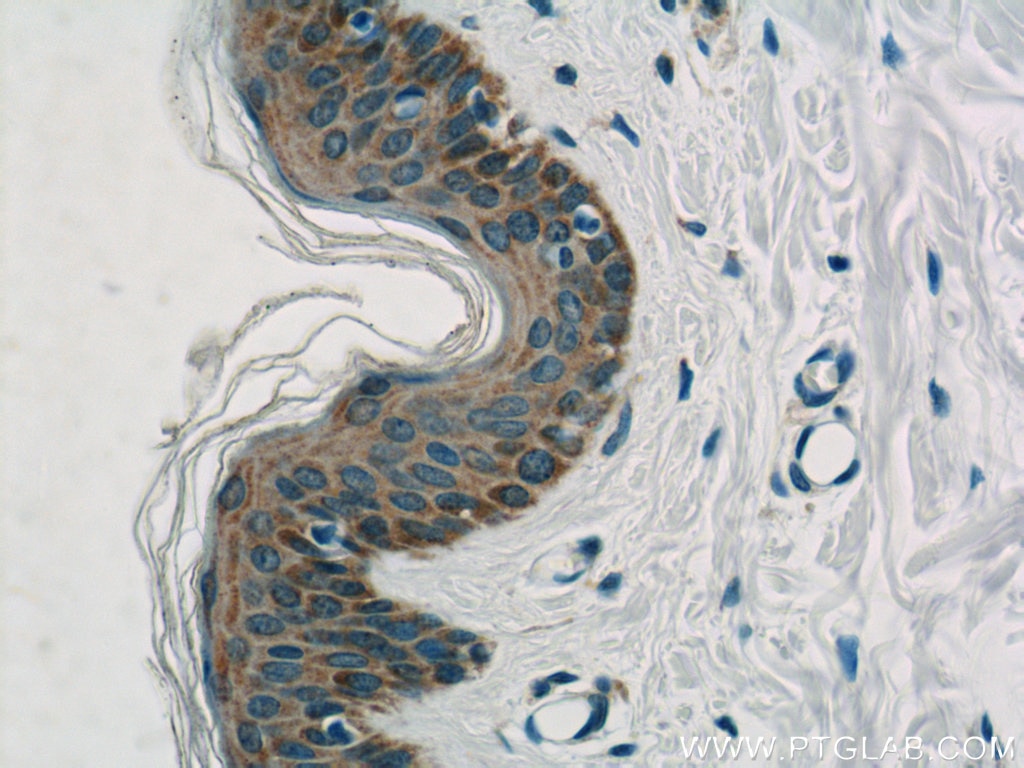
| Positive IHC detected in | human breast cancer tissue, human skin tissue Note: suggested antigen retrieval with TE buffer pH 9.0; (*) Alternatively, antigen retrieval may be performed with citrate buffer pH 6.0 |
| Positive IF/ICC detected in | NIH/3T3 cells |
Recommended dilution
| Application | Dilution |
|---|---|
| Western Blot (WB) | WB : 1:500-1:2000 |
| Immunoprecipitation (IP) | IP : 0.5-4.0 ug for 1.0-3.0 mg of total protein lysate |
| Immunohistochemistry (IHC) | IHC : 1:200-1:800 |
| Immunofluorescence (IF)/ICC | IF/ICC : 1:200-1:800 |
| It is recommended that this reagent should be titrated in each testing system to obtain optimal results. | |
| Sample-dependent, Check data in validation data gallery. | |
Product Information
10518-2-AP targets PACSIN2 in WB, IHC, IF/ICC, IP, ELISA applications and shows reactivity with human, mouse samples.
| Tested Reactivity | human, mouse |
| Cited Reactivity | human, mouse, rat, zebrafish |
| Host / Isotype | Rabbit / IgG |
| Class | Polyclonal |
| Type | Antibody |
| Immunogen |
CatNo: Ag0809 Product name: Recombinant human PACSIN2 protein Source: e coli.-derived, PGEX-4T Tag: GST Domain: 180-486 aa of BC008037 Sequence: PSLNPEQLKKLQDKIEKCKQDVLKTKEKYEKSLKELDQGTPQYMENMEQVFEQCQQFEEKRLRFFREVLLEVQKHLDLSNVAGYKAIYHDLEQSIRAADAVEDLRWFRANHGPGMAMNWPQFEEWSADLNRTLSRREKKKATDGVTLTGINQTGDQSLPSKPSSTLNVPSNPAQSAQSQSSYNPFEDEDDTGSTVSEKDDTKAKNVSSYEKTQSYPTDWSDDESNNPFSSTDANGDSNPFDDDATSGTEVRVRALYDYEGQEHDELSFKAGDELTKMEDEDEQGWCKGRLDNGQVGLYPANYVEAIQ Predict reactive species |
| Full Name | protein kinase C and casein kinase substrate in neurons 2 |
| Calculated Molecular Weight | 56 kDa |
| Observed Molecular Weight | 60-65 kDa |
| GenBank Accession Number | BC008037 |
| Gene Symbol | PACSIN2 |
| Gene ID (NCBI) | 11252 |
| RRID | AB_2161854 |
| Conjugate | Unconjugated |
| Form | Liquid |
| Purification Method | Antigen affinity purification |
| UNIPROT ID | Q9UNF0 |
| Storage Buffer | PBS with 0.02% sodium azide and 50% glycerol, pH 7.3. |
| Storage Conditions | Store at -20°C. Stable for one year after shipment. Aliquoting is unnecessary for -20oC storage. 20ul sizes contain 0.1% BSA. |
Background Information
Peripheral membrane proteins of the Bin/amphiphysin/Rvs (BAR) and Fer-CIP4 homology-BAR (F-BAR) family participate in cellular membrane trafficking (PMID: 19549836). PACSIN2 (also known as syndapin-2) is a member of the protein kinase C and casein kinase substrate in neurons (PACSIN) family, which are membrane-binding proteins characterized by an amino-terminal F-BAR domain. PACSIN2 plays a role in intracellular vesicle-mediated transport. It is involved in the endocytosis of cell-surface receptors like the EGF receptor, contributing to its internalization (PMID: 23129763). PACSIN2 also has a role in caveolae membrane sculpting (PMID: 21610094).
Protocols
| Product Specific Protocols | |
|---|---|
| IF protocol for PACSIN2 antibody 10518-2-AP | Download protocol |
| IHC protocol for PACSIN2 antibody 10518-2-AP | Download protocol |
| IP protocol for PACSIN2 antibody 10518-2-AP | Download protocol |
| WB protocol for PACSIN2 antibody 10518-2-AP | Download protocol |
| Standard Protocols | |
|---|---|
| Click here to view our Standard Protocols |
Publications
What published studies show
The PACSIN2 Polyclonal antibody (Cat# 10518-2-AP) has 10 citations across journals including Nature Communications, Acta Pharmacologica Sinica, Cell Reports, ACS Nano, Journal of Agricultural and Food Chemistry, Journal of Dental Research, Journal of Bacteriology, Molecular Biology of the Cell, and PLoS ONE. Research fields span membrane tubulation and cilia assembly, caveolae structure, synaptic injury and behavioral disorders, mitogenic signaling, blood-brain barrier transport, temporomandibular joint proteomics, bacterial protein translocation, recycling endosome biogenesis, plasma membrane localization, and curvature-sensing apoptosis.
| Species | Application | Title |
|---|---|---|
Nat Commun Investigation of F-BAR domain PACSIN proteins uncovers membrane tubulation function in cilia assembly and transport.
| ||
Nat Commun The molecular organization of differentially curved caveolae indicates bendable structural units at the plasma membrane | ||
Acta Pharmacol Sin PACSIN2-mediated synaptic injury contributes to behavioral disorders caused by chronic stress. | ||
Cell Rep Endogenous Cyclin D1 Promotes the Rate of Onset and Magnitude of Mitogenic Signaling via Akt1 Ser473 Phosphorylation. | ||
J Agric Food Chem Caveolin Regulates the Transport Mechanism of the Walnut-Derived Peptide EVSGPGYSPN to Penetrate the Blood-Brain Barrier | ||
Reviews
What customers say
The single review (rated 1/5) reports a Western Blot on platelets at 1:1000 dilution. The reviewer noted the antibody detected a 55-kDa band in both Pacsin2 wild-type and knockout platelet lysates, whereas PACSIN2 is expected to migrate at ~65 kDa and should be absent in KO samples. This suggests the signal was non-specific rather than true PACSIN2 recognition, indicating a specificity concern for this lot.
The reviews below have been submitted by verified Proteintech customers who received an incentive for providing their feedback.
Herve (Verified Customer) (11-13-2019) | This antibody recognizes a 55-kDa band that is present in both Pacsin2 WT and KO platelet lysates. PACSIN2 runs normally at 65 kDa in human and mouse platelet lysates and is absent in mouse Pacsin2 KO samples.
|












