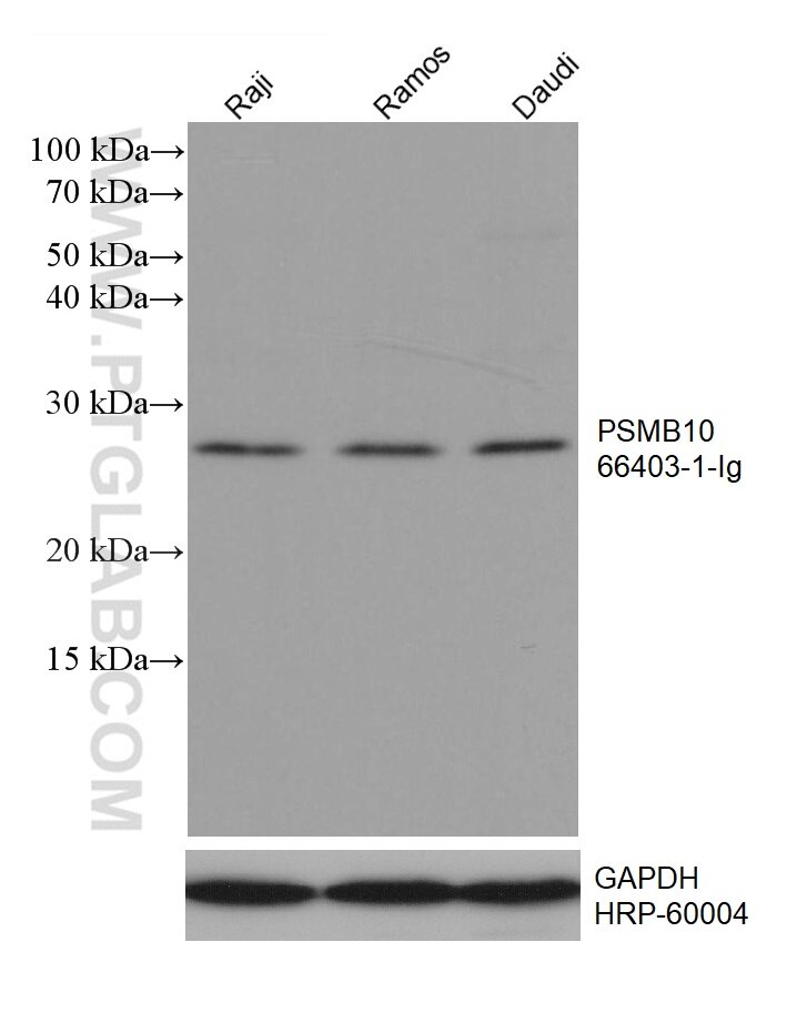
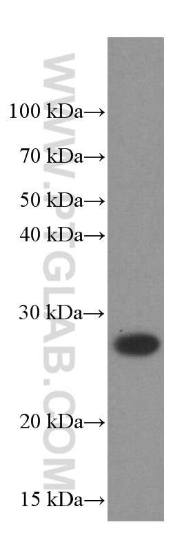
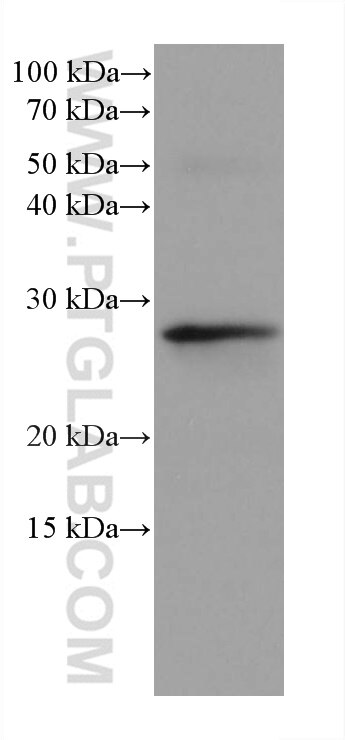
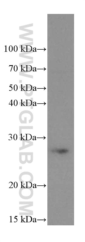
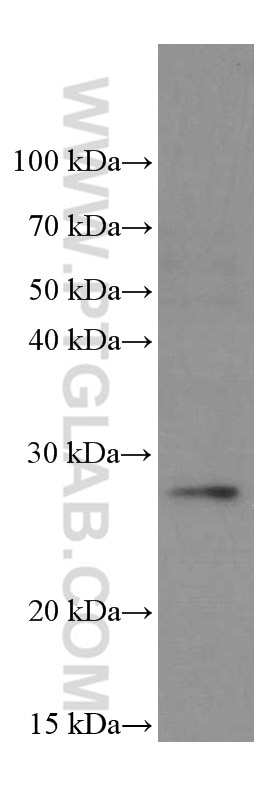
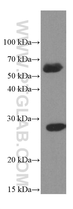
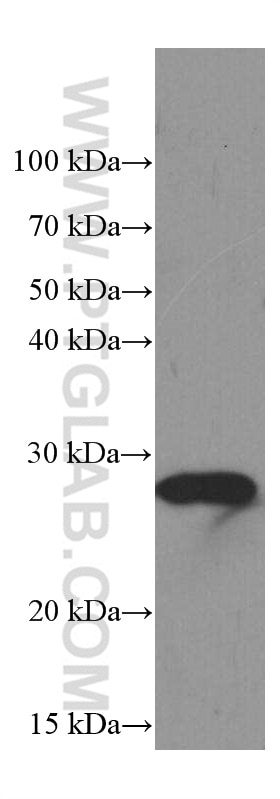
Various lysates were subjected to SDS PAGE followed by western blot with 66403-1-Ig (PSMB10 antibody) at dilution of 1:2000 incubated at room temperature for 1.5 hours. The membrane was stripped and reblotted with HRP-conjugated GAPDH Monoclonal antibody (HRP-60004) as loading control.
WB analysis of Raji using 66403-1-Ig
Raji cells were subjected to SDS PAGE followed by western blot with 66403-1-Ig (PSMB10 Antibody) at dilution of 1:1000 incubated at room temperature for 1.5 hours.
WB analysis of rat thymus using 66403-1-Ig
rat thymus tissue were subjected to SDS PAGE followed by western blot with 66403-1-Ig (PSMB10 antibody) at dilution of 1:2000 incubated at room temperature for 1.5 hours.
WB analysis of Ramos using 66403-1-Ig
Ramos cells were subjected to SDS PAGE followed by western blot with 66403-1-Ig (PSMB10 Antibody) at dilution of 1:1000 incubated at room temperature for 1.5 hours.
WB analysis of U-937 using 66403-1-Ig
U-937 cells were subjected to SDS PAGE followed by western blot with 66403-1-Ig (PSMB10 Antibody) at dilution of 1:1000 incubated at room temperature for 1.5 hours.
WB analysis of human spleen using 66403-1-Ig
human spleen tissue were subjected to SDS PAGE followed by western blot with 66403-1-Ig (PSMB10 Antibody) at dilution of 1:1000 incubated at room temperature for 1.5 hours.
WB analysis of pig thymus using 66403-1-Ig
pig thymus tissue were subjected to SDS PAGE followed by western blot with 66403-1-Ig (PSMB10 Antibody) at dilution of 1:1000 incubated at room temperature for 1.5 hours.
IHC staining of human liver cancer using 66403-1-Ig
Immunohistochemical analysis of paraffin-embedded human liver cancer tissue slide using 66403-1-Ig (PSMB10 antibody) at dilution of 1:300 (under 10x lens. Heat mediated antigen retrieval with Tris-EDTA buffer (pH 9.0).
IHC staining of human liver cancer using 66403-1-Ig
Immunohistochemical analysis of paraffin-embedded human liver cancer tissue slide using 66403-1-Ig (PSMB10 antibody) at dilution of 1:300 (under 40x lens. Heat mediated antigen retrieval with Tris-EDTA buffer (pH 9.0).
IHC staining of mouse kidney using 66403-1-Ig
Immunohistochemical analysis of paraffin-embedded mouse kidney tissue slide using 66403-1-Ig (PSMB10 antibody) at dilution of 1:400 (under 40x lens). Heat mediated antigen retrieval with Tris-EDTA buffer (pH 9.0).
IF Staining of Jurkat using 66403-1-Ig
Immunofluorescent analysis of (4% PFA) fixed Jurkat cells using PSMB10 antibody (66403-1-Ig, Clone: 1G8C1 ) at dilution of 1:800 and CoraLite®488-Conjugated Goat Anti-Mouse IgG(H+L) (SA00013-1).
The Proteintech guarantee covers Proteintech antibodies in any species and any application, including those not listed on the datasheet. If the antibody doesn’t perform, you can receive a hassle-free refund or credit note.
Raji cells, Ramos cells, U-937 cells, rat thymus tissue, human spleen tissue, pig thymus tissue
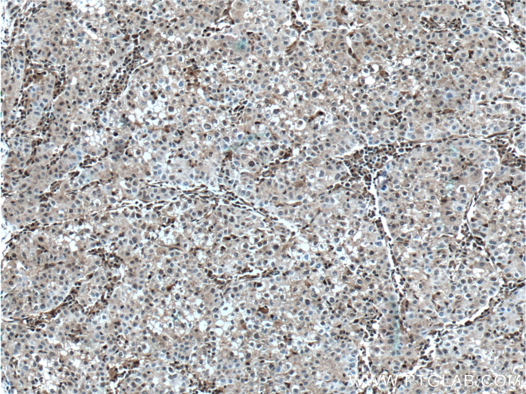
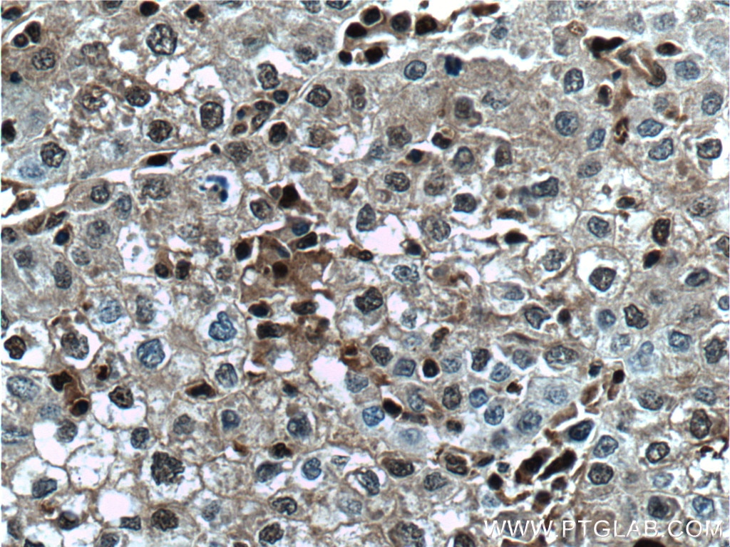
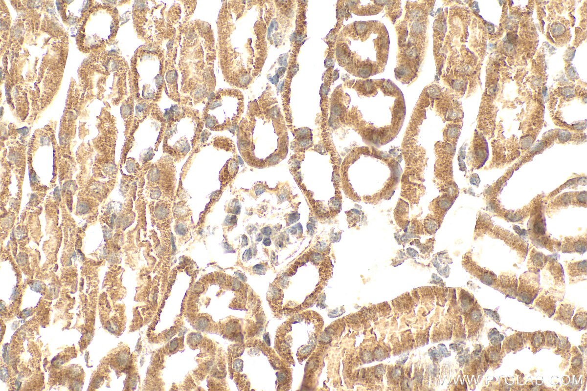
Positive IHC detected in
human liver cancer tissue, mouse kidney tissue Note: suggested antigen retrieval with TE buffer pH 9.0; (*) Alternatively, antigen retrieval may be performed with citrate buffer pH 6.0
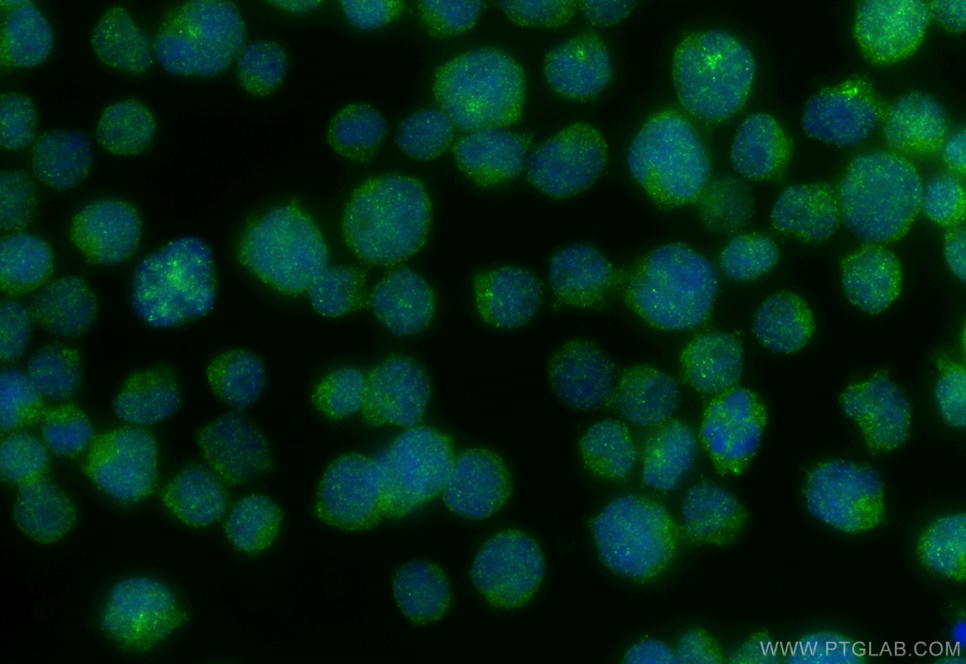
Positive IF/ICC detected in
Jurkat cells
Recommended dilution
Application
Dilution
Western Blot (WB)
WB : 1:1000-1:4000
Immunohistochemistry (IHC)
IHC : 1:150-1:600
Immunofluorescence (IF)/ICC
IF/ICC : 1:400-1:1600
It is recommended that this reagent should be titrated in each testing system to obtain optimal results.
Sample-dependent, Check data in validation data gallery.
Product Information
66403-1-Ig targets PSMB10 in WB, IHC, IF/ICC, ELISA applications and shows reactivity with human, mouse, rat, pig samples.
PBS with 0.02% sodium azide and 50% glycerol pH 7.3.
Storage Conditions
Store at -20°C. Stable for one year after shipment. Aliquoting is unnecessary for -20oC storage. 20ul sizes contain 0.1% BSA.
Background Information
SMB10(Proteasome subunit beta type-10) is also named as LMP10, MECL1.It belongs to the peptidase T1B family and is a multicatalytic proteinase complex which is characterized by its ability to cleave peptides with Arg, Phe, Tyr, Leu, and Glu adjacent to the leaving group at neutral or slightly basic pH.
Various lysates were subjected to SDS PAGE followed by western blot with 66403-1-Ig (PSMB10 antibody) at dilution of 1:2000 incubated at room temperature for 1.5 hours. The membrane was stripped and reblotted with HRP-conjugated GAPDH Monoclonal antibody (HRP-60004) as loading control.
WB analysis of Raji using 66403-1-Ig
Raji cells were subjected to SDS PAGE followed by western blot with 66403-1-Ig (PSMB10 Antibody) at dilution of 1:1000 incubated at room temperature for 1.5 hours.
WB analysis of rat thymus using 66403-1-Ig
rat thymus tissue were subjected to SDS PAGE followed by western blot with 66403-1-Ig (PSMB10 antibody) at dilution of 1:2000 incubated at room temperature for 1.5 hours.
WB analysis of Ramos using 66403-1-Ig
Ramos cells were subjected to SDS PAGE followed by western blot with 66403-1-Ig (PSMB10 Antibody) at dilution of 1:1000 incubated at room temperature for 1.5 hours.
WB analysis of U-937 using 66403-1-Ig
U-937 cells were subjected to SDS PAGE followed by western blot with 66403-1-Ig (PSMB10 Antibody) at dilution of 1:1000 incubated at room temperature for 1.5 hours.
WB analysis of human spleen using 66403-1-Ig
human spleen tissue were subjected to SDS PAGE followed by western blot with 66403-1-Ig (PSMB10 Antibody) at dilution of 1:1000 incubated at room temperature for 1.5 hours.
WB analysis of pig thymus using 66403-1-Ig
pig thymus tissue were subjected to SDS PAGE followed by western blot with 66403-1-Ig (PSMB10 Antibody) at dilution of 1:1000 incubated at room temperature for 1.5 hours.
IHC Figures
IHC staining of human liver cancer using 66403-1-Ig
Immunohistochemical analysis of paraffin-embedded human liver cancer tissue slide using 66403-1-Ig (PSMB10 antibody) at dilution of 1:300 (under 10x lens. Heat mediated antigen retrieval with Tris-EDTA buffer (pH 9.0).
IHC staining of human liver cancer using 66403-1-Ig
Immunohistochemical analysis of paraffin-embedded human liver cancer tissue slide using 66403-1-Ig (PSMB10 antibody) at dilution of 1:300 (under 40x lens. Heat mediated antigen retrieval with Tris-EDTA buffer (pH 9.0).
IHC staining of mouse kidney using 66403-1-Ig
Immunohistochemical analysis of paraffin-embedded mouse kidney tissue slide using 66403-1-Ig (PSMB10 antibody) at dilution of 1:400 (under 40x lens). Heat mediated antigen retrieval with Tris-EDTA buffer (pH 9.0).
IF/ICC Figures
IF Staining of Jurkat using 66403-1-Ig
Immunofluorescent analysis of (4% PFA) fixed Jurkat cells using PSMB10 antibody (66403-1-Ig, Clone: 1G8C1 ) at dilution of 1:800 and CoraLite®488-Conjugated Goat Anti-Mouse IgG(H+L) (SA00013-1).
The species listed in Tested Reactivity are in-house verified and applicable species. For unlisted species, please refer to the homology analysis of the immunogen sequence and related species. For rabbit polyclonal antibodies, homology >70% is recommended. For mouse monoclonal antibodies and rabbit recombinant antibodies, homology >90% is recommended. Generally, the higher the homology, the greater the applicability. However, there will be certain differences in protein expression in different species, tissues or cells. Therefore, the homology analysis results are for reference only and do not serve as a guarantee.
At Proteintech, we pride ourselves on our antibody quality, customer service and transparency. As such, we are comparing our antibodies with other vendors, enabling easy identification and comparisons of key data to help you choose the suitable antibody for your needs.
We have selected the top cited antibodies from these vendors for you to compare.