

Tested Applications
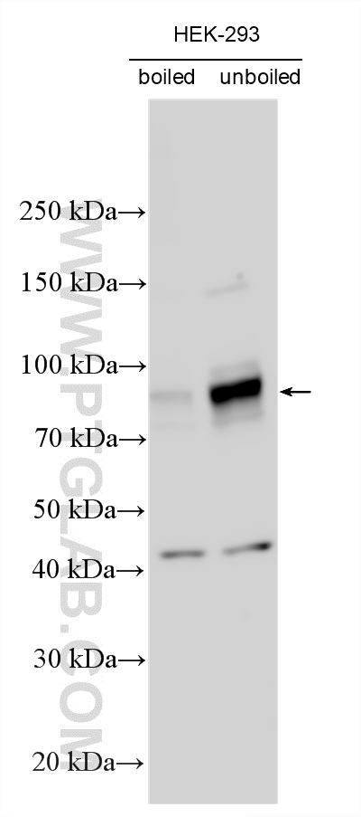
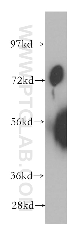
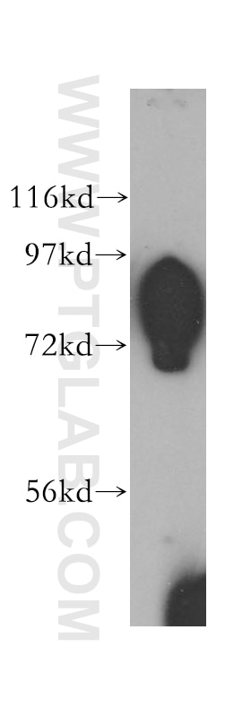
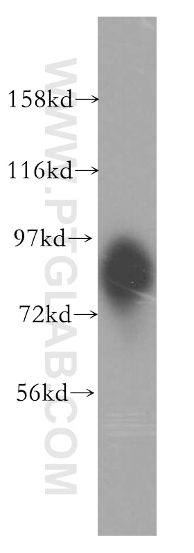
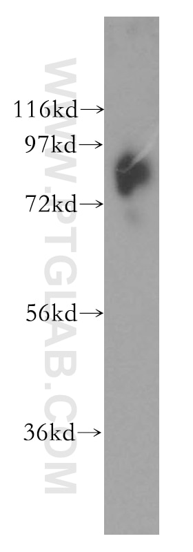
| Positive WB detected in | HEK-293 cells, HeLa cells, human kidney tissue, Jurkat cells, K-562 cells, unboiled HEK-293 cells |
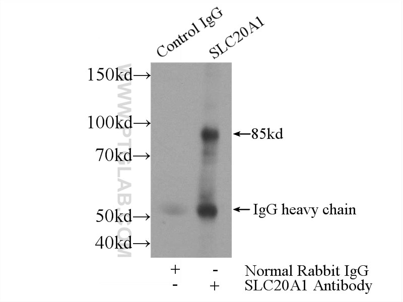
| Positive IP detected in | HEK-293 cells |
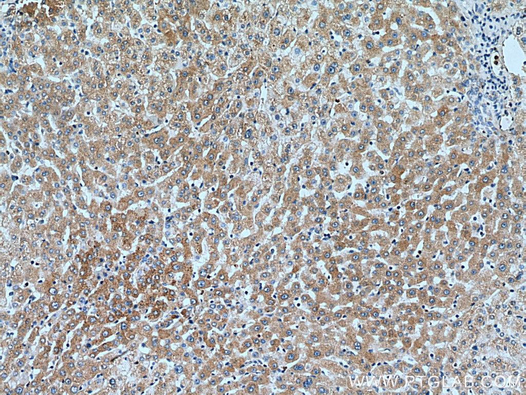
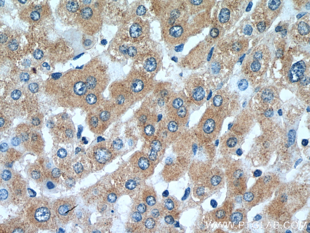
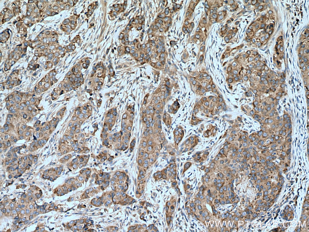
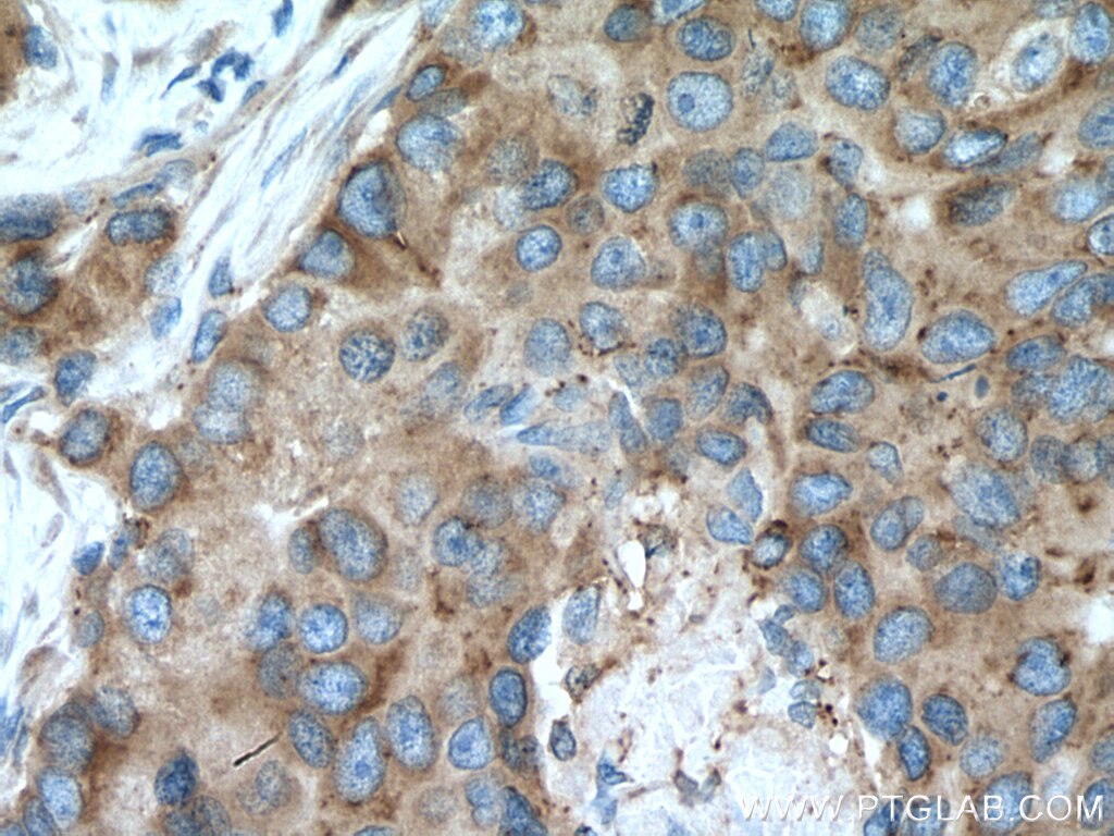
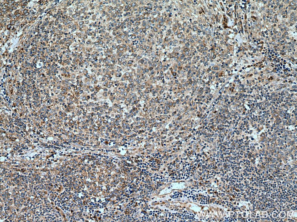
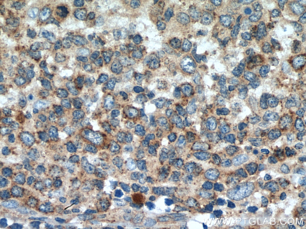
| Positive IHC detected in | human liver tissue, human breast cancer tissue, human lymphoma tissue Note: suggested antigen retrieval with TE buffer pH 9.0; (*) Alternatively, antigen retrieval may be performed with citrate buffer pH 6.0 |
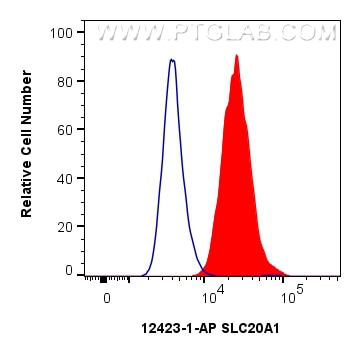
| Positive FC (Intra) detected in | HeLa cells |
Recommended dilution
| Application | Dilution |
|---|---|
| Western Blot (WB) | WB : 1:1000-1:8000 |
| Immunoprecipitation (IP) | IP : 0.5-4.0 ug for 1.0-3.0 mg of total protein lysate |
| Immunohistochemistry (IHC) | IHC : 1:50-1:500 |
| Flow Cytometry (FC) (INTRA) | FC (INTRA) : 0.25 ug per 10^6 cells in a 100 µl suspension |
| It is recommended that this reagent should be titrated in each testing system to obtain optimal results. | |
| Sample-dependent, Check data in validation data gallery. | |
Product Information
12423-1-AP targets SLC20A1 in WB, IHC, IF, FC (Intra), IP, CoIP, ELISA applications and shows reactivity with human samples.
| Tested Reactivity | human |
| Cited Reactivity | human, mouse, rat, zebrafish |
| Host / Isotype | Rabbit / IgG |
| Class | Polyclonal |
| Type | Antibody |
| Immunogen |
CatNo: Ag3084 Product name: Recombinant human SLC20A1 protein Source: e coli.-derived, PGEX-4T Tag: GST Domain: 250-570 aa of BC019944 Sequence: VCPRMKRKIEREIKCSPSESPLMEKKNSLKEDHEETKLSVGDIENKHPVSEVGPATVPLQAVVEERTVSFKLGDLEEAPERERLPSVDLKEETSIDSTVNGAVQLPNGNLVQFSQAVSNQINSSGHYQYHTVHKDSGLYKELLHKLHLAKVGDCMGDSGDKPLRRNNSYTSYTMAICGMPLDSFRAKEGEQKGEEMEKLTWPNADSKKRIRMDSYTSYCNAVSDLHSASEIDMSVKAEMGLGDRKGSNGSLEEWYDQDKPEVSLLFQFLQILTACFGSFAHGGNDVSNAIGPLVALYLVYDTGDVSSKVATPIWLLLYGGV Predict reactive species |
| Full Name | solute carrier family 20 (phosphate transporter), member 1 |
| Calculated Molecular Weight | 679 aa, 74 kDa |
| Observed Molecular Weight | 85 kDa |
| GenBank Accession Number | BC019944 |
| Gene Symbol | SLC20A1 |
| Gene ID (NCBI) | 6574 |
| RRID | AB_2286212 |
| Conjugate | Unconjugated |
| Form | Liquid |
| Purification Method | Antigen affinity purification |
| UNIPROT ID | Q8WUM9 |
| Storage Buffer | PBS with 0.02% sodium azide and 50% glycerol, pH 7.3. |
| Storage Conditions | Store at -20°C. Stable for one year after shipment. Aliquoting is unnecessary for -20oC storage. 20ul sizes contain 0.1% BSA. |
Background Information
SLC20A1 (also known as PiT1) is a high-affinity sodium-dependent phosphate (Pi) import protein. It is widely expressed and plays a fundamental housekeeping role in phosphate transport, such as absorbing phosphate from interstitial fluid for normal cellular functions. SLC20A1 is involved in cell proliferation, tumor growth and cell signaling independently of its transport function. SLC20A1 also functions as a retroviral receptor, causing human cells to be susceptible to infection by gibbon ape leukemia virus, simian sarcoma-associated virus, feline leukemia virus subgroup B, and 10A1 murine leukemia virus.
Protocols
| Product Specific Protocols | |
|---|---|
| FC protocol for SLC20A1 antibody 12423-1-AP | Download protocol |
| IHC protocol for SLC20A1 antibody 12423-1-AP | Download protocol |
| IP protocol for SLC20A1 antibody 12423-1-AP | Download protocol |
| WB protocol for SLC20A1 antibody 12423-1-AP | Download protocol |
| Standard Protocols | |
|---|---|
| Click here to view our Standard Protocols |
Publications
What published studies show
The SLC20A1 (PiT-1) antibody (Cat# 12423-1-AP) has 23 citations across journals including Nature Cancer, Nature Communications, Theranostics, JCI, Cell Death & Disease, Cell Reports, FASEB Journal, JBC, and others. Research fields span vascular calcification, phosphate homeostasis, osteogenesis, mitochondrial function, cancer biology (glioblastoma, hepatocellular carcinoma, head and neck squamous cell carcinoma), viral vector production, placental development, and metabolic reprogramming.
| Species | Application | Title |
|---|---|---|
Nat Cancer GAP43-dependent mitochondria transfer from astrocytes enhances glioblastoma tumorigenicity | ||
Nat Commun Lactobacillus acidophilus potentiates oncolytic virotherapy through modulating gut microbiota homeostasis in hepatocellular carcinoma | ||
Cell Death Dis Slc20a1 and Slc20a2 regulate neuronal plasticity and cognition independently of their phosphate transport ability | ||
Adv Healthc Mater Modulating Temporospatial Phosphate Equilibrium by Nanoparticulate Mineralized Collagen Materials Induces Osteogenesis via PiT-1 and PiT-2 |
Reviews
What customers say
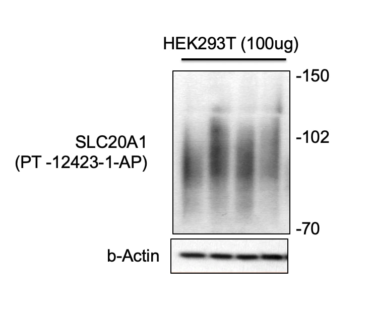
The single review reports a 5-star rating for this antibody used in Western Blot at a 1:500 dilution on HEK293T cells. The reviewer noted the target protein appeared at the expected size as a smear, which they attributed to the protein's heavy glycosylation. No additional background bands were observed within the relevant size range, indicating clean and specific detection. Overall, the feedback is positive, confirming reliable performance and specificity for this application.
The reviews below have been submitted by verified Proteintech customers who received an incentive for providing their feedback.
anwesh (Verified Customer) (07-29-2019) | The protein was found at the right size as a smear. The protein undergoes heavy glycosylation, so that is expected. No additional background bands were observed around the size range.
 |














