

Tested Applications
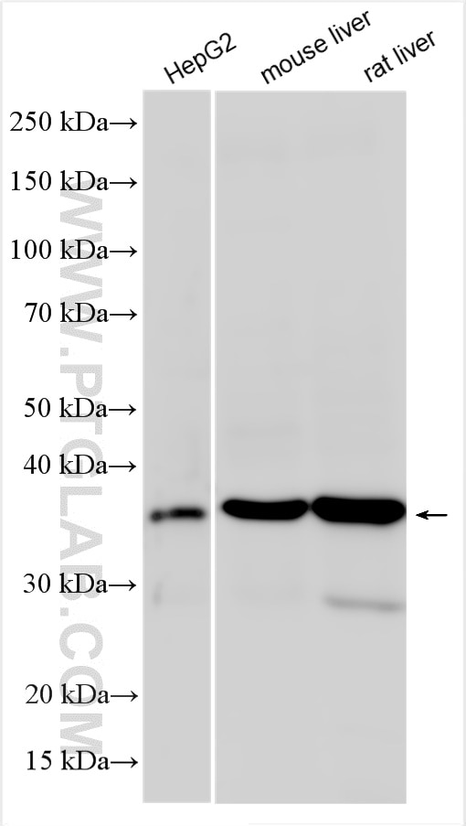
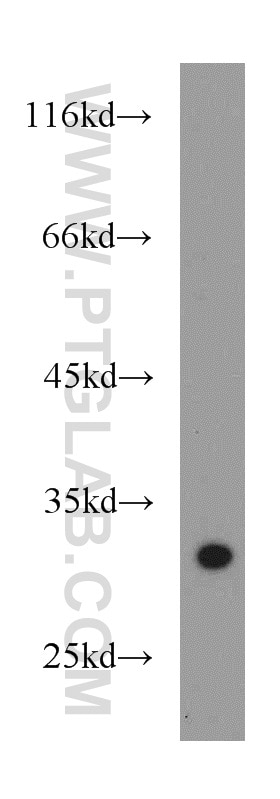
| Positive WB detected in | HepG2 cells, RAW264.7, mouse liver tissue, rat liver tissue |
Recommended dilution
| Application | Dilution |
|---|---|
| Western Blot (WB) | WB : 1:500-1:1000 |
| It is recommended that this reagent should be titrated in each testing system to obtain optimal results. | |
| Sample-dependent, Check data in validation data gallery. | |
Product Information
55100-1-AP targets SPI1 in WB, IHC, IF, ELISA applications and shows reactivity with human, mouse, rat samples.
| Tested Reactivity | human, mouse, rat |
| Cited Reactivity | human, mouse, rat |
| Host / Isotype | Rabbit / IgG |
| Class | Polyclonal |
| Type | Antibody |
| Immunogen |
Peptide Predict reactive species |
| Full Name | spleen focus forming virus (SFFV) proviral integration oncogene spi1 |
| Calculated Molecular Weight | 31 kDa |
| Observed Molecular Weight | 31-42 kDa |
| GenBank Accession Number | NM_003120 |
| Gene Symbol | SPI1 |
| Gene ID (NCBI) | 6688 |
| ENSEMBL Gene ID | ENSG00000066336 |
| RRID | AB_10858486 |
| Conjugate | Unconjugated |
| Form | Liquid |
| Purification Method | Antigen affinity purification |
| UNIPROT ID | P17947 |
| Storage Buffer | PBS with 0.02% sodium azide and 50% glycerol, pH 7.3. |
| Storage Conditions | Store at -20°C. Stable for one year after shipment. Aliquoting is unnecessary for -20oC storage. 20ul sizes contain 0.1% BSA. |
Background Information
SPI1 binds to the PU-box which is a purine-rich DNA sequence (5'-GAGGAA-3') that can act as a lymphoid-specific enhancer. SPI1 is a transcriptional activator that may be specifically involved in the differentiation or activation of macrophages or B-cells. It also binds RNA and may modulate pre-mRNA splicing. The antibody is specific to SPI1. SPI1 can be detected 37-42 kDa (PMID: 28362429, 31586032, 28586009).
Protocols
| Product Specific Protocols | |
|---|---|
| WB protocol for SPI1 antibody 55100-1-AP | Download protocol |
| Standard Protocols | |
|---|---|
| Click here to view our Standard Protocols |
Publications
What published studies show
The anti-SPI1/PU.1 antibody (Cat# 55100-1-AP) has 15 citations published in journals including Cell Reports, Communications Biology, International Immunopharmacology, Scientific Reports, Mucosal Immunology, Medical Oncology, Journal of Cerebral Blood Flow & Metabolism, Experimental Cell Research, Journal of Leukocyte Biology, Frontiers in Genetics, Journal of Cellular Biochemistry, and Hereditas. Research fields span oncology (pancreatic, lung, liver cancer, leukemia), ferroptosis, cardiovascular disease, neuroinflammation, allergic inflammation, rheumatoid arthritis, and inflammatory bowel disease.
| Species | Application | Title |
|---|---|---|
Cell Rep A CLIC1 network coordinates matrix stiffness and the Warburg effect to promote tumor growth in pancreatic cancer | ||
Commun Biol Therapeutic targeting SPI1 in combination with erastin promotes ferroptosis in ccRCC.
| ||
Int Immunopharmacol S100A9 inhibition ameliorates HFpEF by modulating mitochondrial fission and oxidative stress | ||
Int Immunopharmacol Pentraxin 3 promotes microglial M2 polarization and excitatory synapse formation in the hippocampus in depression | ||
Mucosal Immunol IL-33/ST2/IL-9/IL-9R signaling disrupts ocular surface barrier in allergic inflammation. | ||
Reviews
What customers say
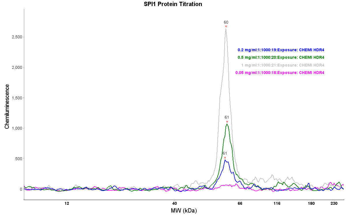
One reviewer (4/5 stars) used this SPI1 antibody in capillary electrophoresis on human brain tissue and observed specific binding to a band around 60 kDa, which they attributed to a dimerized form of the protein. The signal was consistent across lanes and responded as expected when varying sample protein amounts and antibody concentrations, indicating reliable and reproducible performance in their application.
The reviews below have been submitted by verified Proteintech customers who received an incentive for providing their feedback.
Andrew (Verified Customer) (08-09-2021) | When used in our capillary electrophoresis equipment (JESS), the proteintech antibody for SPI1 seemed to show specific binding to a protein at around 60KD. We believe this may be recognising a dimerized form of the protein. This detection was consistent between lanes and responded as expected to varying amounts of sample protein and antibody concentration.
 |



