

Tested Applications
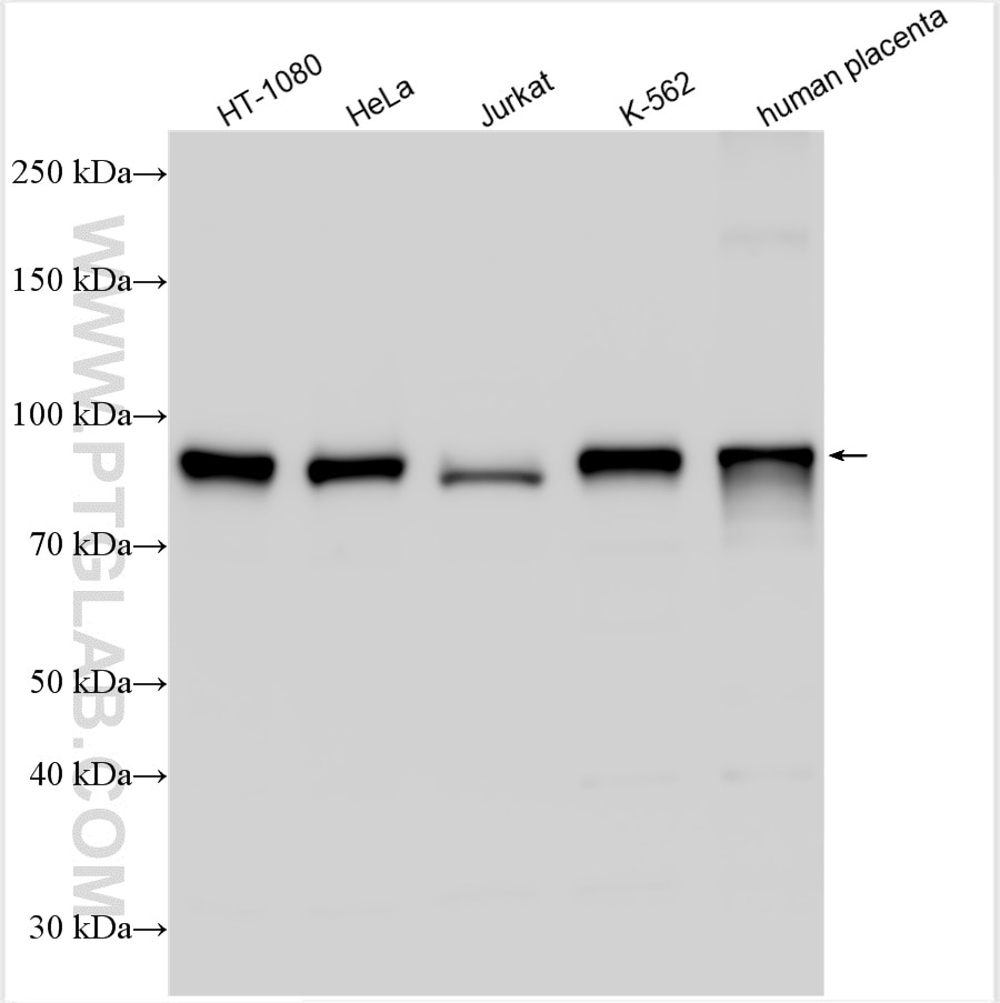
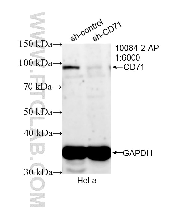
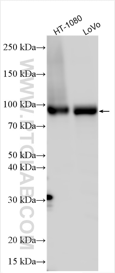
| Positive WB detected in | HT-1080 cells, HeLa cells, LoVo cells, Jurkat cells, K-562 cells, human placenta tissue |
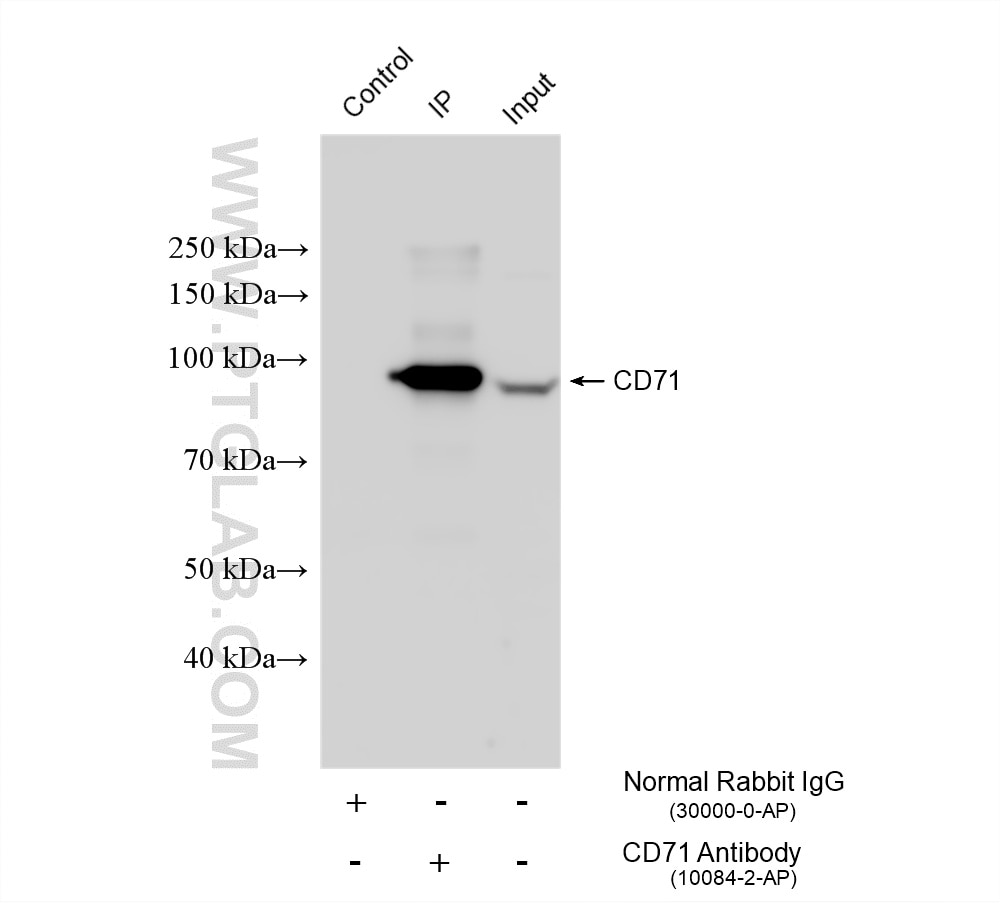
| Positive IP detected in | HeLa cells |
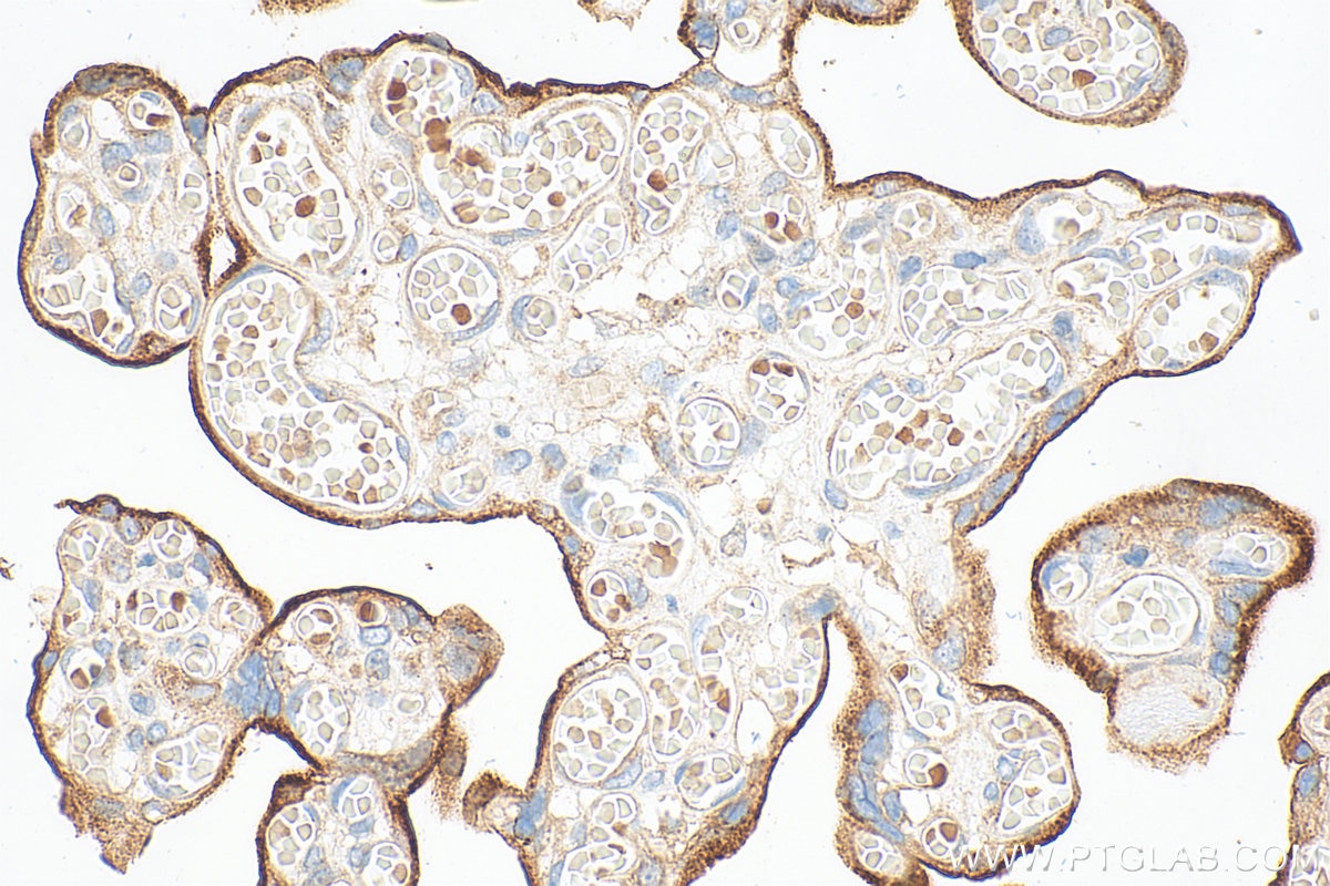
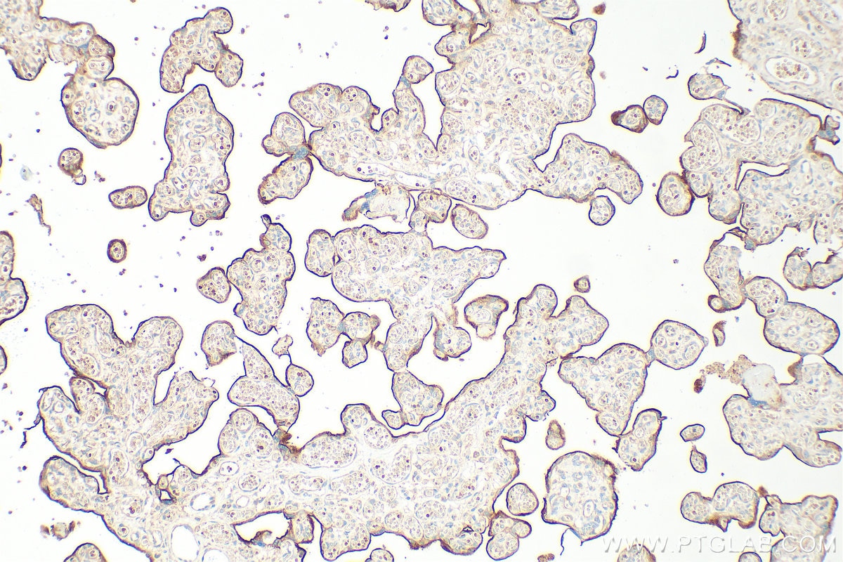
| Positive IHC detected in | human placenta tissue Note: suggested antigen retrieval with TE buffer pH 9.0; (*) Alternatively, antigen retrieval may be performed with citrate buffer pH 6.0 |
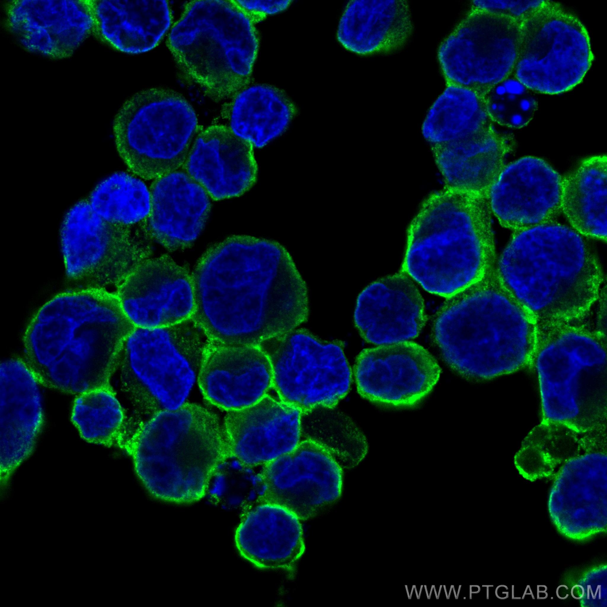
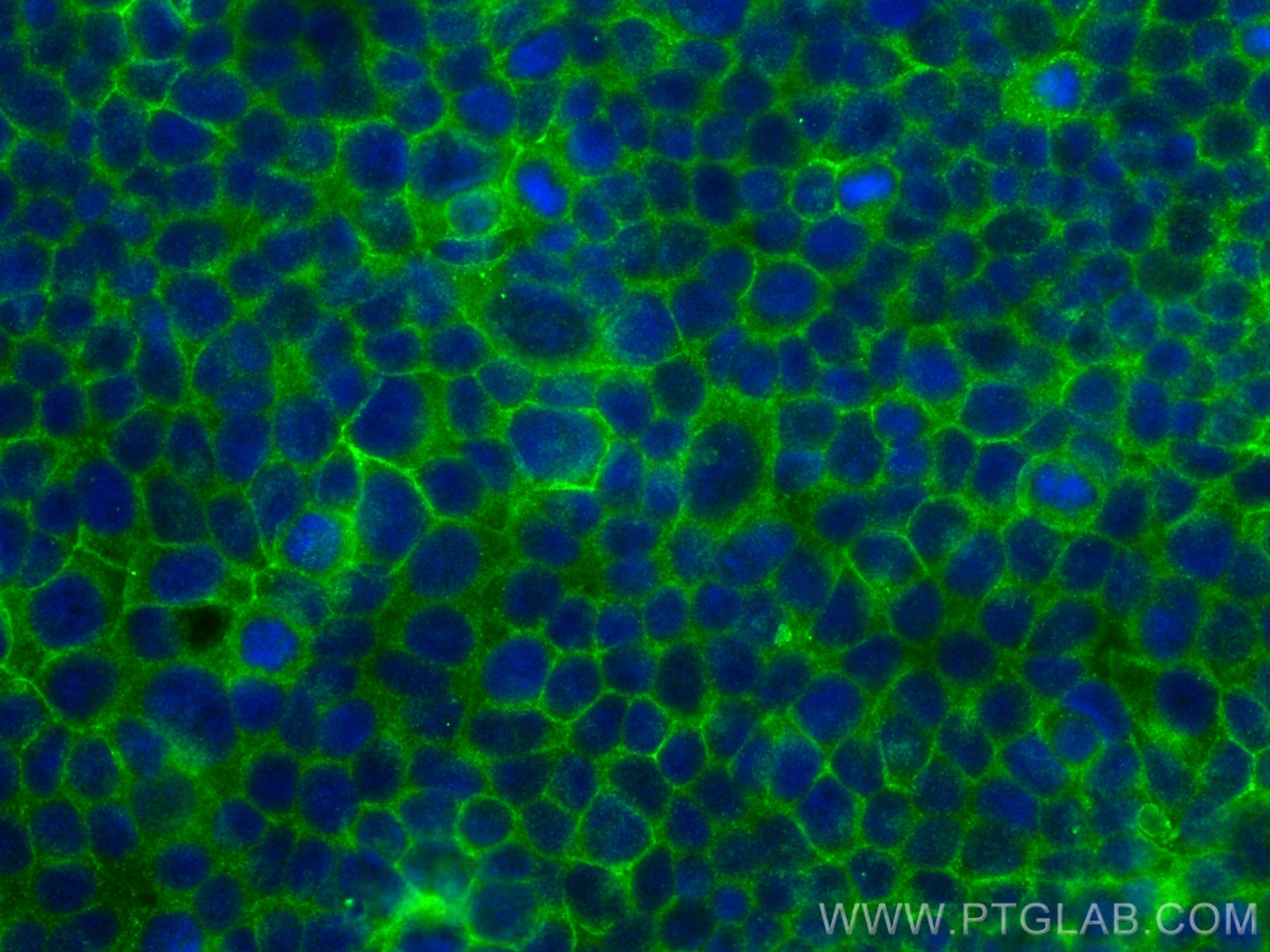
| Positive IF/ICC detected in | K-562 cells, MCF-7 cells |
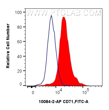
| Positive FC (Intra) detected in | K-562 cells |
Recommended dilution
| Application | Dilution |
|---|---|
| Western Blot (WB) | WB : 1:5000-1:50000 |
| Immunoprecipitation (IP) | IP : 0.5-4.0 ug for 1.0-3.0 mg of total protein lysate |
| Immunohistochemistry (IHC) | IHC : 1:1000-1:4000 |
| Immunofluorescence (IF)/ICC | IF/ICC : 1:200-1:800 |
| Flow Cytometry (FC) (INTRA) | FC (INTRA) : 0.13 ug per 10^6 cells in a 100 µl suspension |
| It is recommended that this reagent should be titrated in each testing system to obtain optimal results. | |
| Sample-dependent, Check data in validation data gallery. | |
Product Information
10084-2-AP targets CD71 in WB, IHC, IF/ICC, IP, CoIP, ELISA, Cell treatment applications and shows reactivity with human samples.
| Tested Reactivity | human |
| Cited Reactivity | human, pig, monkey, chicken, bovine, hamster, duck |
| Host / Isotype | Rabbit / IgG |
| Class | Polyclonal |
| Type | Antibody |
| Immunogen |
CatNo: Ag0123 Product name: Recombinant human CD71 protein Source: e coli.-derived, PGEX-4T Tag: GST Domain: 513-757 aa of BC001188 Sequence: VKHPVTGQFLYQDSNWASKVEKLTLDNAAFPFLAYSGIPAVSFCFCEDTDYPYLGTTMDTYKELIERIPELNKVARAAAEVAGQFVIKLTHDVELNLDYERYNSQLLSFVRDLNQYRADIKEMGLSLQWLYSARGDFFRATSRLTTDFGNAEKTDRFVMKKLNDRVMRVEYHFLSPYVSPKESPFRHVFWGSGSHTLPALLENLKLRKQNNGAFNETLFRNQLALATWTIQGAANALSGDVWDID Predict reactive species |
| Full Name | transferrin receptor (p90, CD71) |
| Calculated Molecular Weight | 85 kDa |
| Observed Molecular Weight | 90 kDa |
| GenBank Accession Number | BC001188 |
| Gene Symbol | CD71 |
| Gene ID (NCBI) | 7037 |
| ENSEMBL Gene ID | ENSG00000072274 |
| RRID | AB_2240403 |
| Conjugate | Unconjugated |
| Form | Liquid |
| Purification Method | Antigen affinity purification |
| UNIPROT ID | P02786 |
| Storage Buffer | PBS with 0.02% sodium azide and 50% glycerol, pH 7.3. |
| Storage Conditions | Store at -20°C. Stable for one year after shipment. Aliquoting is unnecessary for -20oC storage. 20ul sizes contain 0.1% BSA. |
Background Information
CD71, also known as transferrin receptor protein 1 (TfR1), is a transmembrane glycoprotein composed of two disulfide-linked monomers, each of 90 kDa molecular weight. Each monomer binds one holo-transferrin molecule creating an iron-Tf-TfR complex that enters the cell by endocytosis. CD71 is present on actively proliferating cells and is essential for iron transport into proliferating cells.
Protocols
| Product Specific Protocols | |
|---|---|
| FC protocol for CD71 antibody 10084-2-AP | Download protocol |
| IF protocol for CD71 antibody 10084-2-AP | Download protocol |
| IHC protocol for CD71 antibody 10084-2-AP | Download protocol |
| IP protocol for CD71 antibody 10084-2-AP | Download protocol |
| WB protocol for CD71 antibody 10084-2-AP | Download protocol |
| Standard Protocols | |
|---|---|
| Click here to view our Standard Protocols |
Publications
What published studies show
The anti-Transferrin Receptor (TFRC) antibody (Cat# 10084-2-AP) has accumulated 213 total citations across high-impact journals including Nature Communications, Nature Cancer, Cell Reports, Redox Biology, Free Radical Biology and Medicine, Cell Death & Disease, Autophagy, Phytomedicine, Cancer Cell, and Cell Death & Differentiation. Research fields span ferroptosis, iron metabolism, oncology, neuroscience, cardiology, nephrology, hepatology, reproductive biology, toxicology, immunology, aging, and bone/joint diseases.
| Species | Application | Title |
|---|---|---|
Signal Transduct Target Ther Two stress-responsive kinases suppress ferroptosis by activating antioxidant programs under mild oxidative stress. | ||
Cancer Cell Multiomic integration reveals tumoral heterogeneity of lipid dependence within lethal group 3 medulloblastoma. | ||
Cell Res The metabolite α-KG induces GSDMC-dependent pyroptosis through death receptor 6-activated caspase-8. | ||
Nat Cancer ENO1 suppresses cancer cell ferroptosis by degrading the mRNA of iron regulatory protein 1. | ||
Cancer Res Intratumoral payload release influences the potency and bystander-killing effects of antibody-drug conjugates in preclinical models. |
Reviews
What customers say
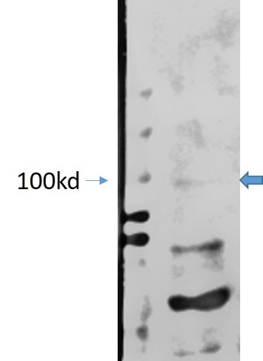
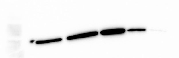
Users report strong Western Blot performance with good signal even at low lysate loads, though one reviewer noted non-specific bands alongside the expected ~100 kDa band in human tissue. Immunofluorescence results were mixed: one user found it clean with no background on HEK cells, while another described the signal as fuzzy and diffuse in mouse spleen B cells. Overall, the antibody is considered functional and specific for WB, with variable IF performance depending on sample type.
The reviews below have been submitted by verified Proteintech customers who received an incentive for providing their feedback.
Sarah (Verified Customer) (07-21-2026) | Functional and specific antibody. No background.
|
Nu (Verified Customer) (10-24-2023) | Although there is a band close to 100 kd, there are other even more stronger non-specific bands.
 |
Sara (Verified Customer) (10-25-2020) | Really good signal in WB, it worked great, even though I loaded 1-2 µg of total cell lysate in some lanes. Not that good for IF, the signal was a bit fuzzy and all over the cell.
 |










