

Tested Applications
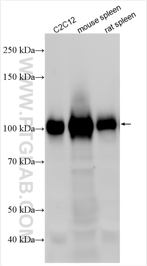
| Positive WB detected in | rat spleen tissue, mouse spleen tissue, C2C12 cells |
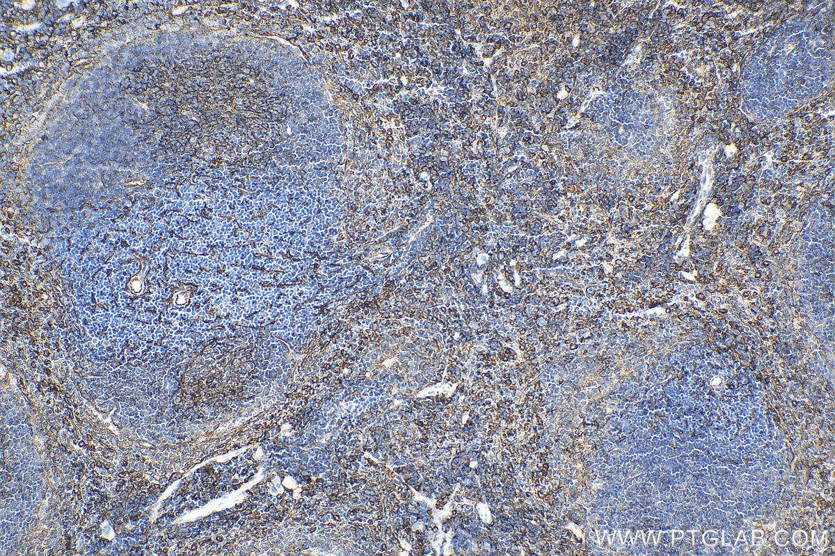
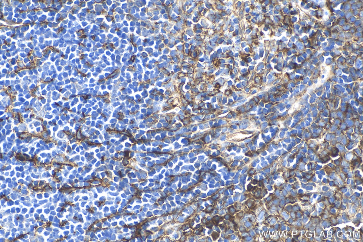
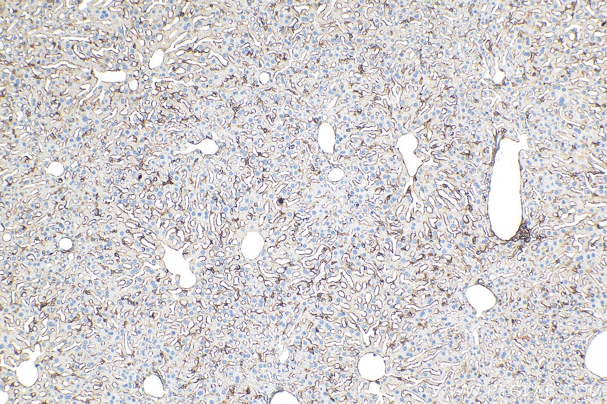
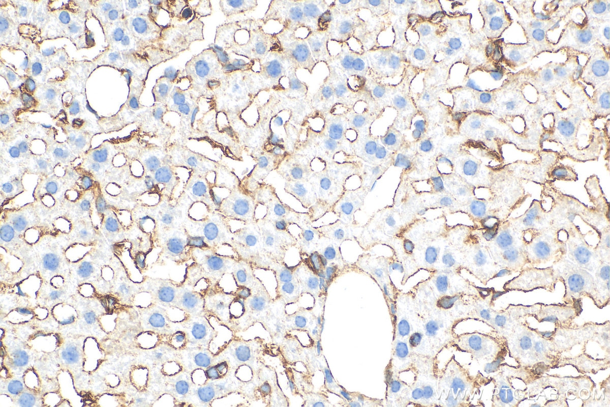
| Positive IHC detected in | mouse spleen tissue, mouse liver tissue Note: suggested antigen retrieval with TE buffer pH 9.0; (*) Alternatively, antigen retrieval may be performed with citrate buffer pH 6.0 |
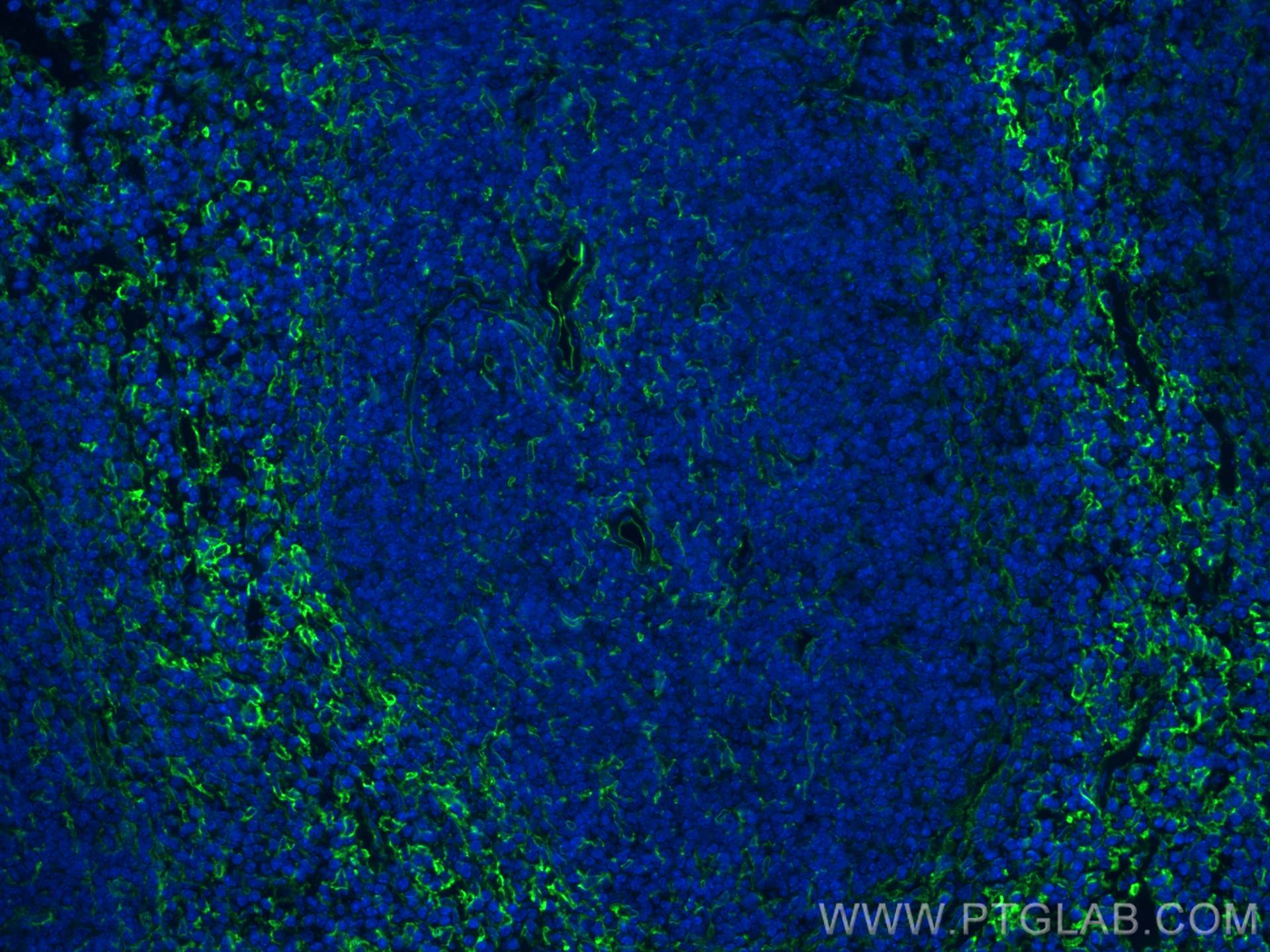
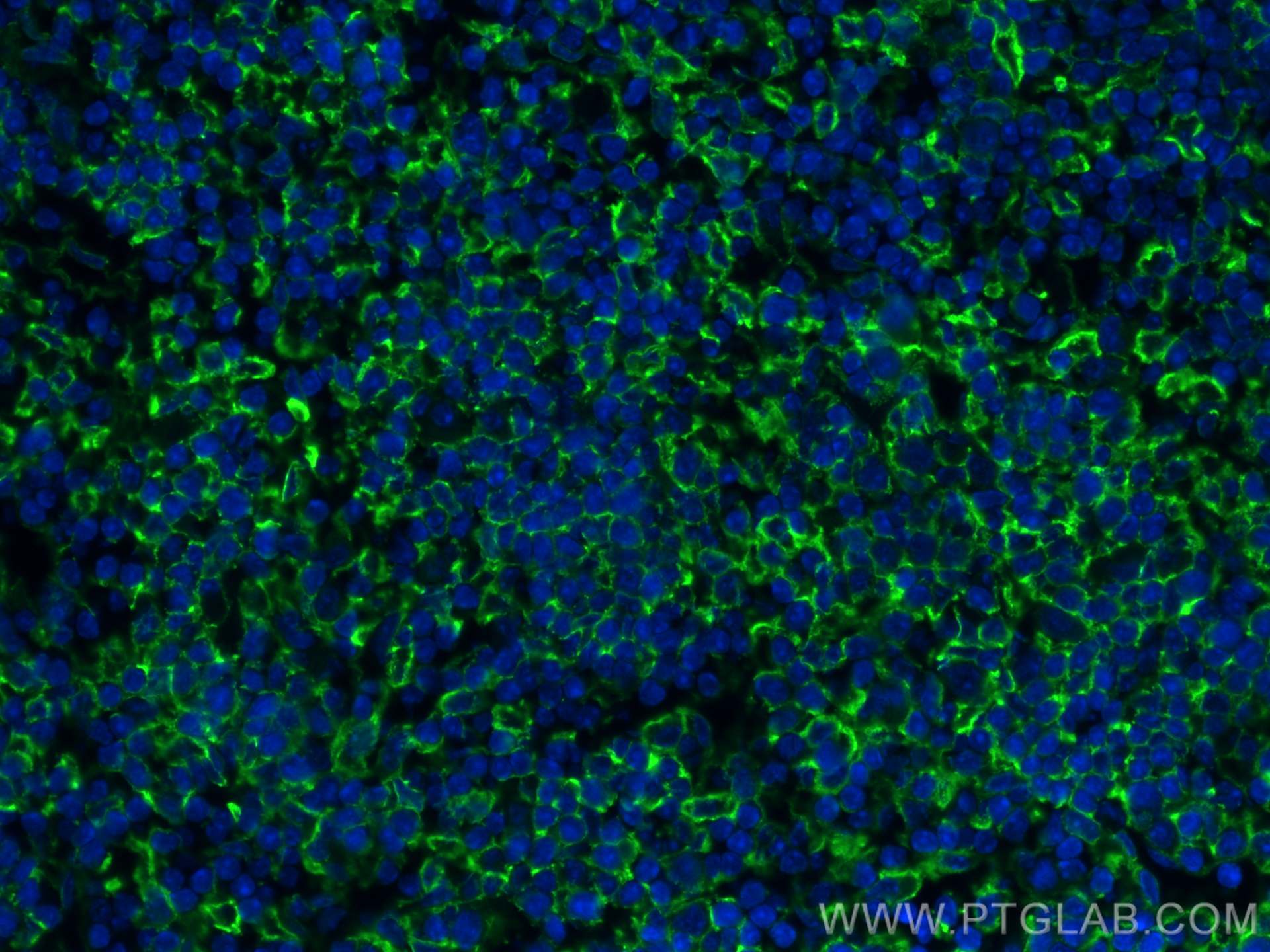
| Positive IF-P detected in | mouse spleen tissue |
Recommended dilution
| Application | Dilution |
|---|---|
| Western Blot (WB) | WB : 1:2000-1:12000 |
| Immunohistochemistry (IHC) | IHC : 1:200-1:800 |
| Immunofluorescence (IF)-P | IF-P : 1:50-1:500 |
| It is recommended that this reagent should be titrated in each testing system to obtain optimal results. | |
| Sample-dependent, Check data in validation data gallery. | |
Product Information
30958-1-AP targets VCAM-1/CD106 in WB, IHC, IF-P, ELISA applications and shows reactivity with mouse, rat samples.
| Tested Reactivity | mouse, rat |
| Cited Reactivity | mouse, rat |
| Host / Isotype | Rabbit / IgG |
| Class | Polyclonal |
| Type | Antibody |
| Immunogen |
CatNo: Eg0639 Product name: Recombinant mouse VCAM-1 protein Source: mammalian cells-derived, pHZ-KIsec-C-6*HIS Tag: C-6*HIS Domain: 25-698 aa of CAA47989.1 Sequence: FKIEISPEYKTIAQIGDSMALTCSTTGCESPLFSWRTQIDSPLNAKVRTEGSKSVLTMEPVSFENEHSYLCTATCGSGKLERSIHVDIYSFPKDPEIQFSGPLEVGKPVTVKCLAPDIYPVYRLEIDLFKGDQLMNRQEFSSEEMTKSLETKSLEVTFTPVIEDIGKALVCRAKLHIDQIDSTLKERETVKELQVYISPRNTTISVHPSTRLQEGGAVTMTCSSEGLPAPEIFWGRKLDNEVLQLLSGNATLTLIAMRMEDSGVYVCEGVNLIGRDKAEVELVVQEKPFIVDISPGSQVAAQVGDSVVLTCAAIGCDSPSFSWRTQTDSPLNGVVRNEGAKSTLVLSSVGFEDEHSYLCAVTCLQRTLEKRTQVEVYSFPEDPVIKMSGPLVHGRPVTVNCTVPNVYPFDHLEIELLKGETTLMKKYFLEEMGIKSLETKILETTFIPTIEDTGKSLVCLARLHSGEMESEPKQRQSVQPLYVNVAPKETTIWVSPSPILEEGSPVNLTCSSDGIPAPKILWSRQLNNGELQPLSENTTLTFMSTKRDDSGIYVCEGINEAGISRKSVELIIQVSPKDIQLTVFPSKSVKEGDTVIISCTCGNVPETWIILKKKAKTGDMVLKSVDGSYTIRQAQLQDAGIYECESKTEVGSQLRSLTLDVKGKEHNKDYFSPE Predict reactive species |
| Full Name | vascular cell adhesion molecule 1 |
| Calculated Molecular Weight | 81 kDa |
| Observed Molecular Weight | 100-110 kDa |
| GenBank Accession Number | CAA47989.1 |
| Gene Symbol | VCAM-1 |
| Gene ID (NCBI) | 22329 |
| RRID | AB_3086429 |
| Conjugate | Unconjugated |
| Form | Liquid |
| Purification Method | Antigen affinity purification |
| UNIPROT ID | P29533 |
| Storage Buffer | PBS with 0.02% sodium azide and 50% glycerol, pH 7.3. |
| Storage Conditions | Store at -20°C. Stable for one year after shipment. Aliquoting is unnecessary for -20oC storage. 20ul sizes contain 0.1% BSA. |
Background Information
Vascular cell adhesion molecule 1 (VCAM1), also known as CD106, is a 110-kDa transmembrane glycoprotein belonging to the immunoglobulin gene superfamily. VCAM1 is expressed by cytokine-activated endothelium, interacts with integrin VLA4 (α4β1) present on the surface of leukocytes, and mediates both adhesion and signal transduction. It is also expressed either constitutively or inducibly in a variety of other cell types, including vascular smooth muscle cells, differentiating skeletal muscle cells, renal and neural epithelial cells, macrophages (Kupffer cells), dendritic cells, and bone marrow stromal cells (PMID: 7507076, 11359843).
Protocols
| Product Specific Protocols | |
|---|---|
| IF protocol for VCAM-1/CD106 antibody 30958-1-AP | Download protocol |
| IHC protocol for VCAM-1/CD106 antibody 30958-1-AP | Download protocol |
| WB protocol for VCAM-1/CD106 antibody 30958-1-AP | Download protocol |
| Standard Protocols | |
|---|---|
| Click here to view our Standard Protocols |
Publications
What published studies show
The VCAM-1/CD106 Polyclonal antibody (Cat# 30958-1-AP) has 9 citations across journals including Advanced Materials, Redox Biology, Advanced Science, ACS Applied Materials & Interfaces, Laboratory Investigation, Microvascular Research, Translational Andrology and Urology, Free Radical Biology and Medicine, and Cell Biology International. Research fields span tumor sonodynamic therapy, vascular inflammation and aneurysm, diabetic cardiomyopathy, heart transplant rejection, lupus nephritis, aging, acute kidney injury, atherosclerosis, and sepsis.
| Species | Application | Title |
|---|---|---|
Adv Mater Ultrasound-Activated Sonophage Synergizes Sonodynamic Therapy and Saltoptosis for Solid Tumor Eradication. | ||
Redox Biol Nox1/PAK1 is required for angiotensin II-induced vascular inflammation and abdominal aortic aneurysm formation | ||
Adv Sci (Weinh) TrxR2 Lactylation Facilitates Mitochondrial Protection and Endothelial Ferroptosis Resistance in Diabetic Cardiomyopathy. | ||
ACS Appl Mater Interfaces Macrophage-Mimicking Biomimetic Microbubbles for Early Ultrasound Molecular Imaging of Acute Rejection in Heart Transplantation. | ||
Lab Invest HMGB1 Encapsulated in Podocyte-Derived Exosomes Plays a Central Role in Glomerular Endothelial Cell Injury in Lupus Nephritis by Regulating TRIM27 Expression | ||
Microvasc Res Characterization of microcirculatory endothelial functions in a D-Galactose-induced aging model |
Reviews
What customers say
The single review for this antibody reports a 5-star rating. The user tested it at a 1:200 dilution on murine inflamed colon tissue for immunofluorescence and found it performed very well, noting that the controls were clean. Overall, the feedback is positive, highlighting strong staining performance and low background in the application tested.
The reviews below have been submitted by verified Proteintech customers who received an incentive for providing their feedback.
Julia (Verified Customer) (05-07-2026) | This antibody worked so well on my samples. The controls were perfect clean.
|








