Tested Applications
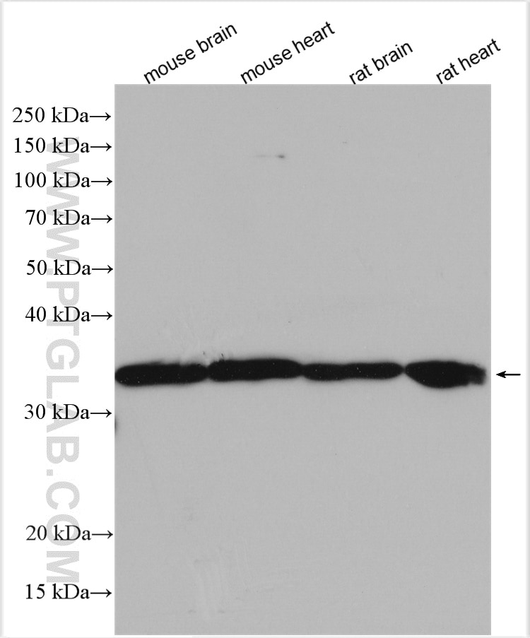
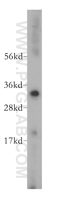
| Positive WB detected in | mouse brain tissue, human heart tissue, mouse heart tissue, rat brain tissue, rat heart tissue |
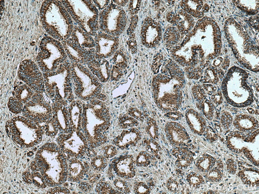
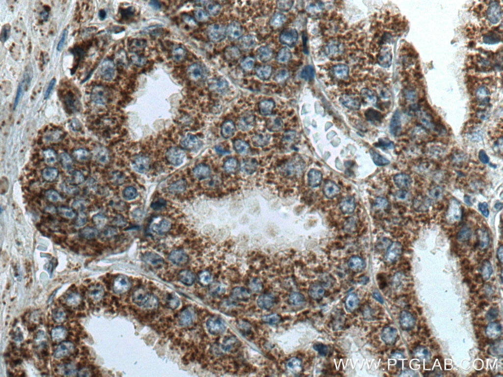
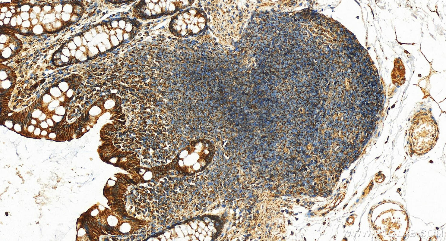
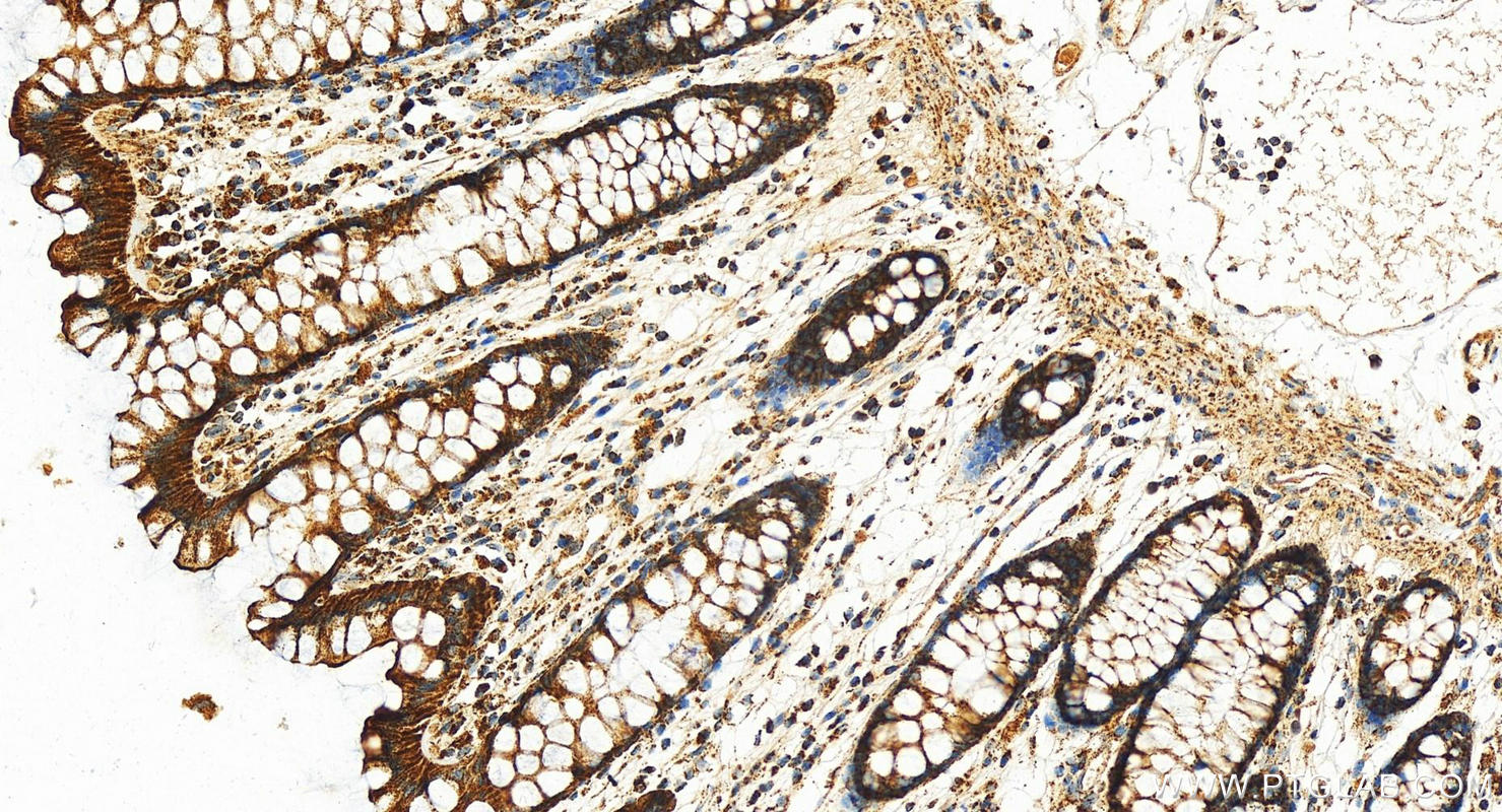
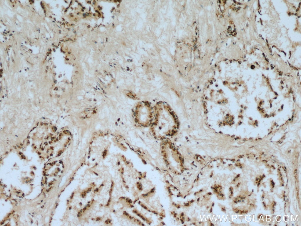
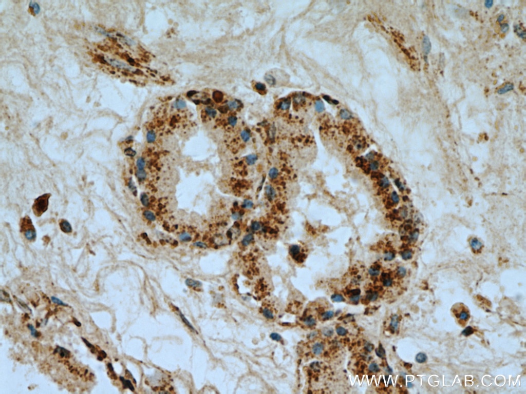
| Positive IHC detected in | human prostate cancer tissue, human normal colon, human prostate tissue Note: suggested antigen retrieval with TE buffer pH 9.0; (*) Alternatively, antigen retrieval may be performed with citrate buffer pH 6.0 |
Recommended dilution
| Application | Dilution |
|---|---|
| Western Blot (WB) | WB : 1:1000-1:6000 |
| Immunohistochemistry (IHC) | IHC : 1:200-1:1200 |
| It is recommended that this reagent should be titrated in each testing system to obtain optimal results. | |
| Sample-dependent, Check data in validation data gallery. | |
Published Applications
| KD/KO | See 1 publications below |
| WB | See 32 publications below |
| IHC | See 3 publications below |
| IF | See 6 publications below |
| IP | See 2 publications below |
| CoIP | See 1 publications below |
Product Information
11663-1-AP targets VDAC1/2/3 in WB, IHC, IF, CoIP, ELISA applications and shows reactivity with human, mouse, rat samples.
| Tested Reactivity | human, mouse, rat |
| Cited Reactivity | human, mouse, rat, duck |
| Host / Isotype | Rabbit / IgG |
| Class | Polyclonal |
| Type | Antibody |
| Immunogen |
CatNo: Ag2266 Product name: Recombinant human VDAC2 protein Source: e coli.-derived, PGEX-4T Tag: GST Domain: 1-294 aa of BC000165 Sequence: MATHGQTCARPMCIPPSYADLGKAARDIFNKGFGFGLVKLDVKTKSCSGVEFSTSGSSNTDTGKVTGTLETKYKWCEYGLTFTEKWNTDNTLGTEIAIEDQICQGLKLTFDTTFSPNTGKKSGKIKSSYKRECINLGCDVDFDFAGPAIHGSAVFGYEGWLAGYQMTFDSAKSKLTRNNFAVGYRTGDFQLHTNVNDGTEFGGSIYQKVCEDLDTSVNLAWTSGTNCTRFGIAAKYQLDPTASISAKVNNSSLIGVGYTQTLRPGVKLTLSALVDGKSINAGGHKVGLALELEA Predict reactive species |
| Full Name | voltage-dependent anion channel 2 |
| Calculated Molecular Weight | 294 aa, 32 kDa |
| Observed Molecular Weight | 32 kDa |
| GenBank Accession Number | BC000165 |
| Gene Symbol | VDAC2 |
| Gene ID (NCBI) | 7417 |
| RRID | AB_2304144 |
| Conjugate | Unconjugated |
| Form | Liquid |
| Purification Method | Antigen affinity purification |
| UNIPROT ID | P45880 |
| Storage Buffer | PBS with 0.02% sodium azide and 50% glycerol, pH 7.3. |
| Storage Conditions | Store at -20°C. Stable for one year after shipment. Aliquoting is unnecessary for -20oC storage. 20ul sizes contain 0.1% BSA. |
Background Information
VDACs (Voltage Dependent Anion selective Channels), also known as mitochondrial porins, are a family of pore-forming proteins discovered in the mitochondrial outer membrane. Mammals show a conserved genetic organization of the VDAC genes. It's reported that the amount of VDAC transcripts in liver is usually lower than in the other tissues. VDAC2 and expecially VDAC3 are highly expressed in testis, while mouse VDAC1 is poorly expressed in this tissue.(PMID: 22020053)
Protocols
| Product Specific Protocols | |
|---|---|
| IHC protocol for VDAC1/2/3 antibody 11663-1-AP | Download protocol |
| WB protocol for VDAC1/2/3 antibody 11663-1-AP | Download protocol |
| Standard Protocols | |
|---|---|
| Click here to view our Standard Protocols |
Publications
| Species | Application | Title |
|---|---|---|
Mol Cell MLKL activity requires a splicing-regulated, druggable intramolecular interaction | ||
Nat Commun Kastor and Polluks polypeptides encoded by a single gene locus cooperatively regulate VDAC and spermatogenesis. | ||
Autophagy MYBL2 guides autophagy suppressor VDAC2 in the developing ovary to inhibit autophagy through a complex of VDAC2-BECN1-BCL2L1 in mammals. | ||
Cell Death Differ SPATA33 is an autophagy mediator for cargo selectivity in germline mitophagy. | ||
Proc Natl Acad Sci U S A SPATA33 localizes calcineurin to the mitochondria and regulates sperm motility in mice. | ||
Cell Death Dis Pathological convergence of APP and SNCA deficiency in hippocampal degeneration of young rats |
Reviews
The reviews below have been submitted by verified Proteintech customers who received an incentive for providing their feedback.
FH Jun (Verified Customer) (06-12-2022) | Works very well.
 |