


Tested Applications
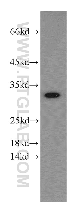
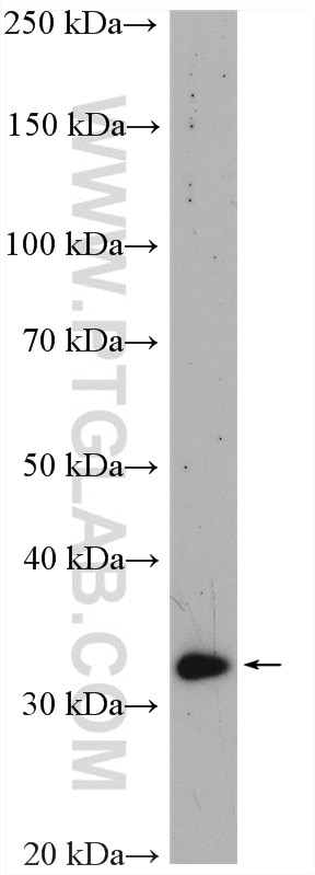
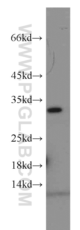
| Positive WB detected in | HepG2 cells, mouse brain tissue, HuH-7 cells |
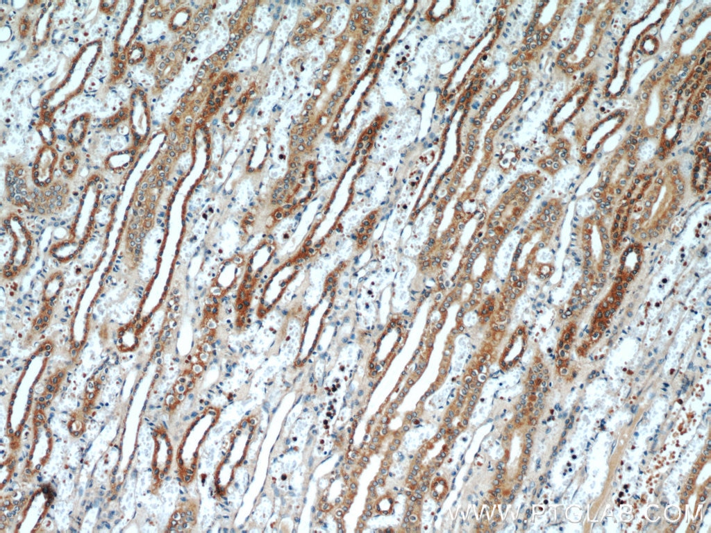
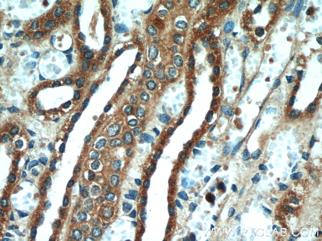
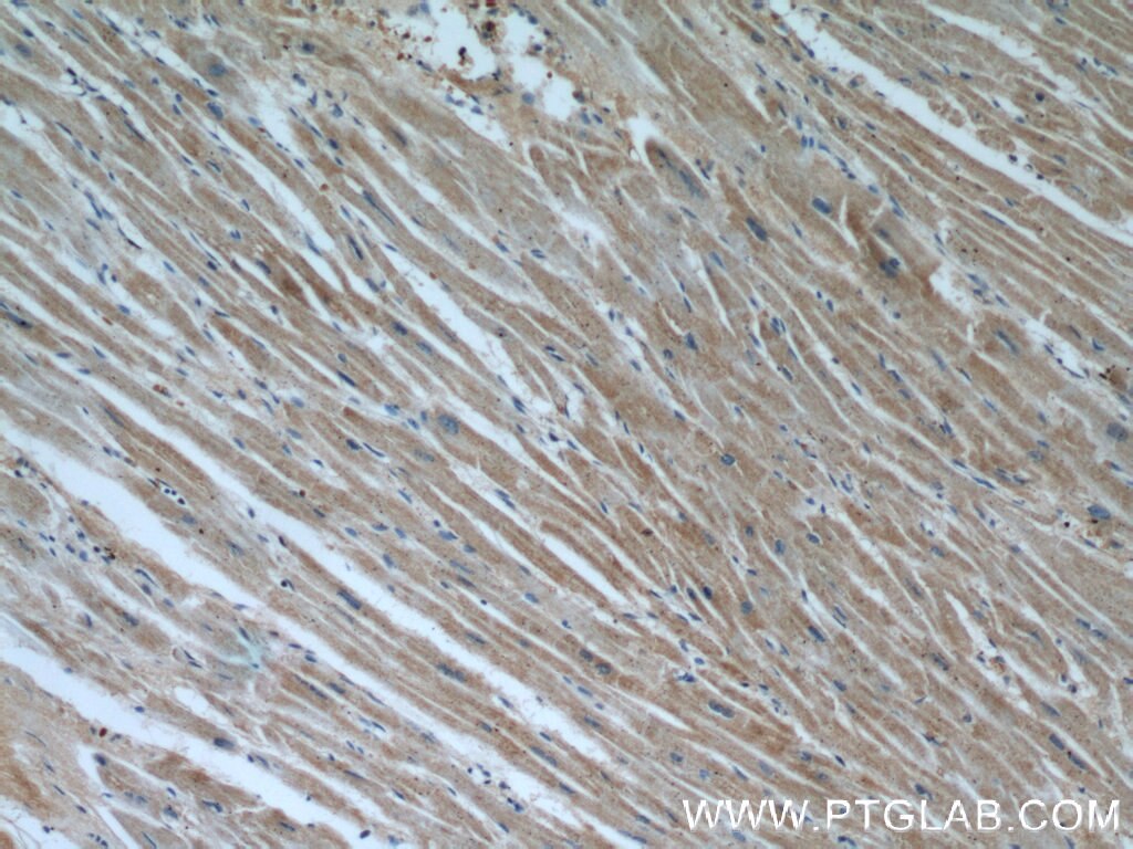
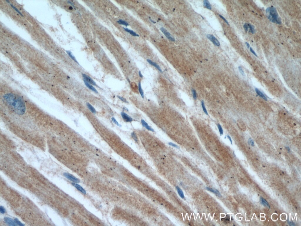
| Positive IHC detected in | human kidney tissue, human heart tissue Note: suggested antigen retrieval with TE buffer pH 9.0; (*) Alternatively, antigen retrieval may be performed with citrate buffer pH 6.0 |
| Positive IF-P detected in | mouse brain tissue |
Recommended dilution
| Application | Dilution |
|---|---|
| Western Blot (WB) | WB : 1:500-1:1000 |
| Immunohistochemistry (IHC) | IHC : 1:20-1:200 |
| Immunofluorescence (IF)-P | IF-P : 1:50-1:500 |
| It is recommended that this reagent should be titrated in each testing system to obtain optimal results. | |
| Sample-dependent, Check data in validation data gallery. | |
Product Information
55261-1-AP targets VDAC2 in WB, IHC, IF-P, ELISA applications and shows reactivity with human, mouse, rat samples.
| Tested Reactivity | human, mouse, rat |
| Cited Reactivity | human, mouse |
| Host / Isotype | Rabbit / IgG |
| Class | Polyclonal |
| Type | Antibody |
| Immunogen |
Peptide Predict reactive species |
| Full Name | voltage-dependent anion channel 2 |
| Calculated Molecular Weight | 33 kDa |
| Observed Molecular Weight | 31-33 kDa |
| GenBank Accession Number | NM_003375 |
| Gene Symbol | VDAC2 |
| Gene ID (NCBI) | 7417 |
| RRID | AB_10852961 |
| Conjugate | Unconjugated |
| Form | Liquid |
| Purification Method | Antigen affinity purification |
| UNIPROT ID | P45880 |
| Storage Buffer | PBS with 0.02% sodium azide and 50% glycerol, pH 7.3. |
| Storage Conditions | Store at -20°C. Stable for one year after shipment. Aliquoting is unnecessary for -20oC storage. 20ul sizes contain 0.1% BSA. |
Background Information
VDAC2 belongs to the eukaryotic mitochondrial porin family. It forms a channel through the mitochondrial outer membrane that allows diffusion of small hydrophilic molecules. This antibody is specific to VDAC2.
Protocols
| Product Specific Protocols | |
|---|---|
| IF protocol for VDAC2 antibody 55261-1-AP | Download protocol |
| IHC protocol for VDAC2 antibody 55261-1-AP | Download protocol |
| WB protocol for VDAC2 antibody 55261-1-AP | Download protocol |
| Standard Protocols | |
|---|---|
| Click here to view our Standard Protocols |
Publications
What published studies show
The VDAC2 Polyclonal antibody (Cat# 55261-1-AP) has 4 citations. These appear in Nature, Cell Death & Differentiation, Journal of Molecular Medicine, and Cell Physiology and Biochemistry. The research fields span gasdermin D-mediated metabolic crosstalk in tissue repair, lipid droplet–mitochondria contacts in microglial neuroinflammation after spinal cord injury, ferroptosis inhibition by cerium oxide nanoparticles, and VDAC1 knockout–induced oxidative stress apoptosis in cardiomyocytes. All citations used the antibody for Western blot in human or mouse samples.
| Species | Application | Title |
|---|---|---|
Cell Death Differ PLIN2-PGAM5-regulated lipid droplet-mitochondria contacts drive microglial neuroinflammation after spinal cord injury via fatty acid metabolic reprogramming | ||
J Mol Med (Berl) Cerium oxide nanoparticles as potent inhibitors of ferroptosis: role of antioxidant activity and protein regulation | ||
Cell Physiol Biochem Knockout of VDAC1 in H9c2 Cells Promotes Oxidative Stress-Induced Cell Apoptosis through Decreased Mitochondrial Hexokinase II Binding and Enhanced Glycolytic Stress. |









