

Tested Applications
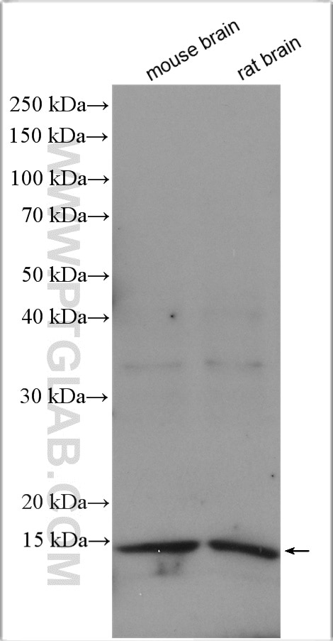
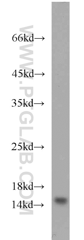
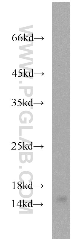
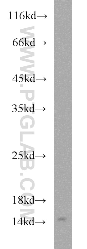
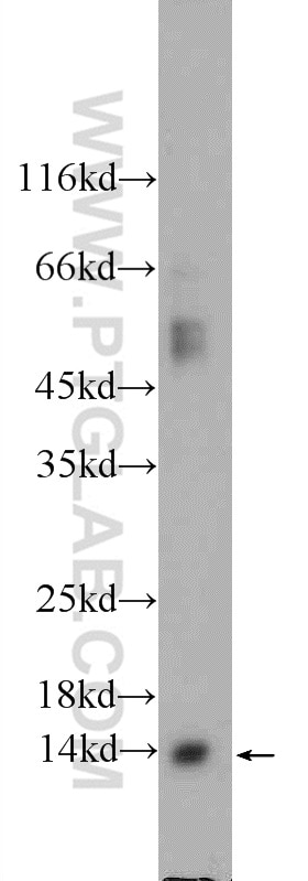
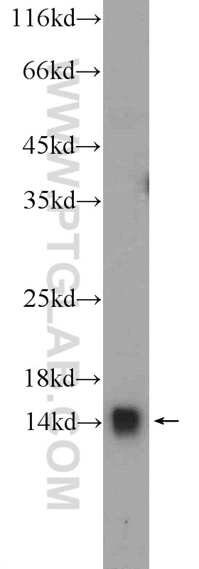
| Positive WB detected in | mouse brain tissue, mouse testis tissue, rat brain tissue |
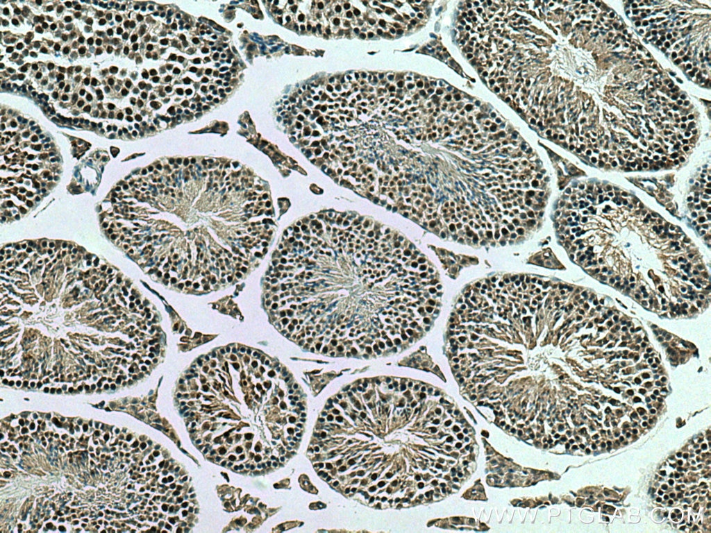
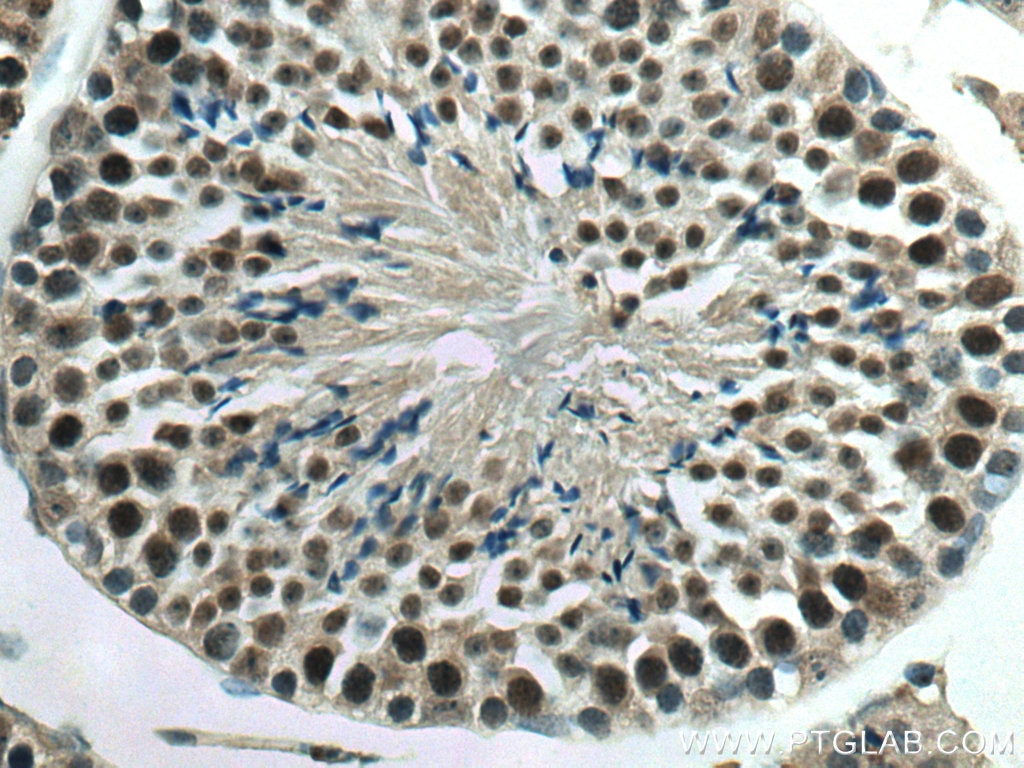
| Positive IHC detected in | mouse testis tissue Note: suggested antigen retrieval with TE buffer pH 9.0; (*) Alternatively, antigen retrieval may be performed with citrate buffer pH 6.0 |
Recommended dilution
| Application | Dilution |
|---|---|
| Western Blot (WB) | WB : 1:1000-1:4000 |
| Immunohistochemistry (IHC) | IHC : 1:50-1:500 |
| It is recommended that this reagent should be titrated in each testing system to obtain optimal results. | |
| Sample-dependent, Check data in validation data gallery. | |
Product Information
11730-1-AP targets YPEL5 in WB, IHC, IF, IP, ELISA applications and shows reactivity with human, mouse, rat samples.
| Tested Reactivity | human, mouse, rat |
| Cited Reactivity | human, mouse, zebrafish |
| Host / Isotype | Rabbit / IgG |
| Class | Polyclonal |
| Type | Antibody |
| Immunogen |
CatNo: Ag2328 Product name: Recombinant human YPEL5 protein Source: e coli.-derived, PGEX-4T Tag: GST Domain: 1-121 aa of BC000836 Sequence: MGRIFLDHIGGTRLFSCANCDTILTNRSELISTRFTGATGRAFLFNKVVNLQYSEVQDRVMLTGRHMVRDVSCKNCNSKLGWIYEFATEDSQRYKEGRVILERALVRESEGFEEHVPSDNS Predict reactive species |
| Full Name | yippee-like 5 (Drosophila) |
| Calculated Molecular Weight | 121 aa, 14 kDa |
| Observed Molecular Weight | 14 kDa |
| GenBank Accession Number | BC000836 |
| Gene Symbol | YPEL5 |
| Gene ID (NCBI) | 51646 |
| RRID | AB_2217465 |
| Conjugate | Unconjugated |
| Form | Liquid |
| Purification Method | Antigen affinity purification |
| UNIPROT ID | P62699 |
| Storage Buffer | PBS with 0.02% sodium azide and 50% glycerol, pH 7.3. |
| Storage Conditions | Store at -20°C. Stable for one year after shipment. Aliquoting is unnecessary for -20oC storage. 20ul sizes contain 0.1% BSA. |
Background Information
YPEL5(Protein yippee-like 5) belongs to the yippee family and is involved in a certain cell division-related function.During cell cycle, YPEL5 protein is detected at different subcellular localizations; at interphase, it is located in the nucleus and centrosome, then it changes location sequentially to spindle poles, mitotic spindle, and spindle midzone during mitosis, and finally transferrs to midbody at cytokinesis(PMID:20580816).
Protocols
| Product Specific Protocols | |
|---|---|
| IHC protocol for YPEL5 antibody 11730-1-AP | Download protocol |
| WB protocol for YPEL5 antibody 11730-1-AP | Download protocol |
| Standard Protocols | |
|---|---|
| Click here to view our Standard Protocols |
Publications
What published studies show
The YPEL5 Polyclonal antibody (catalog number 11730-1-AP) has 6 total citations. These appear in Nature Communications, Neuron, Molecular Oncology, iScience, and bioRxiv. The research fields span tuberculosis-infected macrophage biology and the GID/CTLH complex, basal ganglia vocal-learning circuits, colorectal carcinogenesis via the METTL3/YTHDF2 m6A axis, TRIM24 repressor network regulation, and ribosome biogenesis. Applications cited include Western blot, immunofluorescence, and immunoprecipitation across human, mouse, and zebrafish species.
| Species | Application | Title |
|---|---|---|
Nat Commun Genome-wide screen of Mycobacterium tuberculosis-infected macrophages revealed GID/CTLH complex-mediated modulation of bacterial growth | ||
Neuron Molecular microcircuitry underlies functional specification in a basal ganglia circuit dedicated to vocal learning. | ||
iScience Genome-wide CRISPR-Cas9 screen analyzed by SLIDER identifies network of repressor complexes that regulate TRIM24 | ||
Mol Oncol METTL3/YTHDF2 m6A axis accelerates colorectal carcinogenesis through epigenetically suppressing YPEL5. | ||
bioRxiv Proteome-wide C-degron activity profiling connects conditional regulation of the CTLH E3 ligase complex to ribosome biogenesis. | ||
bioRxiv Genome-wide screen of Mycobacterium tuberculosis- infected macrophages identified the GID/CTLH complex as a determinant of intracellular bacterial growth |
Reviews
What customers say
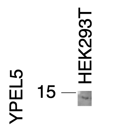
Both reviewers used this antibody for Western Blot on HEK293T cells and reported that the target band appeared at the correct molecular weight but with relatively faint signal. One reviewer noted the signal was detectable yet weak in low-expressing cell lines. The other suggested that using a 0.2 µm pore-size membrane might improve detection, given that YPEL5 is a small protein. Overall, the antibody works but may benefit from optimization for stronger signal.
The reviews below have been submitted by verified Proteintech customers who received an incentive for providing their feedback.
Abigail (Verified Customer) (02-26-2026) | The signal is quite faint in low expressing cell lines, but there.
|
S (Verified Customer) (11-22-2021) | The band was there on the right MW range, but it was a bit weak. It could have been better if I used 0.2 um pore-size membrane, given that YPEL5 is a small protein.
 |









