

Tested Applications
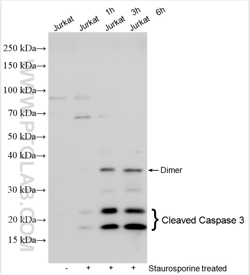
| Positive WB detected in | Jurkat cells |
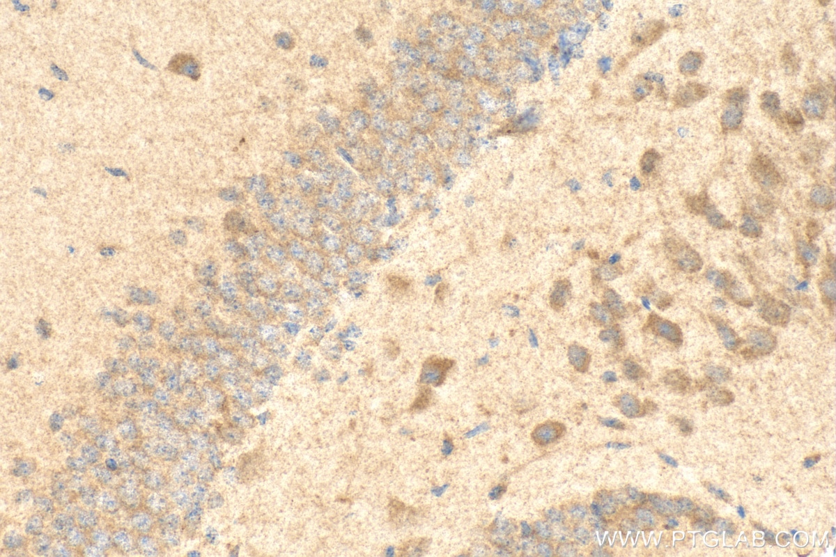
| Positive IHC detected in | mouse brain tissue Note: suggested antigen retrieval with TE buffer pH 9.0; (*) Alternatively, antigen retrieval may be performed with citrate buffer pH 6.0 |
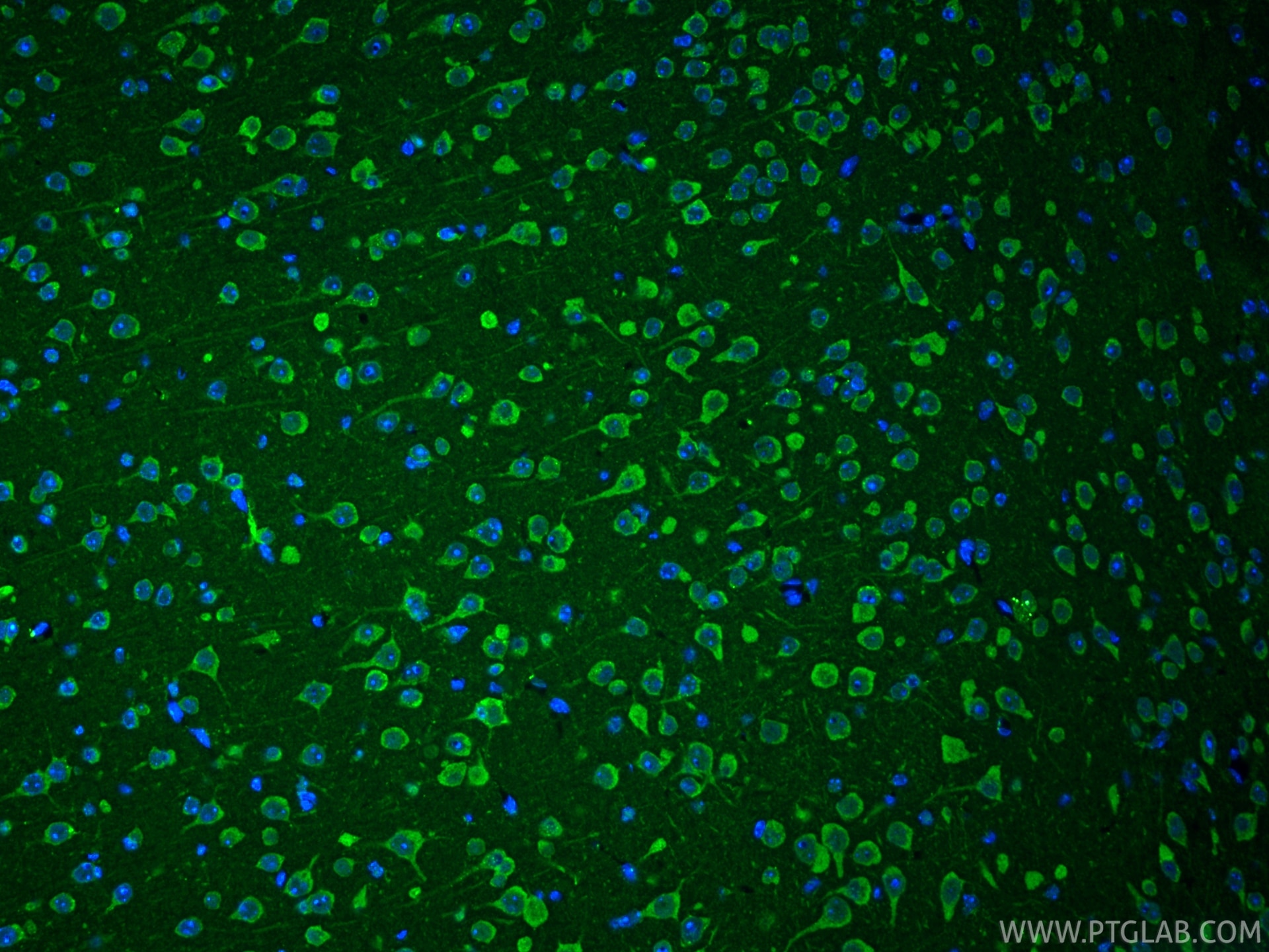
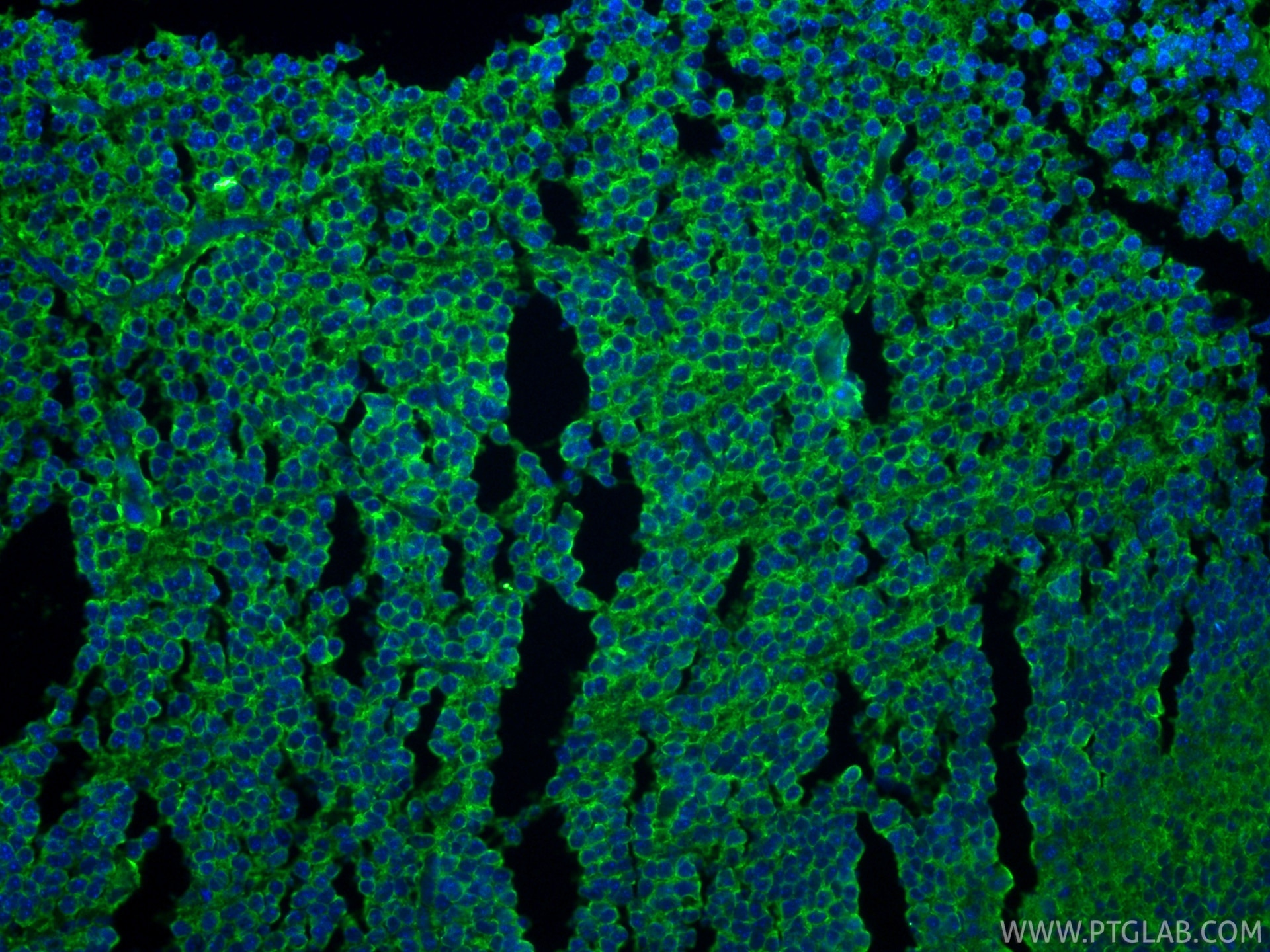
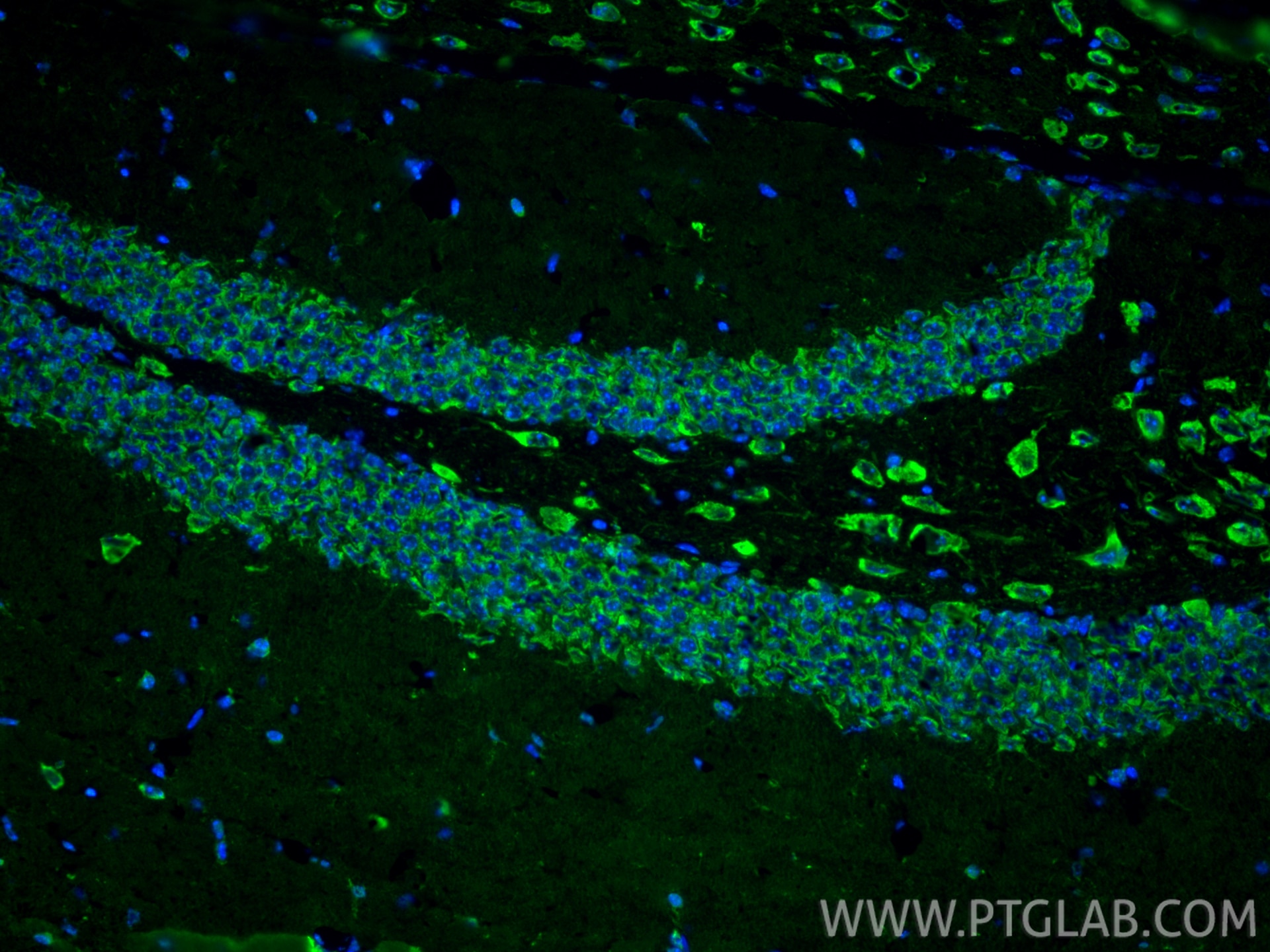
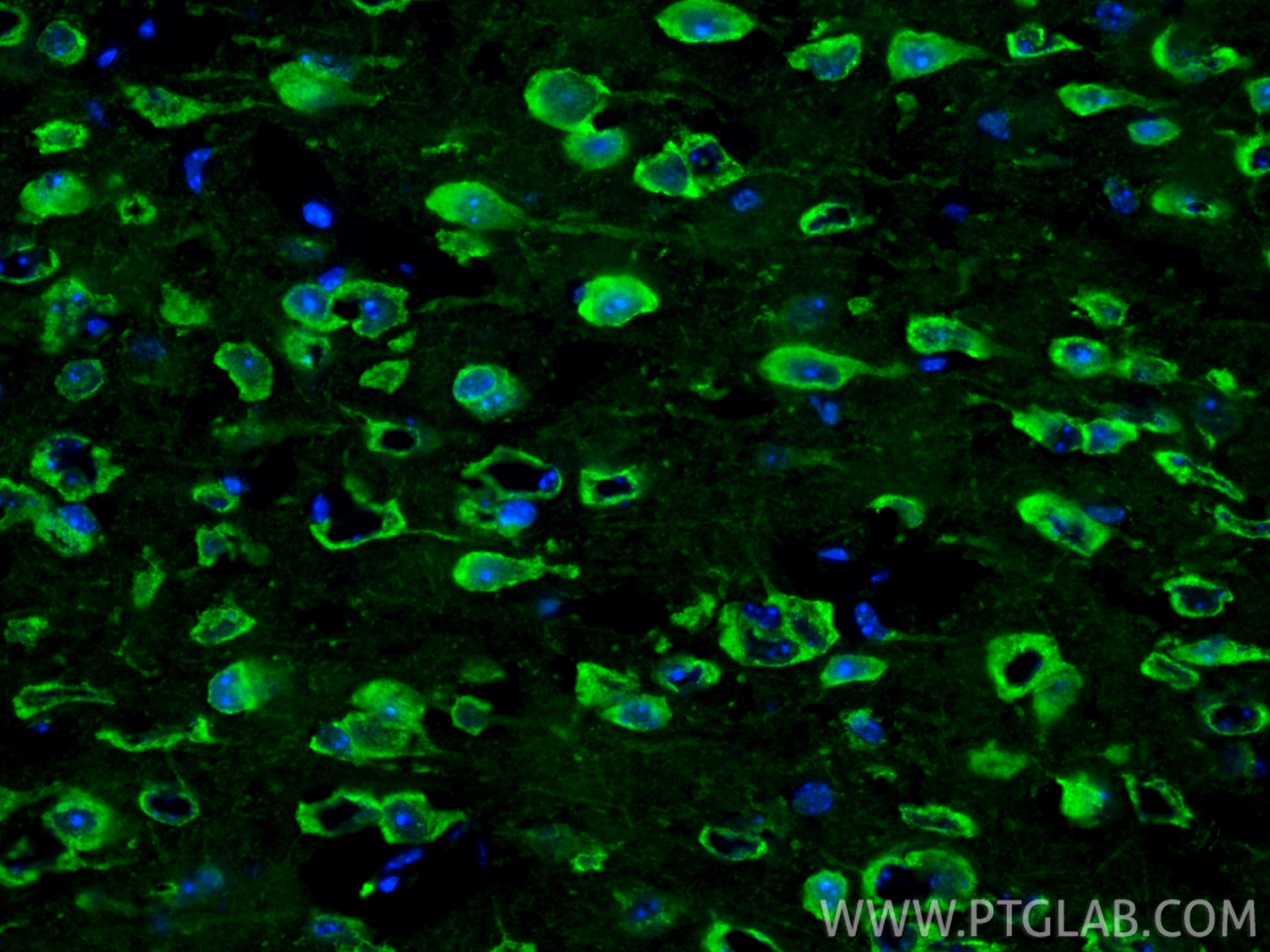
| Positive IF-P detected in | mouse brain tissue, mouse embryo tissue |
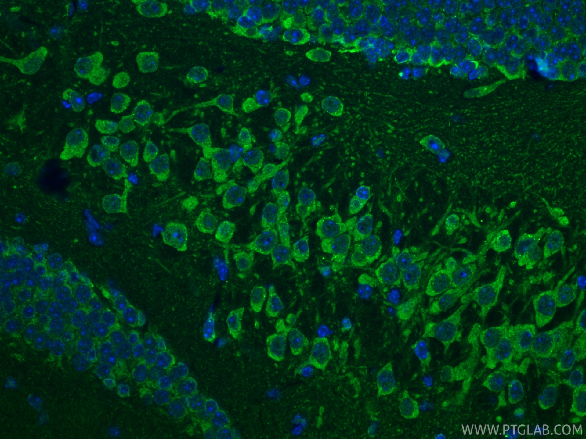
| Positive IF-Fro detected in | mouse brain tissue |
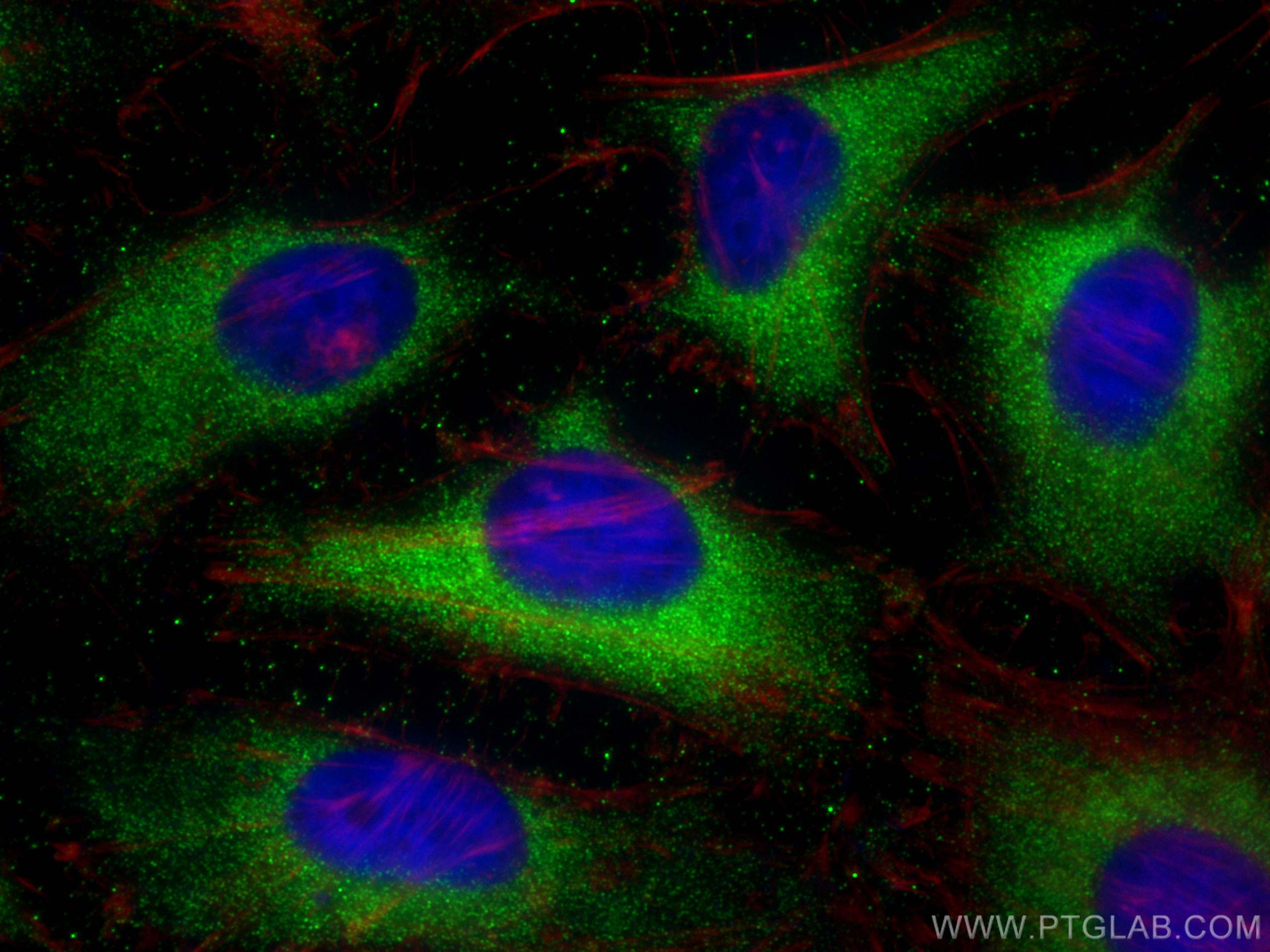
| Positive IF/ICC detected in | HeLa cells |
Recommended dilution
| Application | Dilution |
|---|---|
| Western Blot (WB) | WB : 1:500-1:2000 |
| Immunohistochemistry (IHC) | IHC : 1:50-1:500 |
| Immunofluorescence (IF)-P | IF-P : 1:50-1:500 |
| Immunofluorescence (IF)-FRO | IF-FRO : 1:50-1:500 |
| Immunofluorescence (IF)/ICC | IF/ICC : 1:50-1:500 |
| It is recommended that this reagent should be titrated in each testing system to obtain optimal results. | |
| Sample-dependent, Check data in validation data gallery. | |
Product Information
25128-1-AP targets Cleaved Caspase 3 in WB, IHC, IF/ICC, IF-P, IF-Fro, ELISA, Immunofluorescence, Western Blot applications and shows reactivity with human, mouse samples.
| Tested Reactivity | human, mouse |
| Cited Reactivity | human, mouse, rat, pig, rabbit, canine, chicken, hamster, goat |
| Host / Isotype | Rabbit / IgG |
| Class | Polyclonal |
| Type | Antibody |
| Immunogen |
Peptide Predict reactive species |
| Full Name | caspase 3, apoptosis-related cysteine peptidase |
| Calculated Molecular Weight | 32 kDa |
| Observed Molecular Weight | 17-25 kDa |
| GenBank Accession Number | NM_032991 |
| Gene Symbol | Caspase 3 |
| Gene ID (NCBI) | 836 |
| RRID | AB_3073913 |
| Conjugate | Unconjugated |
| Form | Liquid |
| Purification Method | Antigen affinity purification |
| UNIPROT ID | P42574 |
| Storage Buffer | PBS with 0.02% sodium azide and 50% glycerol, pH 7.3. |
| Storage Conditions | Store at -20°C. Stable for one year after shipment. Aliquoting is unnecessary for -20oC storage. 20ul sizes contain 0.1% BSA. |
Background Information
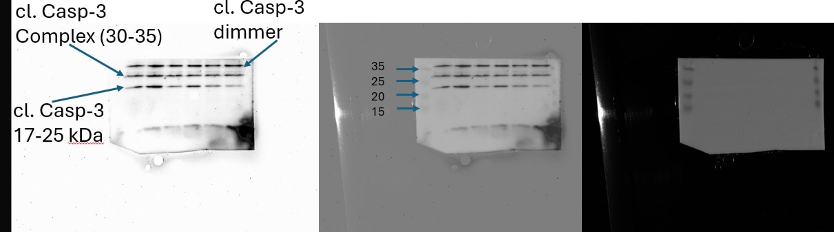
Caspases, a family of endoproteases, are critical players in cell regulatory networks controlling inflammation and cell death. Initiator caspases (caspase-2, -8, -9, -10, -11, and -12) cleave and activate downstream effector caspases (caspase-3, -6, and -7), which in turn execute apoptosis by cleaving targeted cellular proteins. Caspase 3 plays a key role in the activation of sterol regulatory element binding proteins (SREBPs) between the basic helix-loop-helix leucine zipper domain and the membrane attachment domain. Caspase 3 can form heterocomplex with other proteins. This antibody can recognize the cleaved Caspase 3 fragments. This antibody is specific for cleaved caspase 3, and does not recognize full length caspase-3. The cleaved fragment of Caspase 3 might form complex and shows at around 30-35 kDa by western blot (PMID: 25501826).
Protocols
| Product Specific Protocols | |
|---|---|
| IF protocol for Cleaved Caspase 3 antibody 25128-1-AP | Download protocol |
| IHC protocol for Cleaved Caspase 3 antibody 25128-1-AP | Download protocol |
| WB protocol for Cleaved Caspase 3 antibody 25128-1-AP | Download protocol |
| Standard Protocols | |
|---|---|
| Click here to view our Standard Protocols |
Publications
What published studies show
The Cleaved Caspase 3 Polyclonal antibody (Cat# 25128-1-AP) has 694 total citations. Key journals include Nature Communications, Advanced Science, Cell Reports, Autophagy, Neuron, ACS Nano, Biomaterials, Redox Biology, Cancer Research, and Signal Transduction and Targeted Therapy. Research fields encompass oncology (liver, lung, breast, colorectal, renal cancers), programmed cell death (apoptosis, ferroptosis, cuproptosis, PANoptosis), autophagy/mitophagy, neuroscience, cardiology, nephrology, inflammation, oxidative stress, and nanomedicine.
| Species | Application | Title |
|---|---|---|
Signal Transduct Target Ther Activation of Nerve Growth Factor signaling limits the response to lenvatinib in hepatocellular carcinoma | ||
Signal Transduct Target Ther Single-cell identifies and validates human circulating Treg subtype/state Treg(fci) in non-small cell lung cancer. | ||
Adv Mater Piezoelectric Ceramic Nanofiber Aerogels Direct Neutrophil Fate for Diabetic Tissue Regeneration. | ||
Bioact Mater Integrated apoptotic extracellular vesicle-recruitment peptide coating reprograms the diabetic bone microenvironment and orchestrates enhanced implant osseointegration. | ||
Cancer Res Tumor-Resident Streptococcus pneumoniae Promotes Malignant Progression and Pazopanib Resistance in Clear Cell Renal Cell Carcinoma | ||
Drug Resist Updat ATP-binding cassette subfamily A member 5 suppresses pancreatic ductal adenocarcinoma progression and chemoresistance by promoting β-catenin ubiquitin-dependent degradation. |
Reviews
What customers say
Users rate this antibody highly (average ~4.8/5 across 4 reviews). It performs well in Western Blot, IHC, and IF applications. One user noted it outperformed a comparable CTK antibody, yielding clear signal at 1:1000 dilution where the competitor required 1:250. Staining quality was described as "nice" for IHC/IF on glioblastoma cells at 1/200. WB was validated on mouse liver, HK-2, and MCF-231 cells at dilutions ranging from 1:500 to 1:10000. No negative feedback was reported.
The reviews below have been submitted by verified Proteintech customers who received an incentive for providing their feedback.
Audrey (Verified Customer) (02-10-2026) | Works well, nice staining
|
Kamal (Verified Customer) (01-22-2026) | Liver lysates were subjected to SDS PAGE followed by western blot with 25128-1-AP (Cleaved caspase polyclonal antibody) at dilution of 1:1000 in 1XTBST incubated overnight.
|
Antonijo (Verified Customer) (11-11-2025) | Previously, we used Cl. Casp3 (Asp175) from Cell Signaling Technology and had trouble getting a signal, and only at a dilution of 1:250 were we able to get a signal. Since we use Cl. Casp3 from Proteintech, we are able to get a signal at a dilution of 1:1000 and this antibody gives the quality result we need for our research on HK-2 cell line.
 |
Vignesh (Verified Customer) (09-19-2025) |
|









