Tested Applications
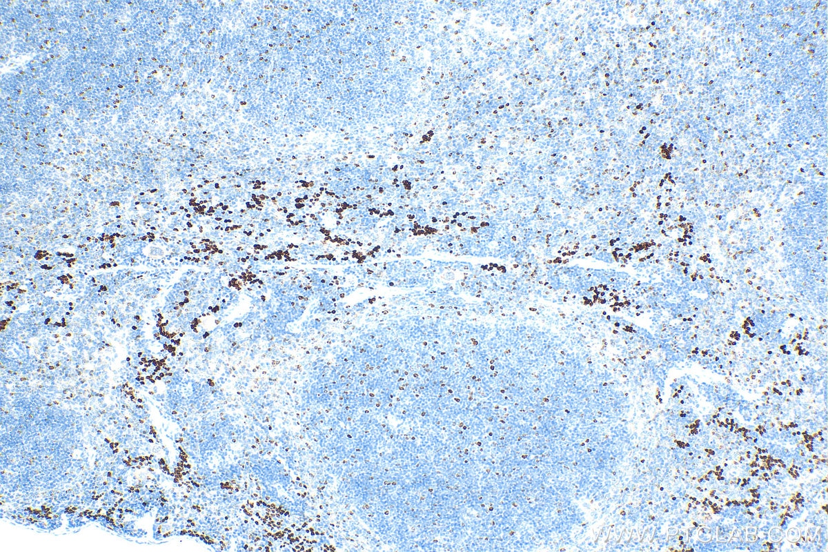
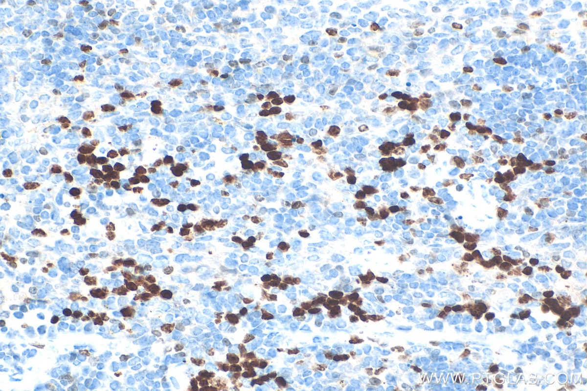
| Positive IHC detected in | mouse spleen tissue Note: suggested antigen retrieval with TE buffer pH 9.0; (*) Alternatively, antigen retrieval may be performed with citrate buffer pH 6.0 |
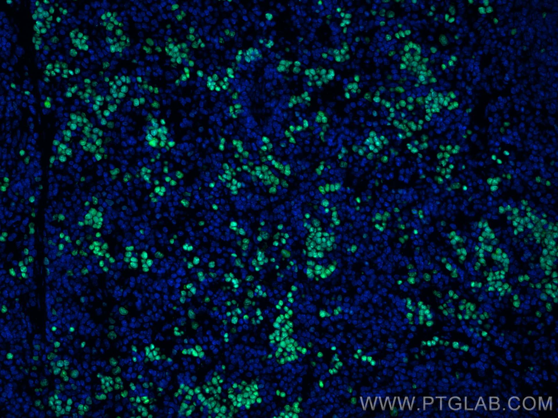
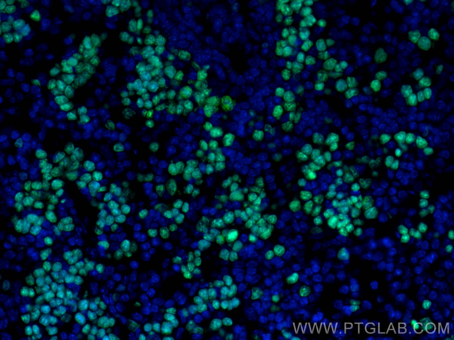
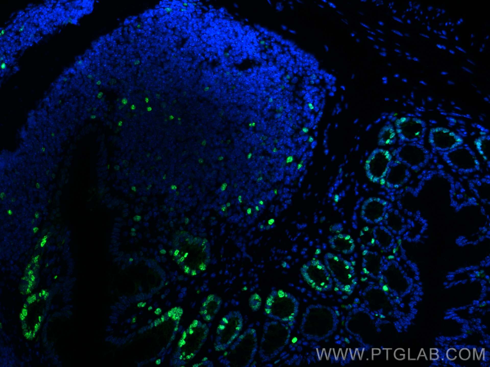
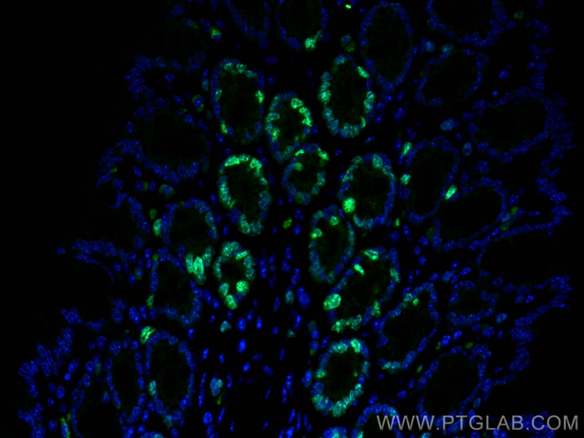
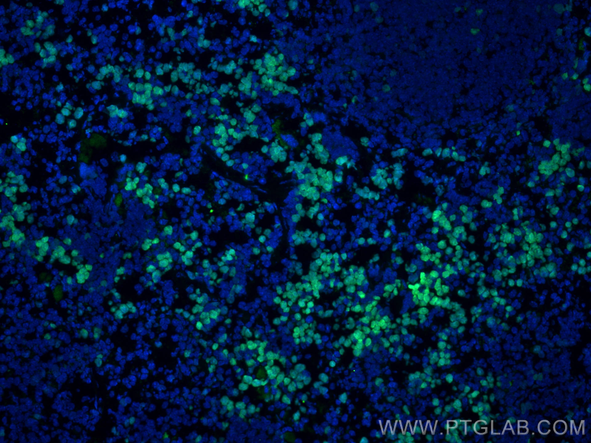
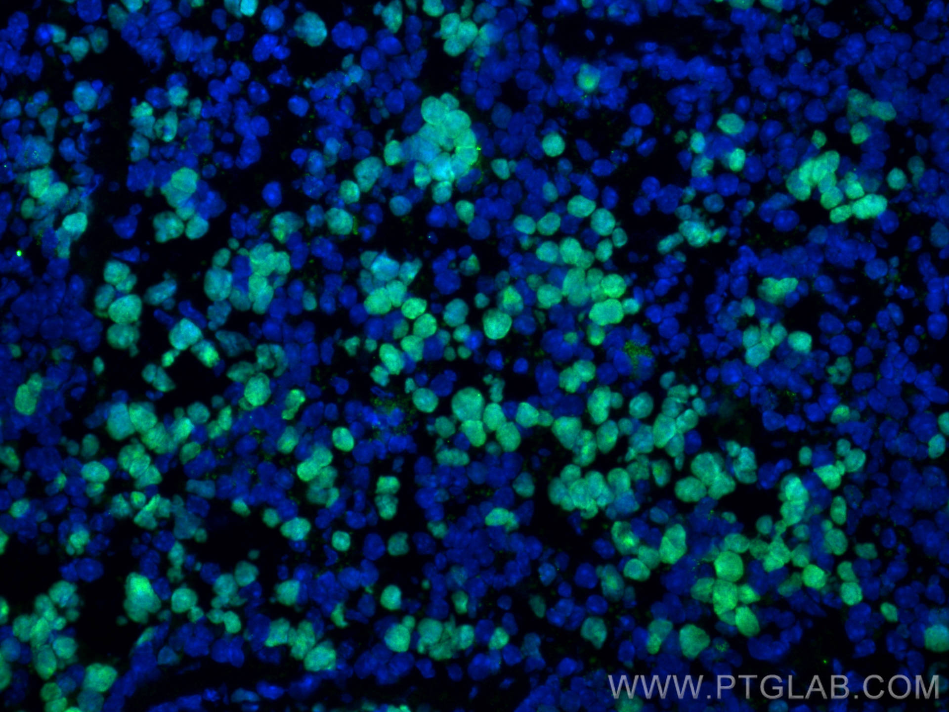
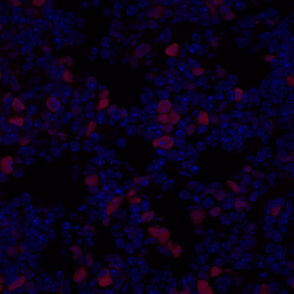
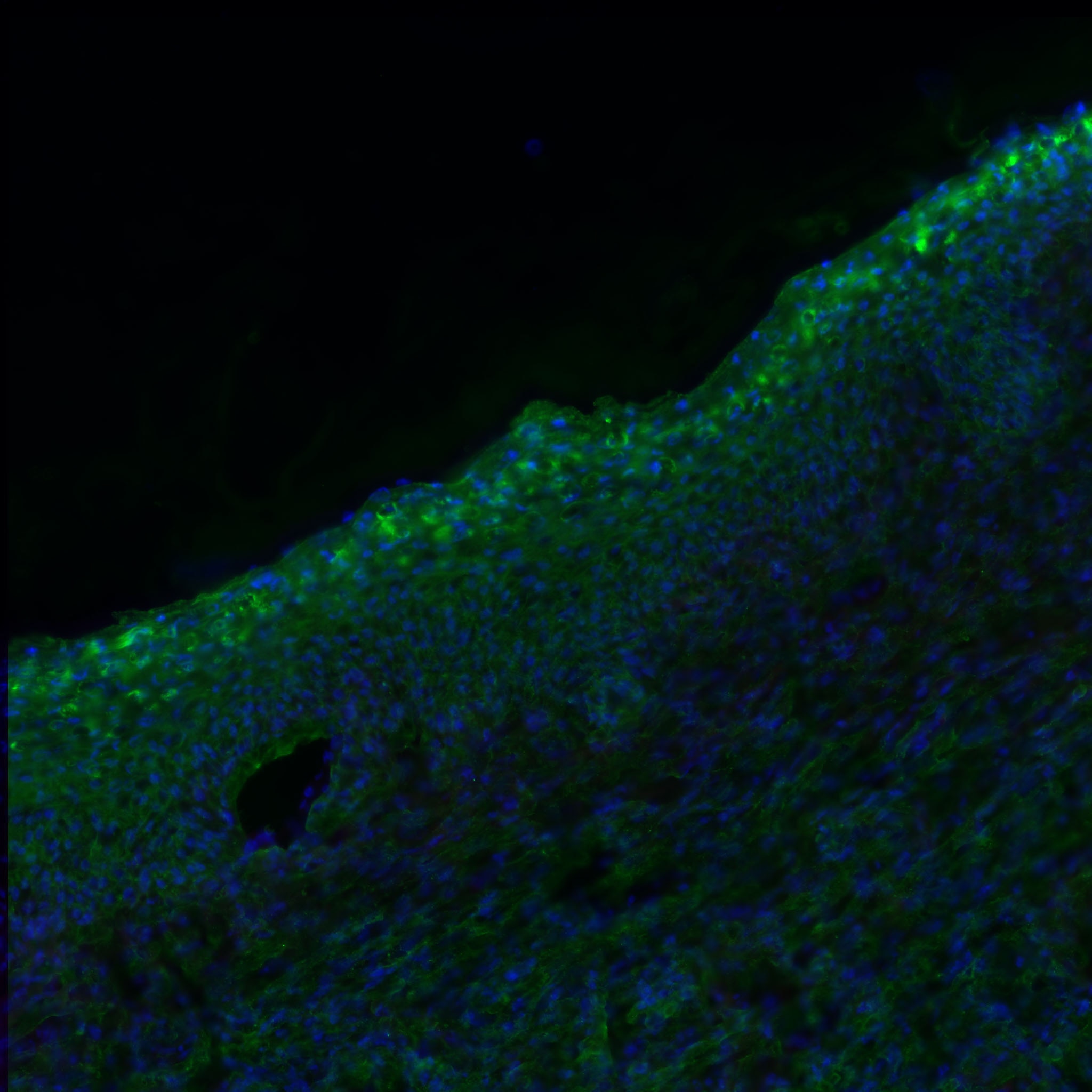
| Positive IF-P detected in | mouse colon tissue, mouse spleen tissue |
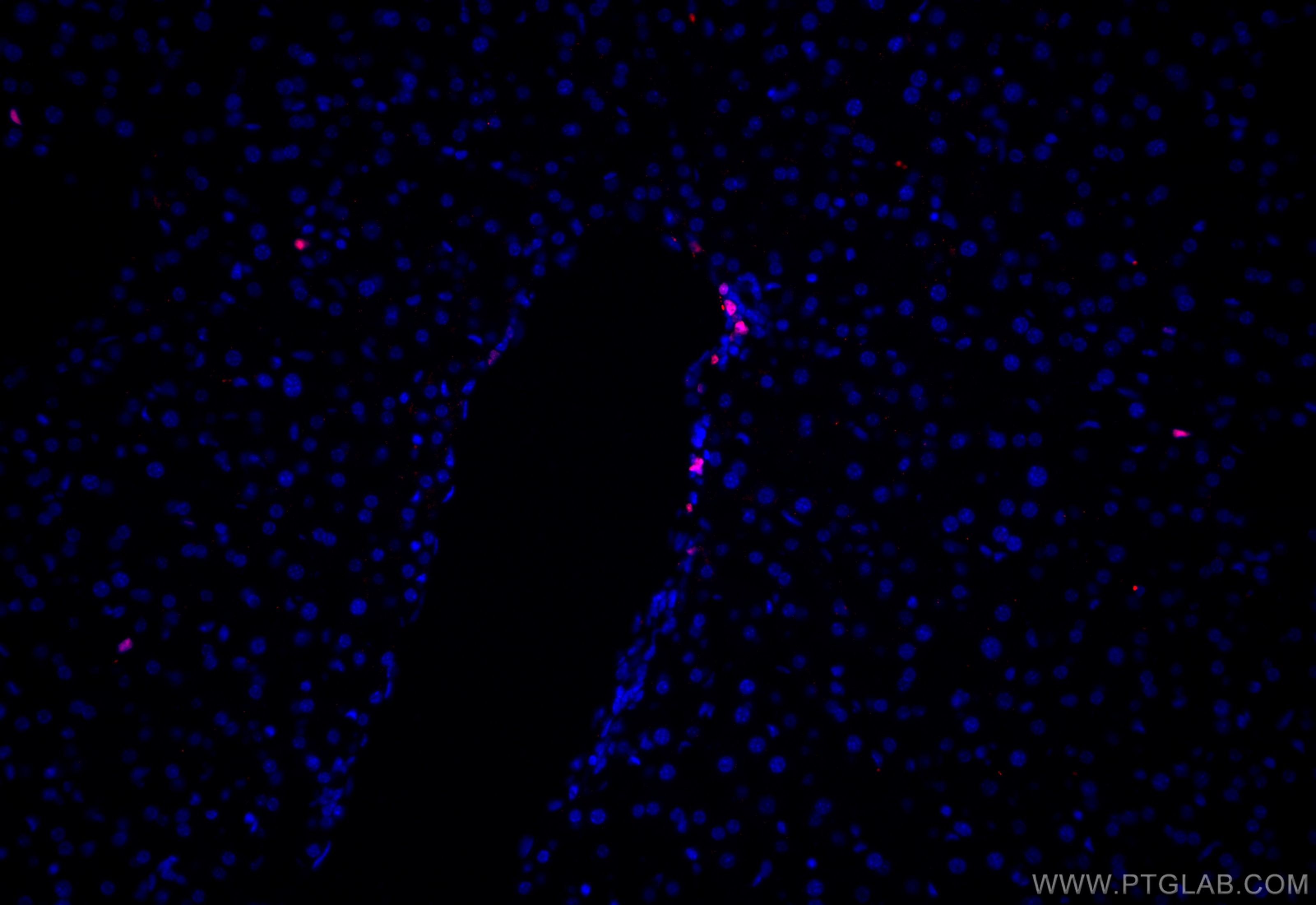
| Positive IF-Fro detected in | mouse spleen tissue, mouse liver tissue |
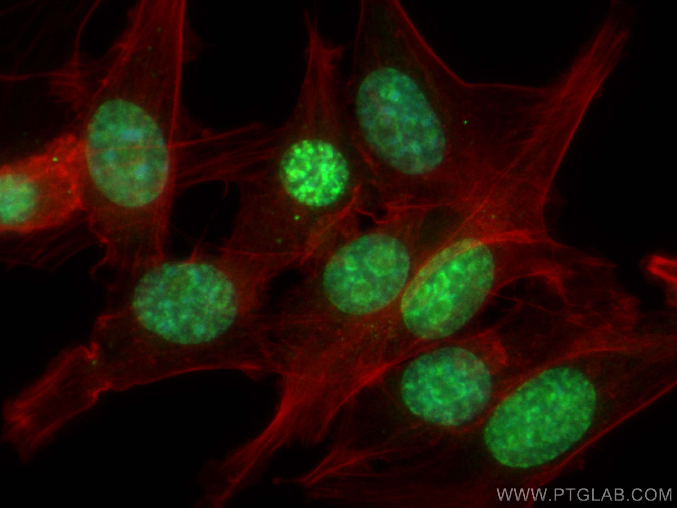
| Positive IF/ICC detected in | NIH/3T3 cells |
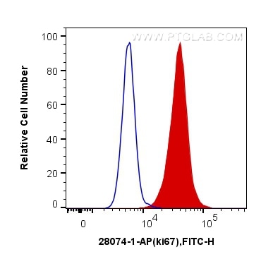
| Positive FC (Intra) detected in | NIH/3T3 cells |
Recommended dilution
| Application | Dilution |
|---|---|
| Immunohistochemistry (IHC) | IHC : 1:2000-1:8000 |
| Immunofluorescence (IF)-P | IF-P : 1:1500-1:6000 |
| Immunofluorescence (IF)-FRO | IF-FRO : 1:50-1:500 |
| Immunofluorescence (IF)/ICC | IF/ICC : 1:400-1:1600 |
| Flow Cytometry (FC) (INTRA) | FC (INTRA) : 0.20 ug per 10^6 cells in a 100 µl suspension |
| It is recommended that this reagent should be titrated in each testing system to obtain optimal results. | |
| Sample-dependent, Check data in validation data gallery. | |
Published Applications
| WB | See 32 publications below |
| IHC | See 197 publications below |
| IF | See 75 publications below |
Product Information
28074-1-AP targets Ki-67 in WB, IHC, IF/ICC, IF-P, IF-Fro, FC (Intra), ELISA, Cell treatment applications and shows reactivity with mouse samples.
| Tested Reactivity | mouse |
| Cited Reactivity | mouse, rat |
| Host / Isotype | Rabbit / IgG |
| Class | Polyclonal |
| Type | Antibody |
| Immunogen |
CatNo: Ag27894 Product name: Recombinant mouse ki67 protein Source: e coli.-derived, PGEX-4T Tag: GST Domain: 2777-3005 aa of NM_001081117 Sequence: MFQSSPRRPRTRRGKVEADEEPSAVRKTVSTSRQTMRSRKVPEIGNNGTQVSKASIKQTLDTVAKVTGSRRQLRTHKDGVQPLEVLGDSKEITQISDHSEKLAHDTSILKSTQQQKPDSVKPLRTCRRVLRASKEDPKEVLVDTRDHATLQSKSNPLLSPKRKSARDGSIVRTRALRSLAPKQEATDEKPVPEKKRAASSKRHVSPEPVKMKHLKIVSNKLESVEEQVST Predict reactive species |
| Full Name | antigen identified by monoclonal antibody Ki 67 |
| Calculated Molecular Weight | 351 kDa |
| GenBank Accession Number | NM_001081117 |
| Gene Symbol | ki67 |
| Gene ID (NCBI) | 17345 |
| RRID | AB_2918145 |
| Conjugate | Unconjugated |
| Form | Liquid |
| Purification Method | Antigen affinity purification |
| UNIPROT ID | E9PVX6 |
| Storage Buffer | PBS with 0.02% sodium azide and 50% glycerol, pH 7.3. |
| Storage Conditions | Store at -20°C. Stable for one year after shipment. Aliquoting is unnecessary for -20oC storage. 20ul sizes contain 0.1% BSA. |
Background Information
The Ki-67 protein (also known as MKI67) is a cellular marker for proliferation. Ki67 is present during all active phases of the cell cycle (G1, S, G2 and M), but is absent in resting cells (G0). Cellular content of Ki-67 protein markedly increases during cell progression through S phase of the cell cycle. Therefore, the nuclear expression of Ki67 can be evaluated to assess tumor proliferation by immunohistochemistry.
Protocols
| Product Specific Protocols | |
|---|---|
| FC protocol for Ki-67 antibody 28074-1-AP | Download protocol |
| IF protocol for Ki-67 antibody 28074-1-AP | Download protocol |
| IHC protocol for Ki-67 antibody 28074-1-AP | Download protocol |
| Standard Protocols | |
|---|---|
| Click here to view our Standard Protocols |
Publications
| Species | Application | Title |
|---|---|---|
Drug Resist Updat Lansoprazole (LPZ) reverses multidrug resistance (MDR) in cancer through impeding ATP-binding cassette (ABC) transporter-mediated chemotherapeutic drug efflux and lysosomal sequestration | ||
Cell Mol Immunol Targeting PHGDH reverses the immunosuppressive phenotype of tumor-associated macrophages through α-ketoglutarate and mTORC1 signaling | ||
Bioact Mater Biomimetic gender-specific human skin model based on gonads/epidermis-on-a-chip | ||
Neuron C5aR1+ microglia exacerbate neuroinflammation and cerebral edema in acute brain injury | ||
Biosens Bioelectron 3D co-culture of macrophages and fibroblasts in a sessile drop array for unveiling the role of macrophages in skin wound-healing | ||
Adv Healthc Mater A Multifunctional Nanodrug Increases the Therapeutic Sensitivity of Lenvatinib to Hepatocellular Carcinoma by Inhibiting the Stemness of Hepatic Cancer Stem Cells |
Reviews
The reviews below have been submitted by verified Proteintech customers who received an incentive for providing their feedback.
FH Tatiana (Verified Customer) (06-02-2026) | Used for PFA-fixed frozen murine lymph node sections. Incubation was 15 min at room temperature, followed by 1 h incubation with the secondary antibody. The staining worked well.
 |
FH Kamal (Verified Customer) (01-22-2026) | Antibody was diluted 1:200 in 1X PBS.
|
FH YINGJIAN (Verified Customer) (11-15-2025) | The antibody produced clear signals with negligible background and high target specificity.
 |
FH Ruchi (Verified Customer) (06-08-2023) | Stained Ki67 on murine diabetic skin
 |