"Alpha Tubulin Antibodies" Comparison
View side-by-side comparison of Alpha Tubulin antibodies from other vendors to find the one that best suits your research needs.
Tested Applications
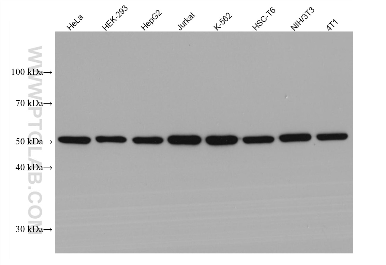
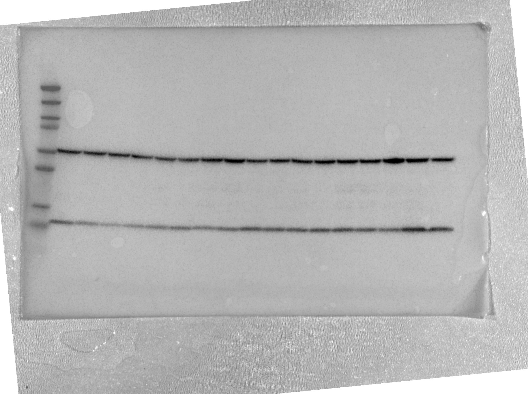
| Positive WB detected in | HeLa cells, HEK-293 cells, HepG2 cells, Jurkat cells, K-562 cells, HSC-T6 cells, NIH/3T3 cells, 4T1 cells |
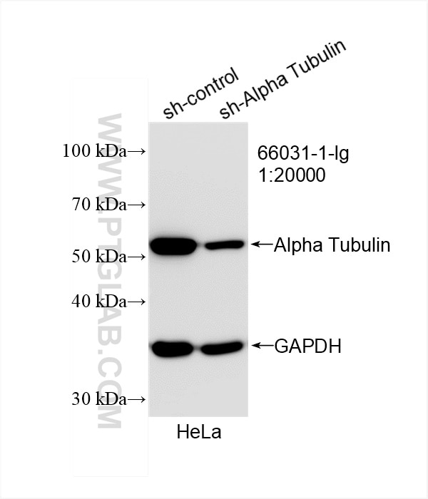
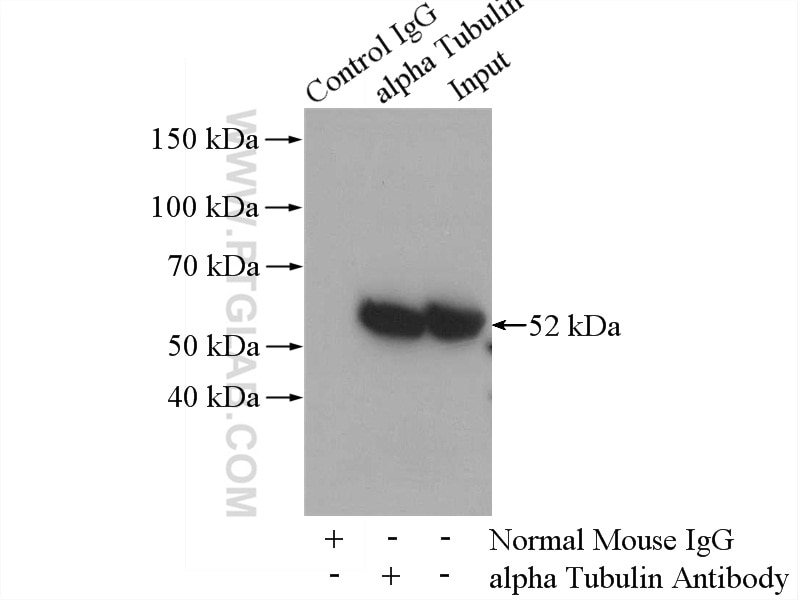
| Positive IP detected in | HeLa cells |
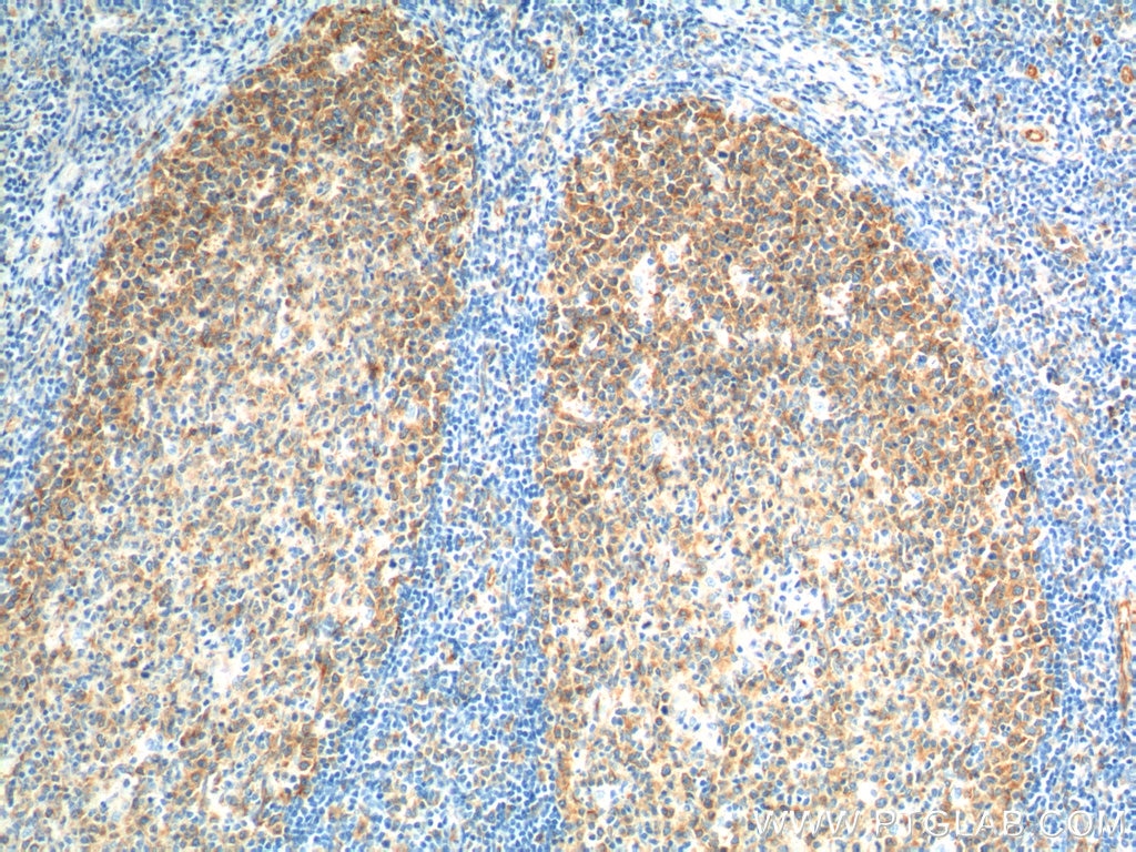
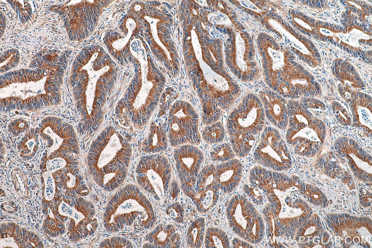
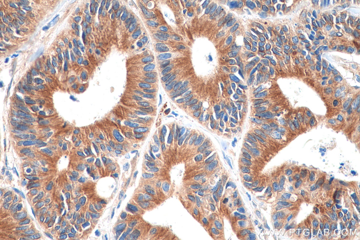
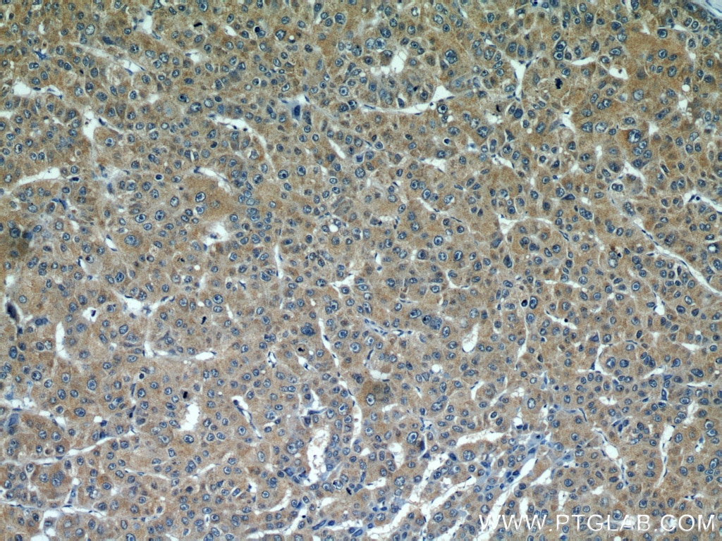
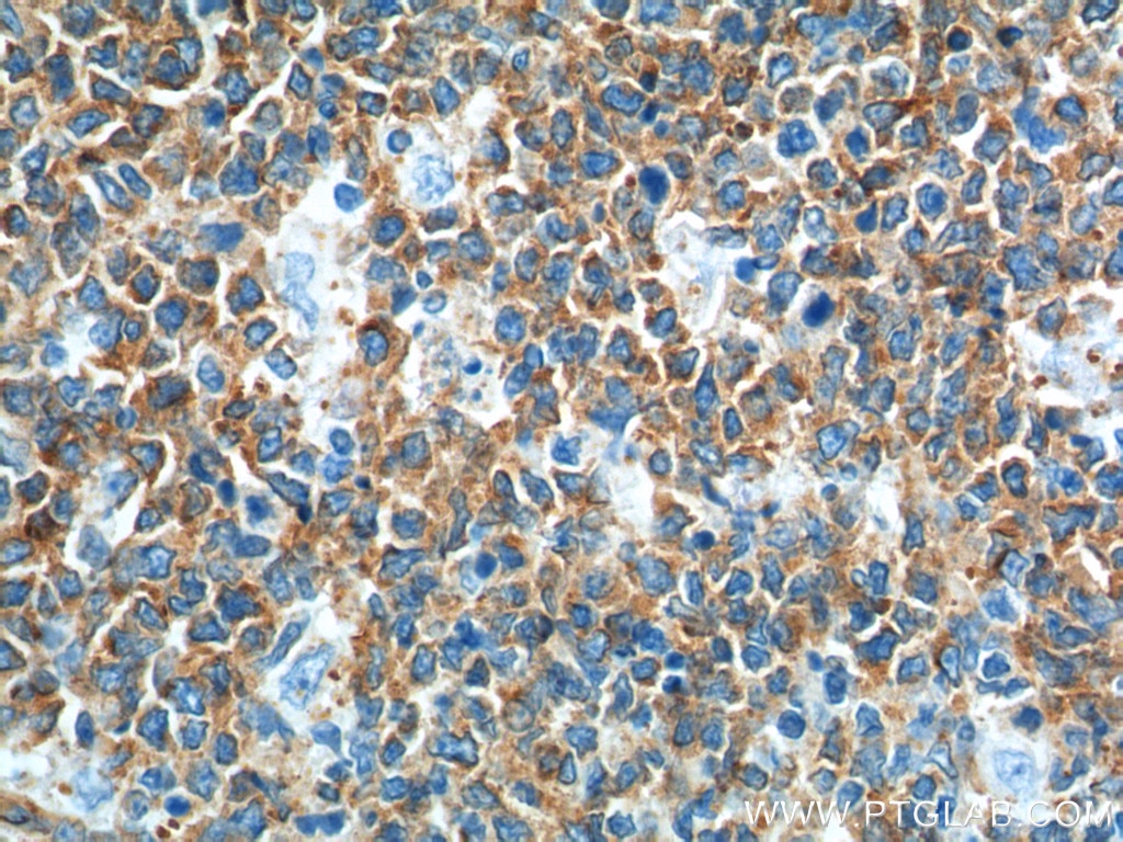
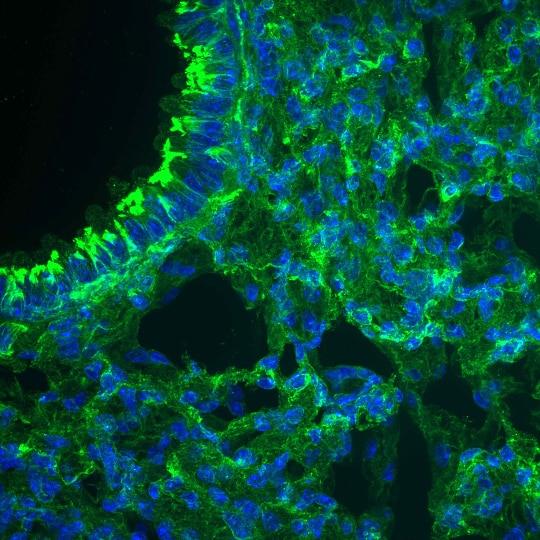
| Positive IHC detected in | human tonsillitis tissue, human liver cancer tissue, human colon cancer tissue Note: suggested antigen retrieval with TE buffer pH 9.0; (*) Alternatively, antigen retrieval may be performed with citrate buffer pH 6.0 |
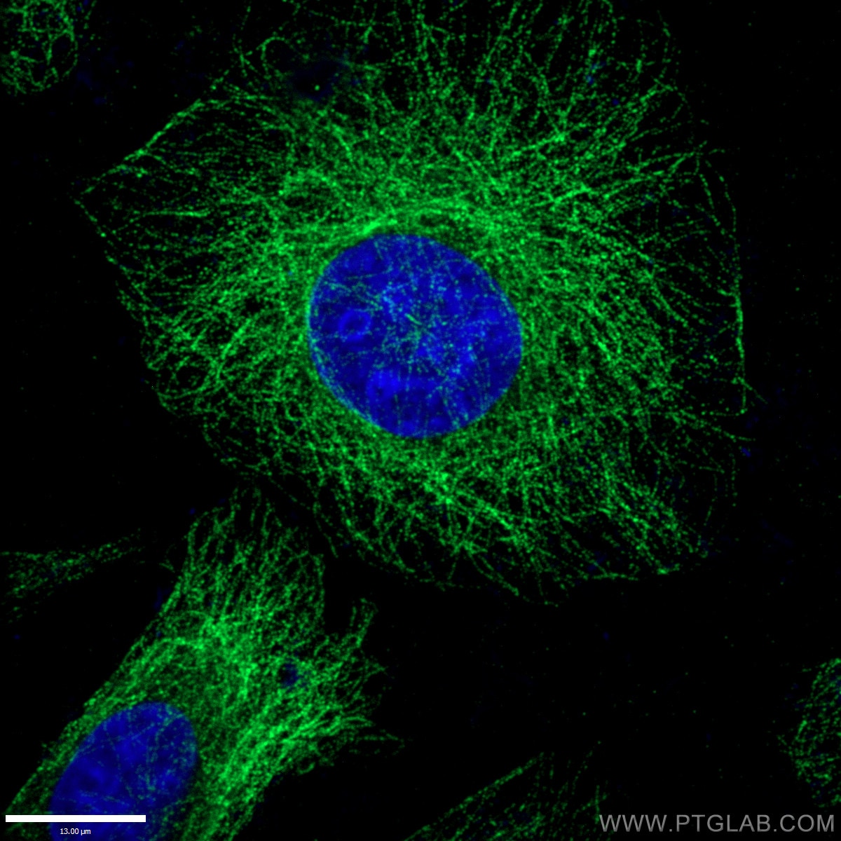
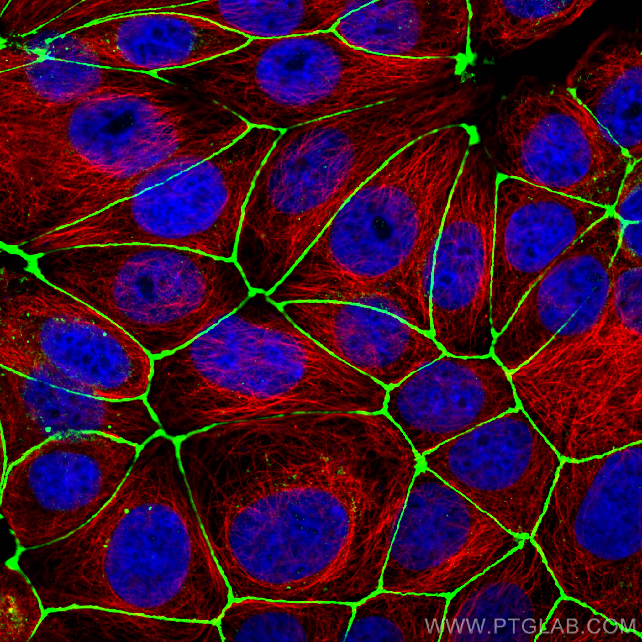
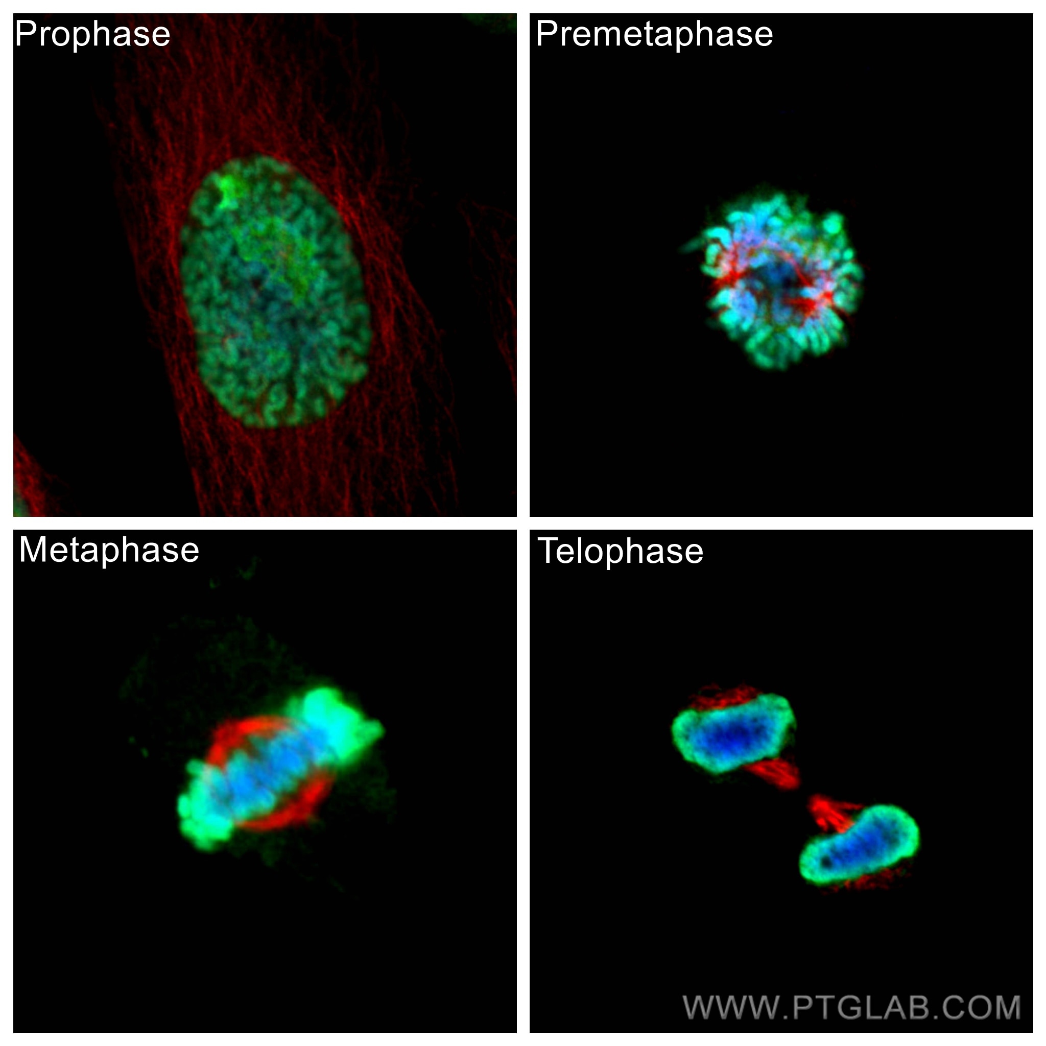
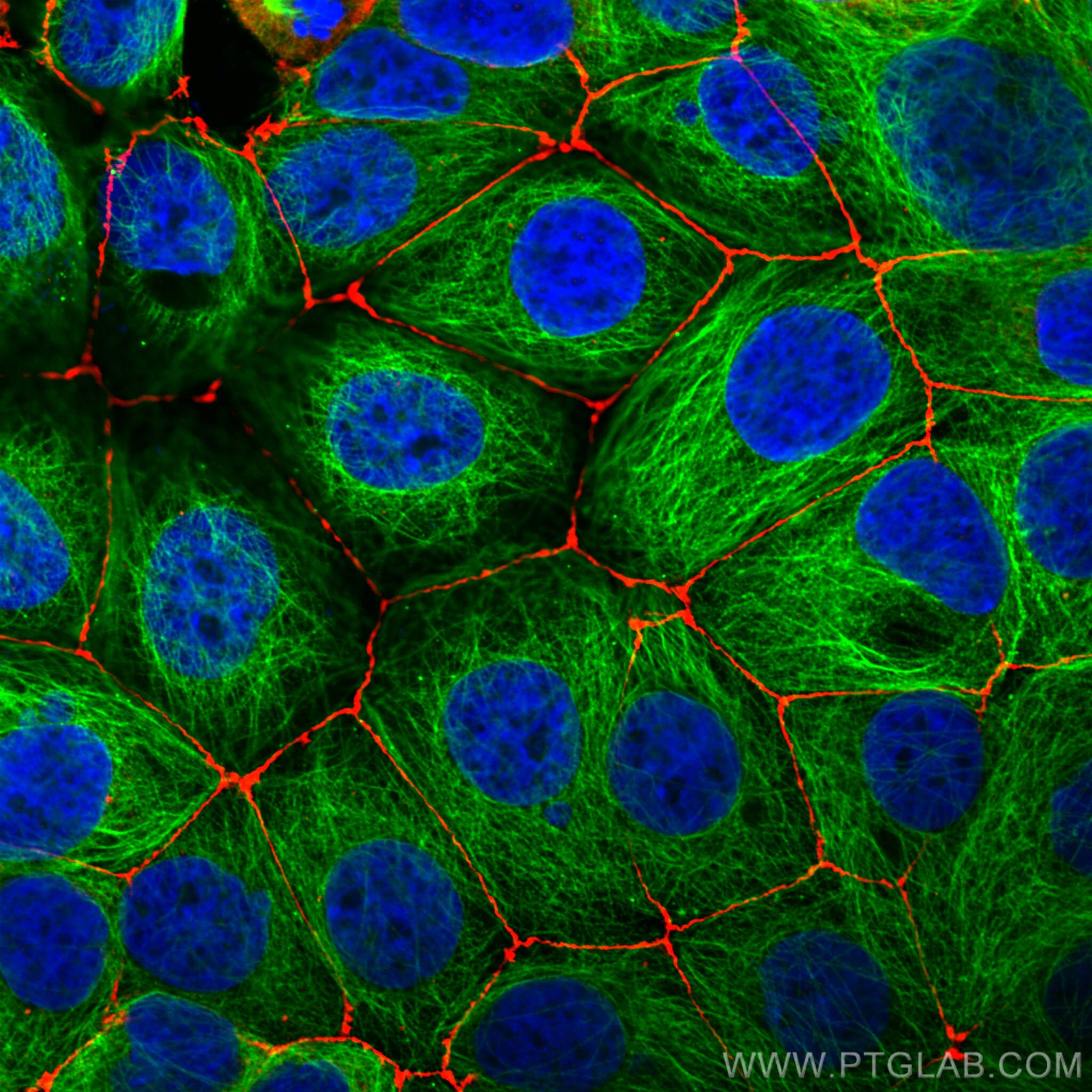
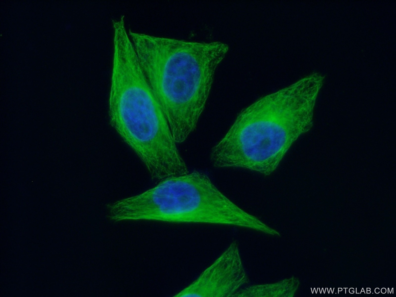
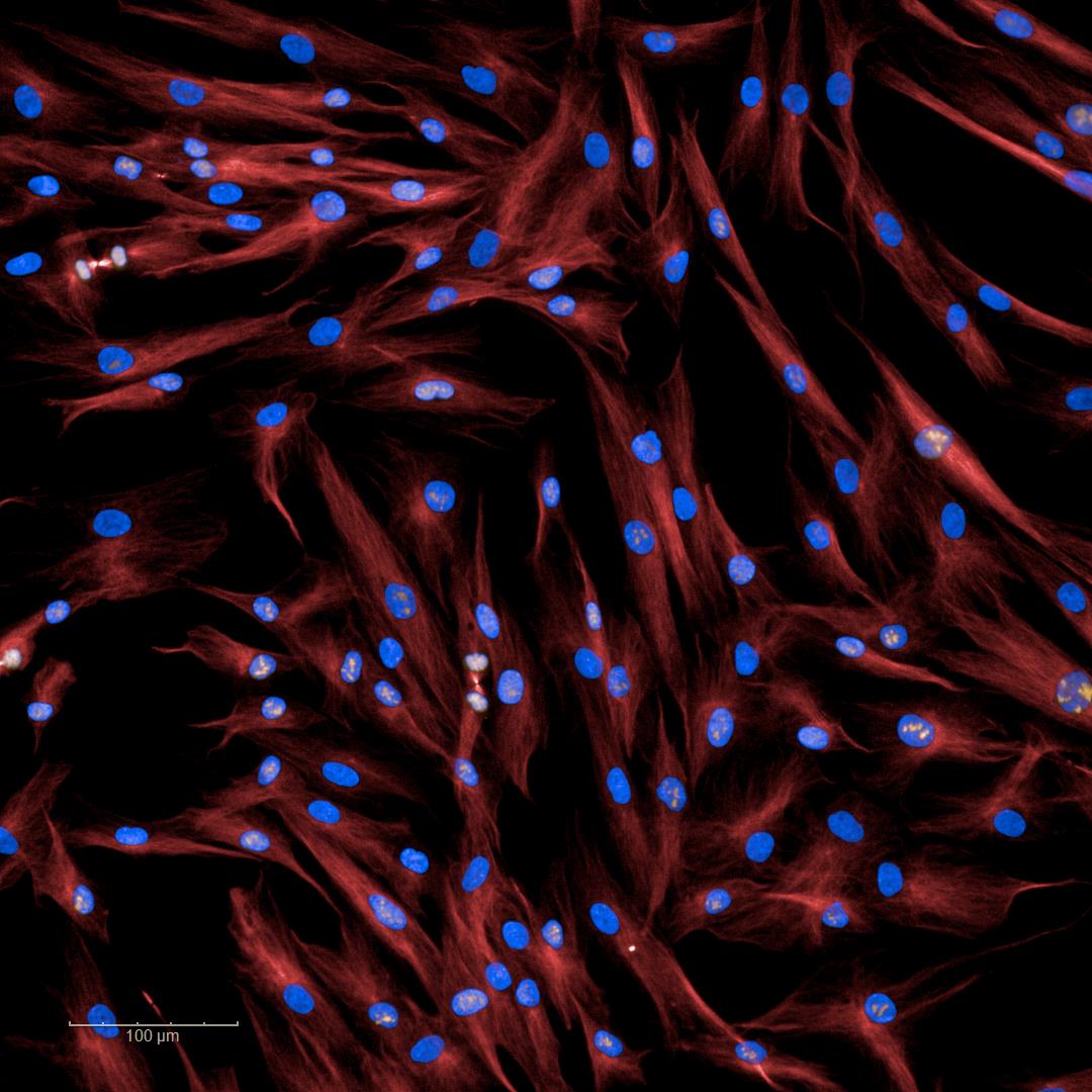
| Positive IF/ICC detected in | HeLa cells, HepG2 cells, MCF-7 cells |
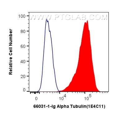
| Positive FC (Intra) detected in | HeLa cells |
Recommended dilution
| Application | Dilution |
|---|---|
| Western Blot (WB) | WB : 1:20000-1:100000 |
| Immunoprecipitation (IP) | IP : 0.5-4.0 ug for 1.0-3.0 mg of total protein lysate |
| Immunohistochemistry (IHC) | IHC : 1:200-1:1000 |
| Immunofluorescence (IF)/ICC | IF/ICC : 1:500-1:2000 |
| Flow Cytometry (FC) (INTRA) | FC (INTRA) : 0.40 ug per 10^6 cells in a 100 µl suspension |
| It is recommended that this reagent should be titrated in each testing system to obtain optimal results. | |
| Sample-dependent, Check data in validation data gallery. | |
Published Applications
| KD/KO | See 3 publications below |
| WB | See 1553 publications below |
| IHC | See 1 publications below |
| IF | See 133 publications below |
| IP | See 10 publications below |
| CoIP | See 5 publications below |
Product Information
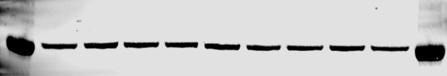
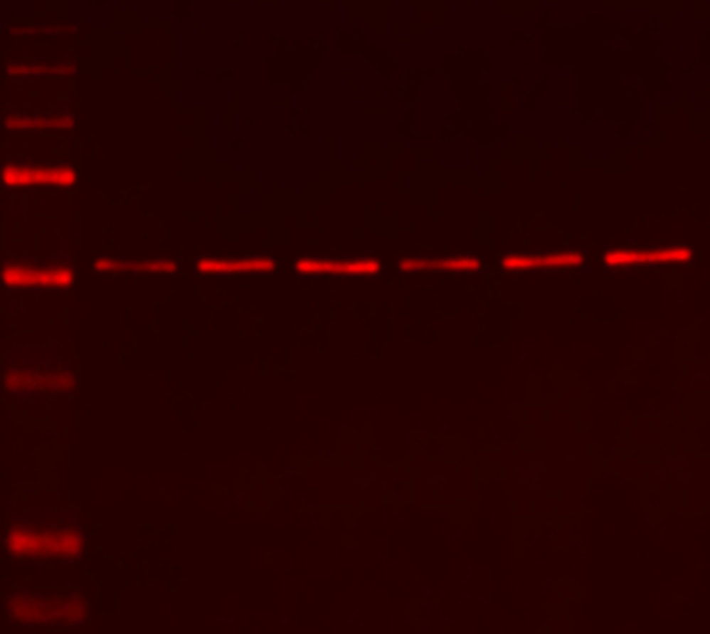
66031-1-Ig targets Alpha Tubulin in WB, IHC, IF/ICC, FC (Intra), IP, CoIP, ELISA applications and shows reactivity with human, mouse, rat, canine samples.
| Tested Reactivity | human, mouse, rat, canine |
| Cited Reactivity | human, mouse, rat, rabbit, monkey, chicken, sheep, goat, tick |
| Host / Isotype | Mouse / IgG2b |
| Class | Monoclonal |
| Type | Antibody |
| Immunogen |
CatNo: Ag18034 Product name: Recombinant human Tubulin-Alpha protein Source: e coli.-derived, PET28a Tag: 6*His Domain: 1-451 aa of BC009314 Sequence: MRECISIHVGQAGVQIGNACWELYCLEHGIQPDGQMPSDKTIGGGDDSFNTFFSETGAGKHVPRAVFVDLEPTVIDEVRTGTYRQLFHPEQLITGKEDAANNYARGHYTIGKEIIDLVLDRIRKLADQCTGLQGFLVFHSFGGGTGSGFTSLLMERLSVDYGKKSKLEFSIYPAPQVSTAVVEPYNSILTTHTTLEHSDCAFMVDNEAIYDICRRNLDIERPTYTNLNRLISQIVSSITASLRFDGALNVDLTEFQTNLVPYPRIHFPLATYAPVISAEKAYHEQLSVAEITNACFEPANQMVKCDPRHGKYMACCLLYRGDVVPKDVNAAIATIKTKRSIQFVDWCPTGFKVGINYQPPTVVPGGDLAKVQRAVCMLSNTTAIAEAWARLDHKFDLMYAKRAFVHWYVGEGMEEGEFSEAREDMAALEKDYEEVGVDSVEGEGEEEGEEY Predict reactive species |
| Full Name | tubulin, alpha 1b |
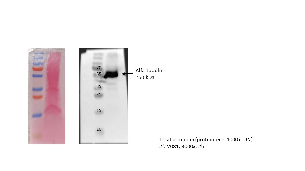
| Calculated Molecular Weight | 50 kDa |
| Observed Molecular Weight | 50-55 kDa |
| GenBank Accession Number | BC009314 |
| Gene Symbol | Alpha Tubulin |
| Gene ID (NCBI) | 10376 |
| RRID | AB_11042766 |
| Conjugate | Unconjugated |
| Form | Liquid |
| Purification Method | Protein A purification |
| UNIPROT ID | P68363 |
| Storage Buffer | PBS with 0.02% sodium azide and 50% glycerol, pH 7.3. |
| Storage Conditions | Store at -20°C. Stable for one year after shipment. Aliquoting is unnecessary for -20oC storage. 20ul sizes contain 0.1% BSA. |
Background Information
What is the function of alpha tubulin?
Alpha-tubulin belongs to a large superfamily of tubulin proteins. There are a number of different subtypes that have a molecular weight of ~50kDa and are able to bind to beta-tubulin, forming a heterodimer that polymerises to microtubules as part of the cytoskeleton. These maintain cell structure, provide platforms for intracellular transport and are also involved in cell division.
Where is alpha-tubulin expressed?
Alpha tubulin is highly conserved and is present in nearly all eukaryotic cells as one of the building blocks of microtubules. The ubiquitous nature of this protein has led to its common use as a control protein for many tissue types as well as highlighting the structure of the cytoskeleton.
What are the post-translational modifications of alpha tubulin?
The function and properties of microtubules are drastically affected by the post-translational modifications undergone by tubulin, which may occur to the tubulin dimer directly or to the polymerised mictotubule. For example, the first modification to be identified was detyrosination1, as most alpha-tubulins have a tyrosine at their terminus. This process affects microtubules more than dimers and leads to patches of detyronisation along the structure, regulating protein interactions and allowing subcellular compartments to be defined.2,3 Polyglutamylation also occurs on several sites within the carboxy-terminal tails. However, to date, the most-studied alpha tubulin modification is related to acetylation of lysine 40 (K40).
1. Gundersen, G. G., Khawaja, S. & Bulinski, J. C. Postpolymerization detyrosination of alpha-tubulin: a mechanism for subcellular differentiation of microtubules. J. Cell Biol. 105, 251-64 (1987).
2. Galjart, N. Plus-End-Tracking Proteins and Their Interactions at Microtubule Ends. Curr. Biol. 20, R528-R537 (2010).
3. Jiang, K. & Akhmanova, A. Microtubule tip-interacting proteins: a view from both ends. Curr. Opin. Cell Biol. 23, 94-101 (2011).
Protocols
| Product Specific Protocols | |
|---|---|
| FC protocol for Alpha Tubulin antibody 66031-1-Ig | Download protocol |
| IF protocol for Alpha Tubulin antibody 66031-1-Ig | Download protocol |
| IHC protocol for Alpha Tubulin antibody 66031-1-Ig | Download protocol |
| IP protocol for Alpha Tubulin antibody 66031-1-Ig | Download protocol |
| WB protocol for Alpha Tubulin antibody 66031-1-Ig | Download protocol |
| Standard Protocols | |
|---|---|
| Click here to view our Standard Protocols |
Publications
| Species | Application | Title |
|---|---|---|
Nat Biotechnol Magnify is a universal molecular anchoring strategy for expansion microscopy | ||
Cell Res Mitochondria-localized cGAS suppresses ferroptosis to promote cancer progression | ||
Nature Activity-based E3 ligase profiling uncovers an E3 ligase with esterification activity. |
Reviews
The reviews below have been submitted by verified Proteintech customers who received an incentive for providing their feedback.
FH MALLIKARJUNA (Verified Customer) (10-24-2025) | works well for western blot
|
FH Matthieu (Verified Customer) (09-24-2025) | Band is very clear at the expected size
|
FH Bruna (Verified Customer) (07-17-2025) | worked very nice
 |
FH Jianhua (Verified Customer) (05-28-2025) | Works very well for western blot
|
FH Paula (Verified Customer) (02-06-2025) | Works well. Not affected by treatments, therefore reliable loading control. (The band below is TSPO)
 |
FH Hannah (Verified Customer) (12-02-2024) | Fantastic specific signal with a 1h primary inclubtion and Licor secondary antibody and imaging.
 |
FH Scott (Verified Customer) (10-22-2024) | 20µg of protein was loaded and antibody was incubated overnight at 4oC following a total protein stain. The band appeared at the expected size. Precision plus protein standard ladder #1610373.
 |
FH Bartosz (Verified Customer) (08-05-2024) | Great detection, clear bands, and no problem with these antibodies, I strongly recommend them!
|
FH Alexandra (Verified Customer) (03-07-2024) | Extremely efficient!
 |
FH Chiara (Verified Customer) (10-02-2023) | This antibody worked quite well in our immunofluorescence experiments
 |
FH Udesh (Verified Customer) (08-16-2023) | Worked well for WB at 1:2000
|
FH Amy (Verified Customer) (08-10-2023) | Clean strong bands for western blot on HEK293T lysates.
|
FH Kalin (Verified Customer) (03-12-2022) | I think this antibody works great for western blots.
|
FH Silvia (Verified Customer) (02-18-2022) | This antibody worked quite well in our western blotting and it comes at a great price.
|
FH Azita (Verified Customer) (06-02-2021) | Immunohistochemistry labelling of (4% PFA) fixed mice spinal cord tissues using Alpha Tubulin Monoclonal Antibody at dilution of 1:50 showed strong labelling.
|
FH Scooby (Verified Customer) (10-05-2020) | happy
|
FH Lauren (Verified Customer) (10-05-2020) | thanks
|
FH Chun (Verified Customer) (12-05-2019) | An excellent antibody
|