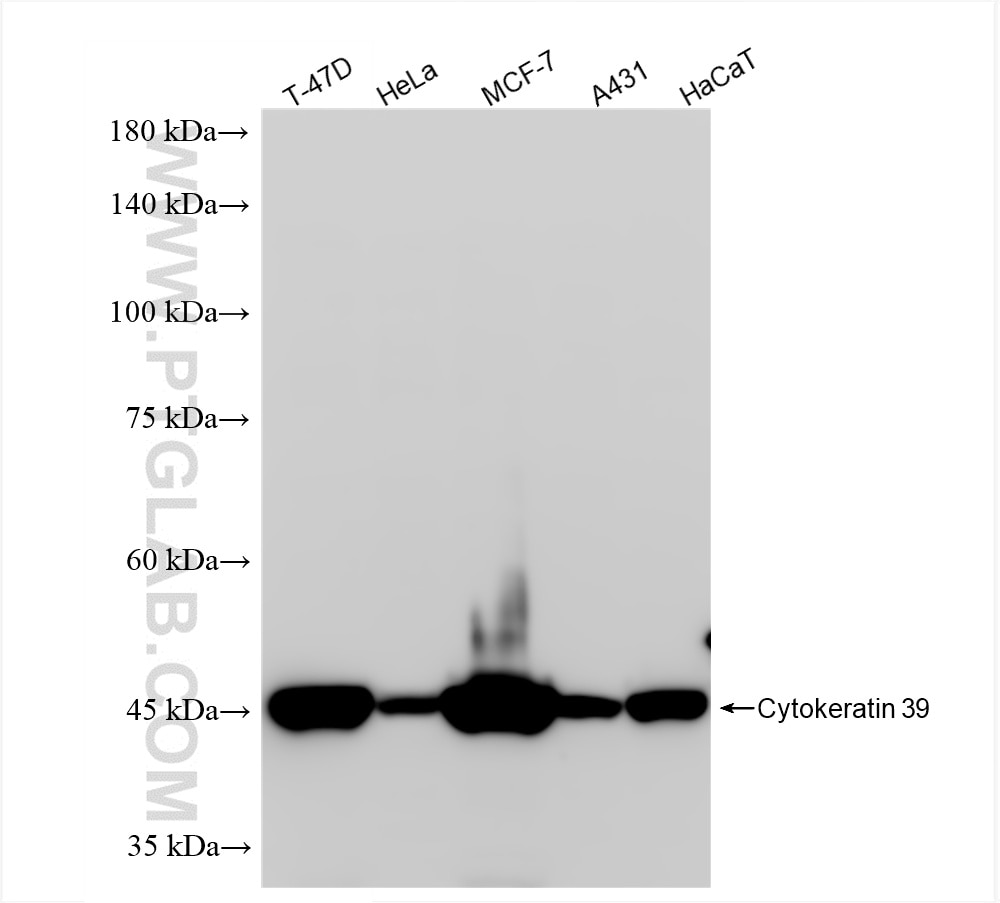
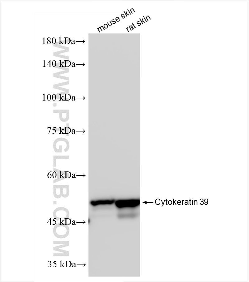
Various lysates were subjected to SDS PAGE followed by western blot with 86277-2-RR (KRT39 antibody) at dilution of 1:5000 incubated at room temperature for 1.5 hours.
Various lysates were subjected to SDS PAGE followed by western blot with 86277-2-RR (KRT39 antibody) at dilution of 1:5000 incubated at room temperature for 1.5 hours.
WB analysis using 86277-2-RR
Various lysates were subjected to SDS PAGE followed by western blot with 86277-2-RR (KRT39 antibody) at dilution of 1:5000 incubated at room temperature for 1.5 hours.
Various lysates were subjected to SDS PAGE followed by western blot with 86277-2-RR (KRT39 antibody) at dilution of 1:5000 incubated at room temperature for 1.5 hours.
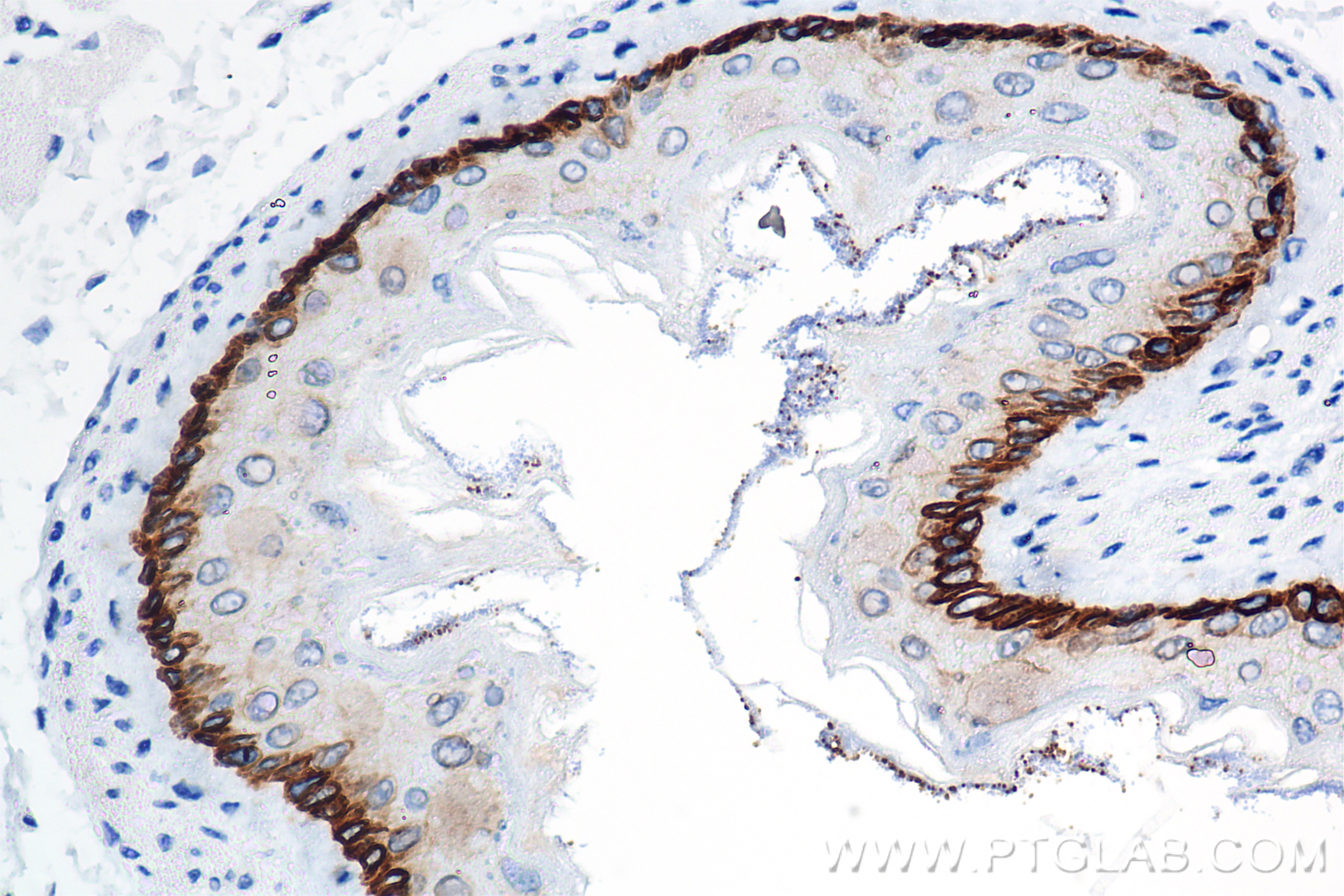
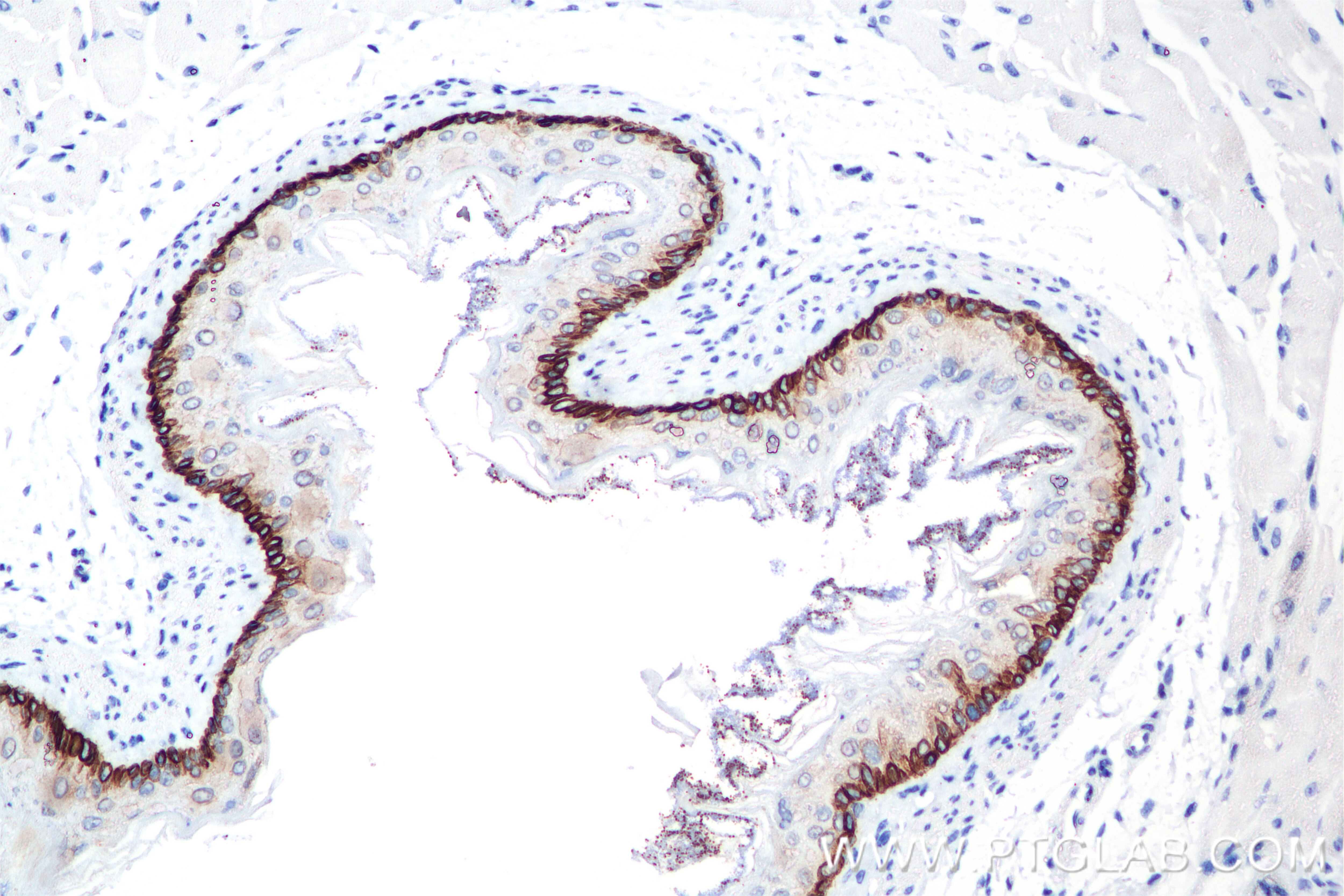
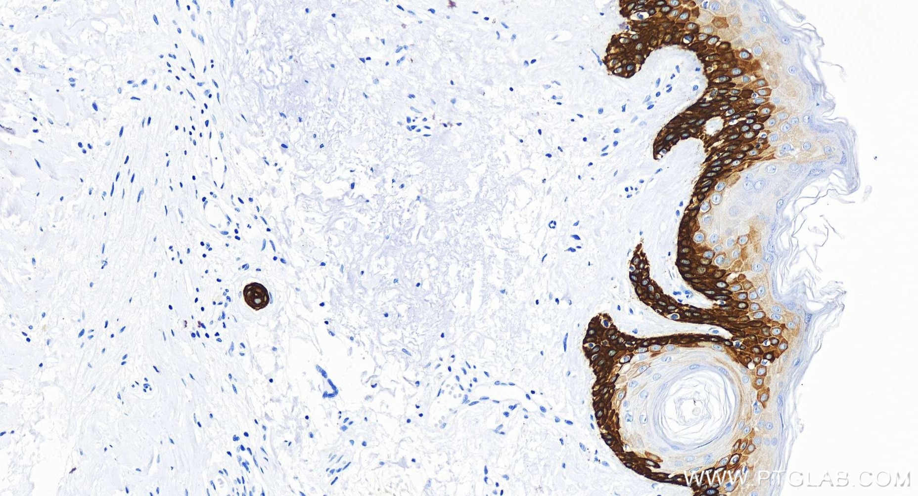
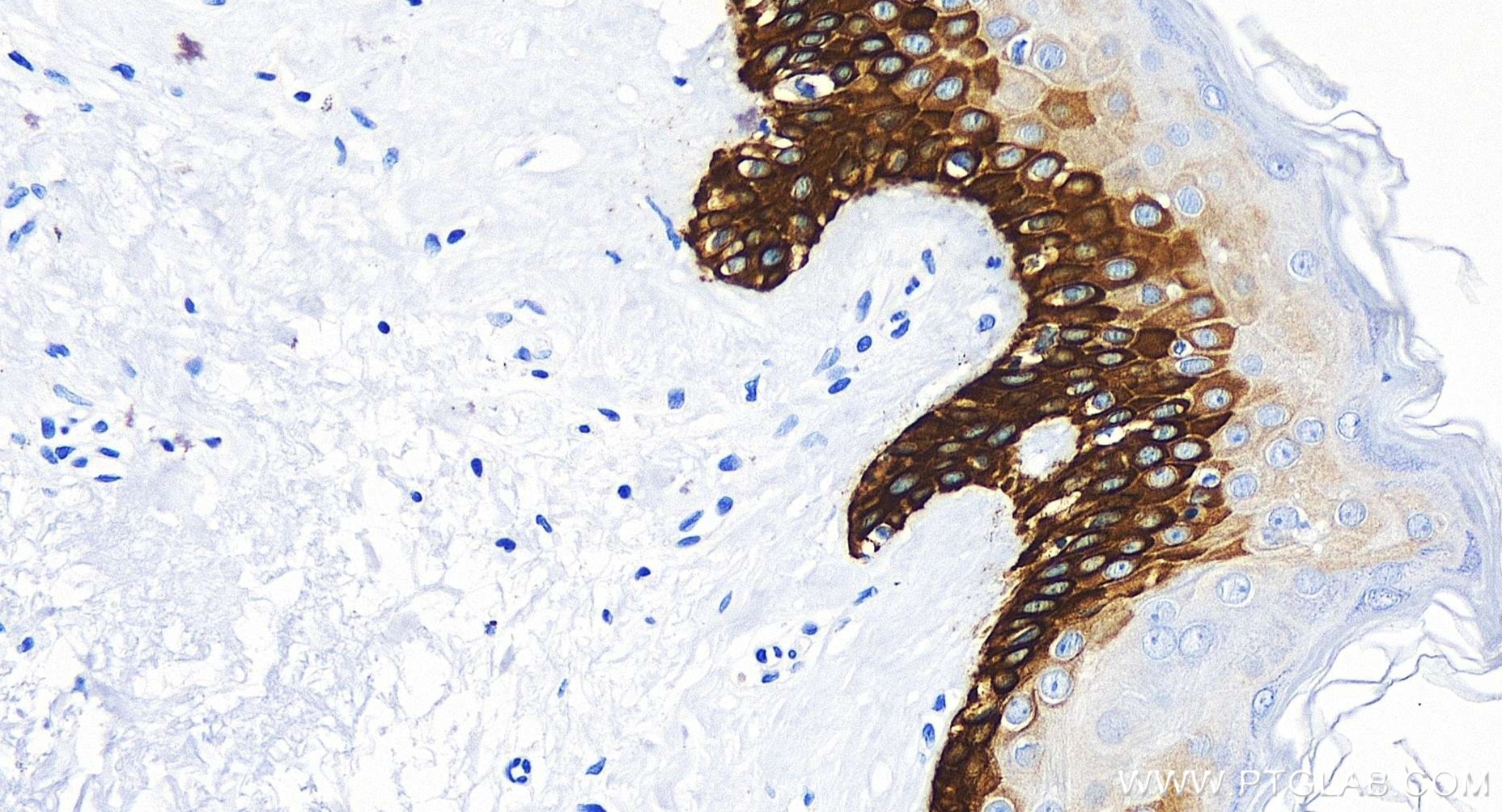
IHC staining of mouse esophagus using 86277-2-RR
Immunohistochemical analysis of paraffin-embedded mouse esophagus tissue slide using 86277-2-RR (Cytokeratin 39 antibody) at dilution of 1:2000 (under 40x lens). Heat mediated antigen retrieval with Tris-EDTA buffer (pH 9.0).
Immunohistochemical analysis of paraffin-embedded mouse esophagus tissue slide using 86277-2-RR (Cytokeratin 39 antibody) at dilution of 1:2000 (under 20x lens). Heat mediated antigen retrieval with Tris-EDTA buffer (pH 9.0).
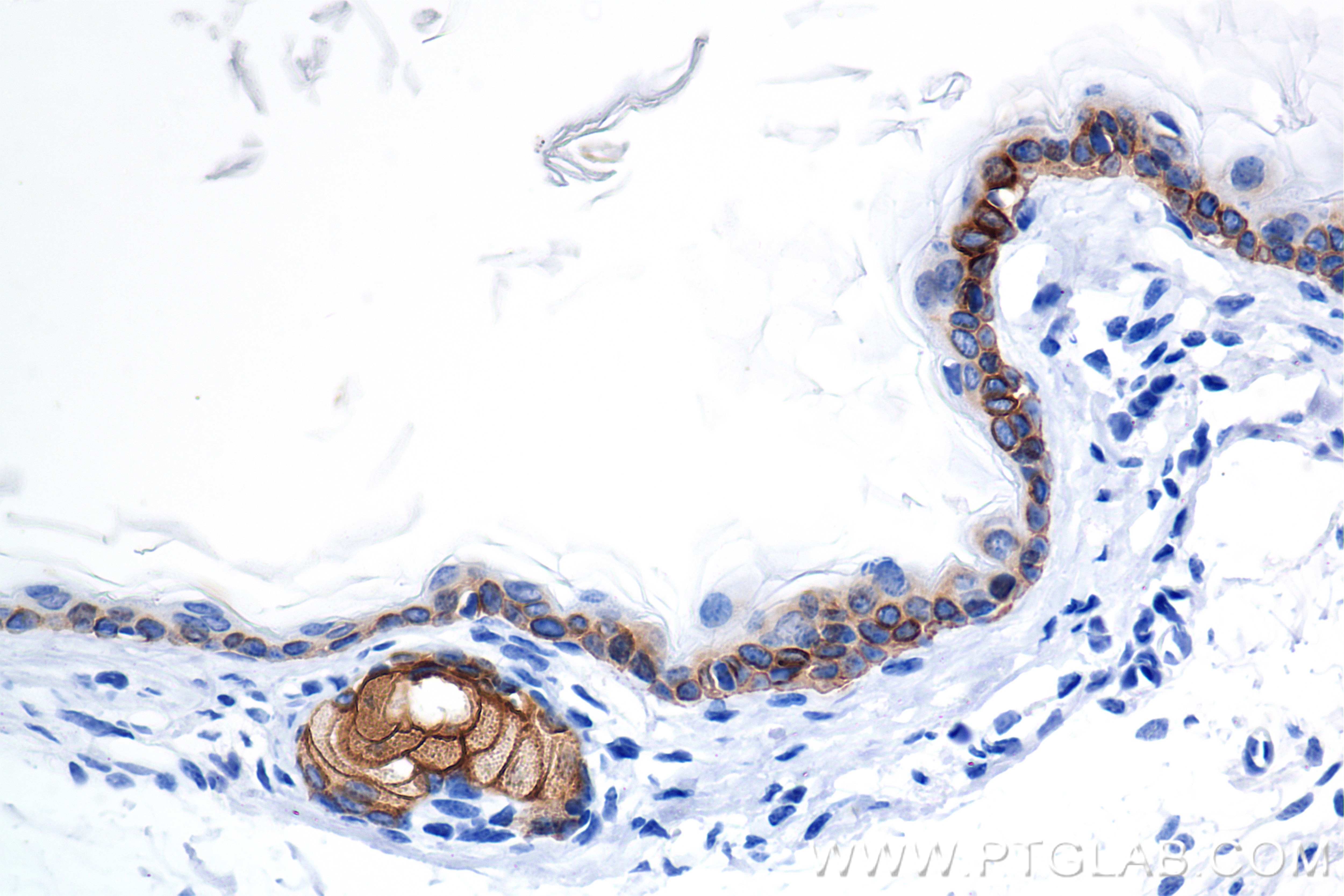
IHC staining of human skin using 86277-2-RR
Immunohistochemical analysis of paraffin-embedded human skin tissue slide using 86277-2-RR (Cytokeratin 39 antibody) at dilution of 1:2000 (under 20x lens). Heat mediated antigen retrieval with Tris-EDTA buffer (pH 9.0).
Immunohistochemical analysis of paraffin-embedded human skin tissue slide using 86277-2-RR (Cytokeratin 39 antibody) at dilution of 1:2000 (under 20x lens). Heat mediated antigen retrieval with Tris-EDTA buffer (pH 9.0).
IHC staining of mouse skin using 86277-2-RR
Immunohistochemical analysis of paraffin-embedded mouse skin tissue slide using 86277-2-RR (Cytokeratin 39 antibody) at dilution of 1:5000 (under 10x lens). Heat mediated antigen retrieval with Tris-EDTA buffer (pH 9.0).
The Proteintech guarantee covers Proteintech antibodies in any species and any application, including those not listed on the datasheet. If the antibody doesn’t perform, you can receive a hassle-free refund or credit note.
T-47D cells, mouse skin tissue, rat skin tissue, HeLa cells, MCF-7 cells, A431 cells, HaCaT cells
Positive IHC detected in
mouse esophagus tissue, human skin tissue, mouse skin tissue Note: suggested antigen retrieval with TE buffer pH 9.0; (*) Alternatively, antigen retrieval may be performed with citrate buffer pH 6.0
Positive IF-P detected in
mouse skin tissue
Positive IF/ICC detected in
MCF-7 cells
Recommended dilution
Application
Dilution
Western Blot (WB)
WB : 1:2000-1:10000
Immunohistochemistry (IHC)
IHC : 1:1000-1:4000
Immunofluorescence (IF)-P
IF-P : 1:50-1:500
Immunofluorescence (IF)/ICC
IF/ICC : 1:50-1:500
It is recommended that this reagent should be titrated in each testing system to obtain optimal results.
Sample-dependent, Check data in validation data gallery.
Product Information
86277-2-RR targets Cytokeratin 39 in WB, IHC, IF/ICC, IF-P, ELISA applications and shows reactivity with human, mouse, rat samples.
PBS with 0.02% sodium azide and 50% glycerol, pH 7.3.
Storage Conditions
Store at -20°C. Stable for one year after shipment. Aliquoting is unnecessary for -20oC storage. 20ul sizes contain 0.1% BSA.
Background Information
Cytokeratin 39 (CK-39) is a member of the cytokeratin family, a group of intermediate filament proteins primarily expressed in epithelial cells. Cytokeratins are classified into Type I (acidic) and Type II (basic) subfamilies, which pair to form heteropolymers essential for maintaining cellular structural integrity, mechanical stability, and stress resistance. CK-39 is predominantly expressed in stratified and glandular epithelia, such as those found in the prostate, bladder, and respiratory tract. Its expression pattern may vary depending on tissue differentiation states.
Protocols
Product Specific Protocols
IF protocol for Cytokeratin 39 antibody 86277-2-RR
Various lysates were subjected to SDS PAGE followed by western blot with 86277-2-RR (KRT39 antibody) at dilution of 1:5000 incubated at room temperature for 1.5 hours.
WB analysis using 86277-2-RR
Various lysates were subjected to SDS PAGE followed by western blot with 86277-2-RR (KRT39 antibody) at dilution of 1:5000 incubated at room temperature for 1.5 hours.
IHC Figures
IHC staining of mouse esophagus using 86277-2-RR
Immunohistochemical analysis of paraffin-embedded mouse esophagus tissue slide using 86277-2-RR (Cytokeratin 39 antibody) at dilution of 1:2000 (under 40x lens). Heat mediated antigen retrieval with Tris-EDTA buffer (pH 9.0).
IHC staining of mouse esophagus using 86277-2-RR
Immunohistochemical analysis of paraffin-embedded mouse esophagus tissue slide using 86277-2-RR (Cytokeratin 39 antibody) at dilution of 1:2000 (under 20x lens). Heat mediated antigen retrieval with Tris-EDTA buffer (pH 9.0).
IHC staining of human skin using 86277-2-RR
Immunohistochemical analysis of paraffin-embedded human skin tissue slide using 86277-2-RR (Cytokeratin 39 antibody) at dilution of 1:2000 (under 20x lens). Heat mediated antigen retrieval with Tris-EDTA buffer (pH 9.0).
IHC staining of human skin using 86277-2-RR
Immunohistochemical analysis of paraffin-embedded human skin tissue slide using 86277-2-RR (Cytokeratin 39 antibody) at dilution of 1:2000 (under 20x lens). Heat mediated antigen retrieval with Tris-EDTA buffer (pH 9.0).
IHC staining of mouse skin using 86277-2-RR
Immunohistochemical analysis of paraffin-embedded mouse skin tissue slide using 86277-2-RR (Cytokeratin 39 antibody) at dilution of 1:5000 (under 10x lens). Heat mediated antigen retrieval with Tris-EDTA buffer (pH 9.0).
IHC staining of mouse skin using 86277-2-RR
Immunohistochemical analysis of paraffin-embedded mouse skin tissue slide using 86277-2-RR (Cytokeratin 39 antibody) at dilution of 1:5000 (under 40x lens). Heat mediated antigen retrieval with Tris-EDTA buffer (pH 9.0).
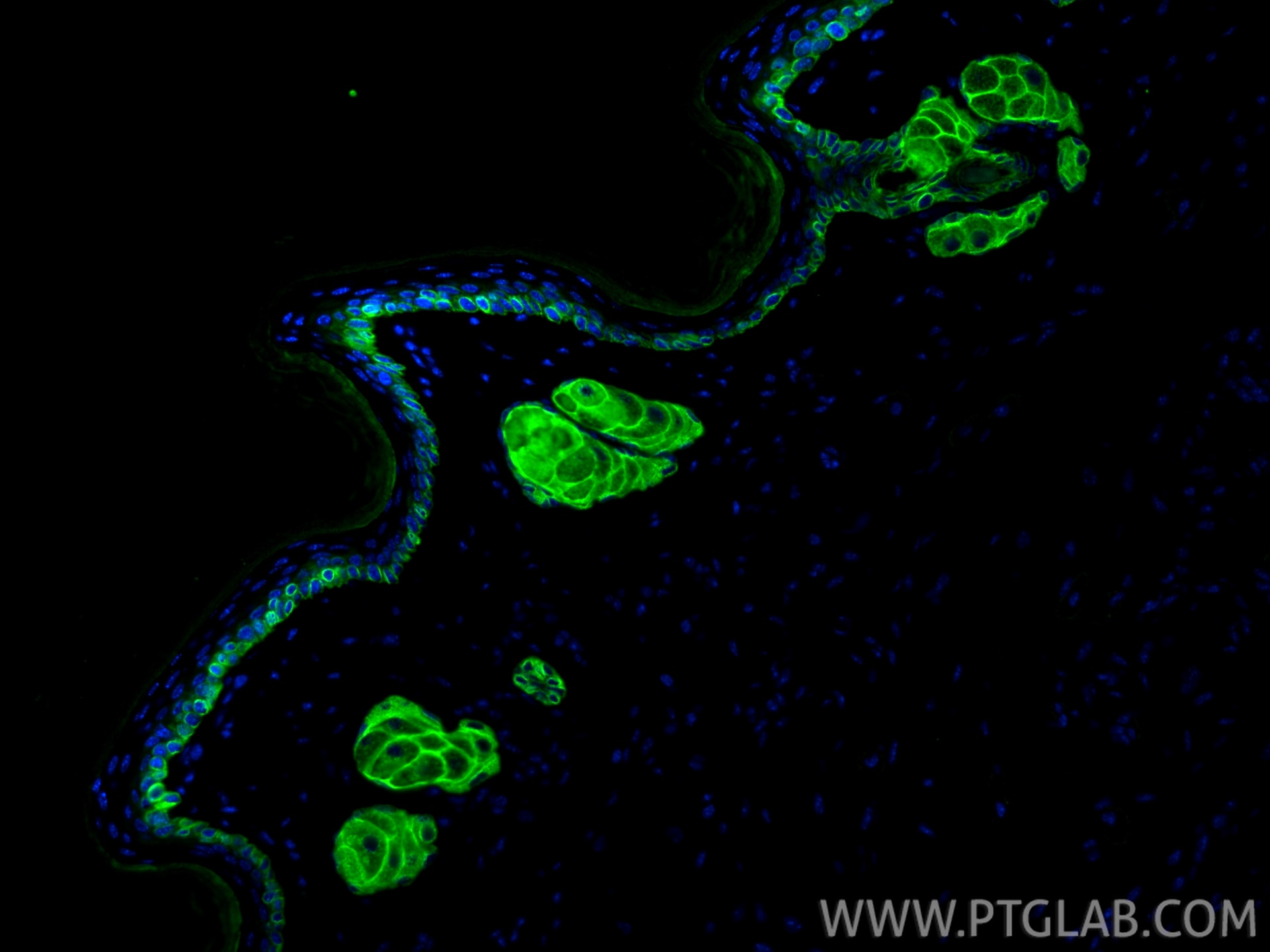
IF-P Figures
IF Staining of mouse skin using 86277-2-RR
Immunofluorescent analysis of (4% PFA) fixed paraffin-embedded mouse skin tissue using Cytokeratin 39 antibody (86277-2-RR, Clone: 250891F5 ) at dilution of 1:200 and CoraLite®488-Conjugated Goat Anti-Rabbit IgG(H+L) (SA00013-2). Heat mediated antigen retrieval with Tris-EDTA buffer (pH 9.0).
IF/ICC Figures
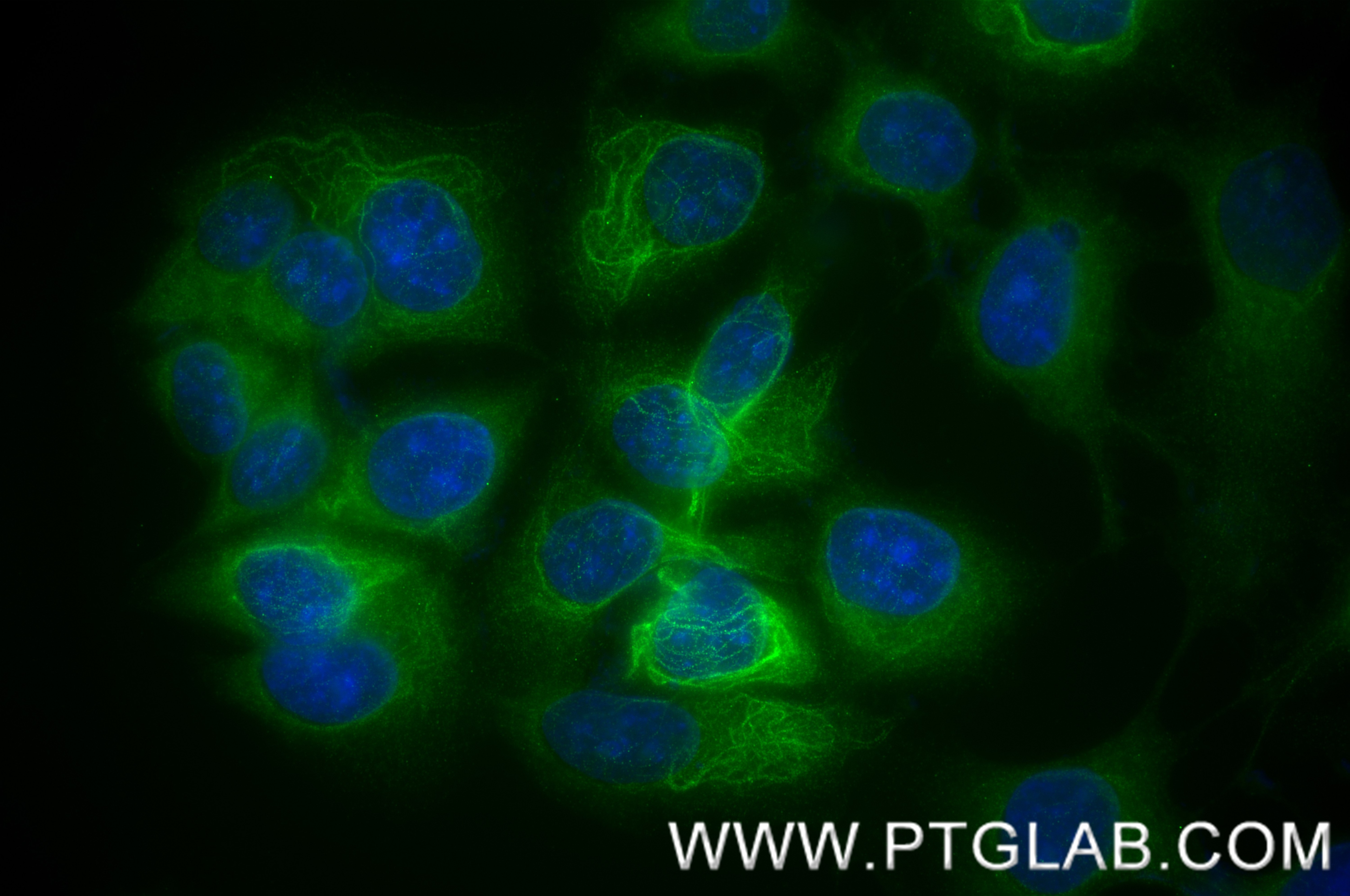
IF Staining of MCF-7 using 86277-2-RR
Immunofluorescent analysis of (-20°C Methanol) fixed MCF-7 cells using Cytokeratin 39 antibody (86277-2-RR, Clone: 250891F5 ) at dilution of 1:200 and CoraLite®488-Conjugated Goat Anti-Rabbit IgG(H+L) (SA00013-2).
The species listed in Tested Reactivity are in-house verified and applicable species. For unlisted species, please refer to the homology analysis of the immunogen sequence and related species. For rabbit polyclonal antibodies, homology >70% is recommended. For mouse monoclonal antibodies and rabbit recombinant antibodies, homology >90% is recommended. Generally, the higher the homology, the greater the applicability. However, there will be certain differences in protein expression in different species, tissues or cells. Therefore, the homology analysis results are for reference only and do not serve as a guarantee.
At Proteintech, we pride ourselves on our antibody quality, customer service and transparency. As such, we are comparing our antibodies with other vendors, enabling easy identification and comparisons of key data to help you choose the suitable antibody for your needs.
We have selected the top cited antibodies from these vendors for you to compare.

 at dilution of 1:5000 incubated at room temperature for 1.5 hours.")
 at dilution of 1:5000 incubated at room temperature for 1.5 hours.")
 at dilution of 1:2000 (under 40x lens). Heat mediated antigen retrieval with Tris-EDTA buffer (pH 9.0).")
 at dilution of 1:2000 (under 20x lens). Heat mediated antigen retrieval with Tris-EDTA buffer (pH 9.0).")
 at dilution of 1:2000 (under 20x lens). Heat mediated antigen retrieval with Tris-EDTA buffer (pH 9.0).")
 at dilution of 1:2000 (under 20x lens). Heat mediated antigen retrieval with Tris-EDTA buffer (pH 9.0).")
 at dilution of 1:5000 (under 10x lens). Heat mediated antigen retrieval with Tris-EDTA buffer (pH 9.0).")
 at dilution of 1:5000 (under 40x lens). Heat mediated antigen retrieval with Tris-EDTA buffer (pH 9.0).")
 fixed paraffin-embedded mouse skin tissue using Cytokeratin 39 antibody (86277-2-RR, Clone: 250891F5 ) at dilution of 1:200 and CoraLite®488-Conjugated Goat Anti-Rabbit IgG(H+L) (SA00013-2). Heat mediated antigen retrieval with Tris-EDTA buffer (pH 9.0).")
 fixed MCF-7 cells using Cytokeratin 39 antibody (86277-2-RR, Clone: 250891F5 ) at dilution of 1:200 and CoraLite®488-Conjugated Goat Anti-Rabbit IgG(H+L) (SA00013-2).")