

Tested Applications
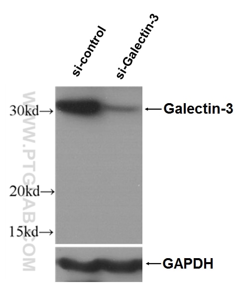
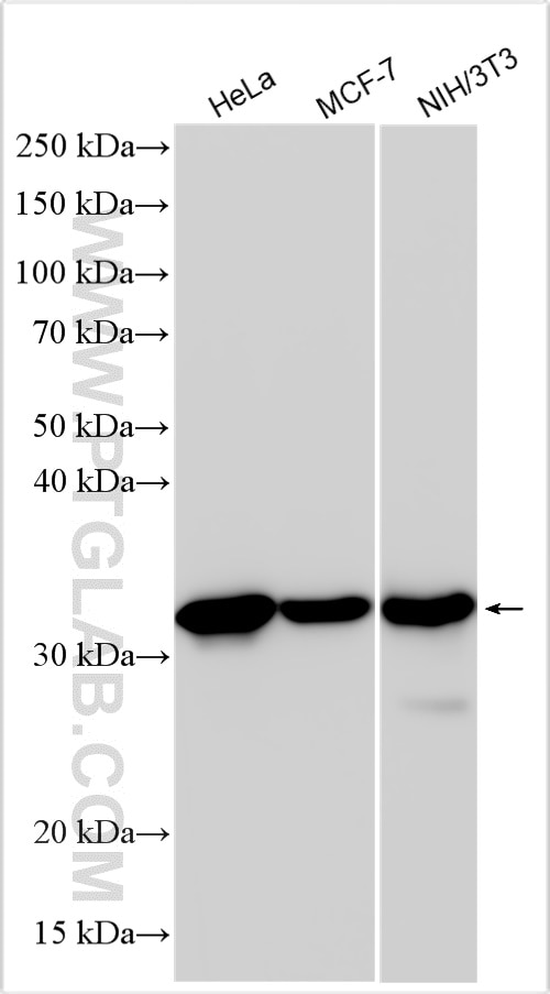
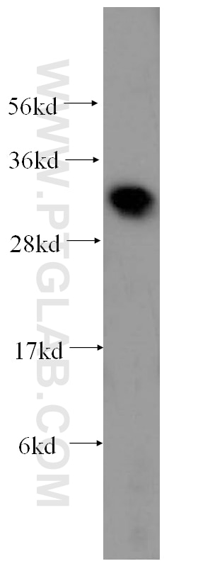
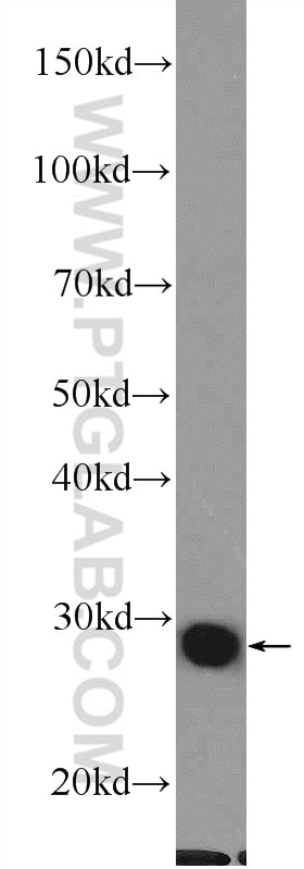
| Positive WB detected in | HeLa cells, human heart tissue, rat colon tissue, MCF-7 cells, NIH/3T3 cells |
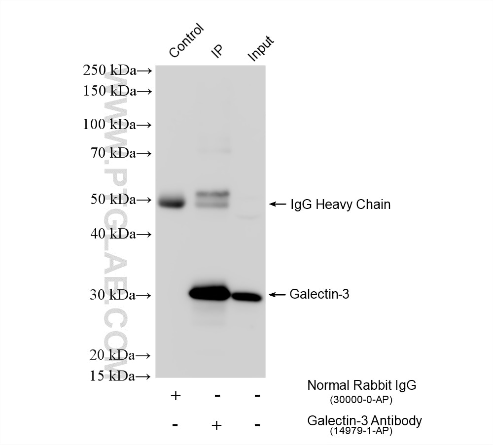
| Positive IP detected in | MCF-7 cells |
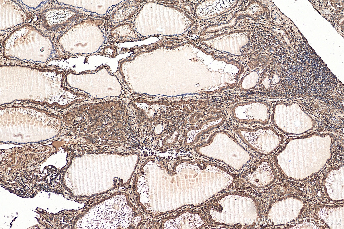
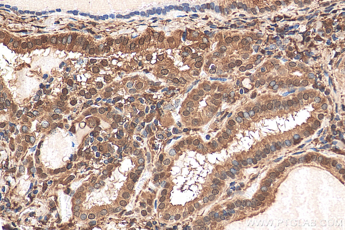
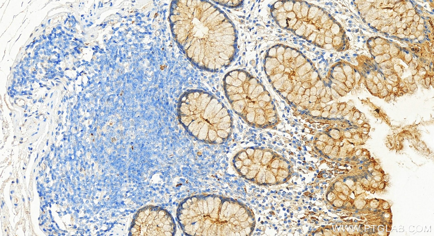
| Positive IHC detected in | human thyroid cancer tissue, human colon tissue Note: suggested antigen retrieval with TE buffer pH 9.0; (*) Alternatively, antigen retrieval may be performed with citrate buffer pH 6.0 |
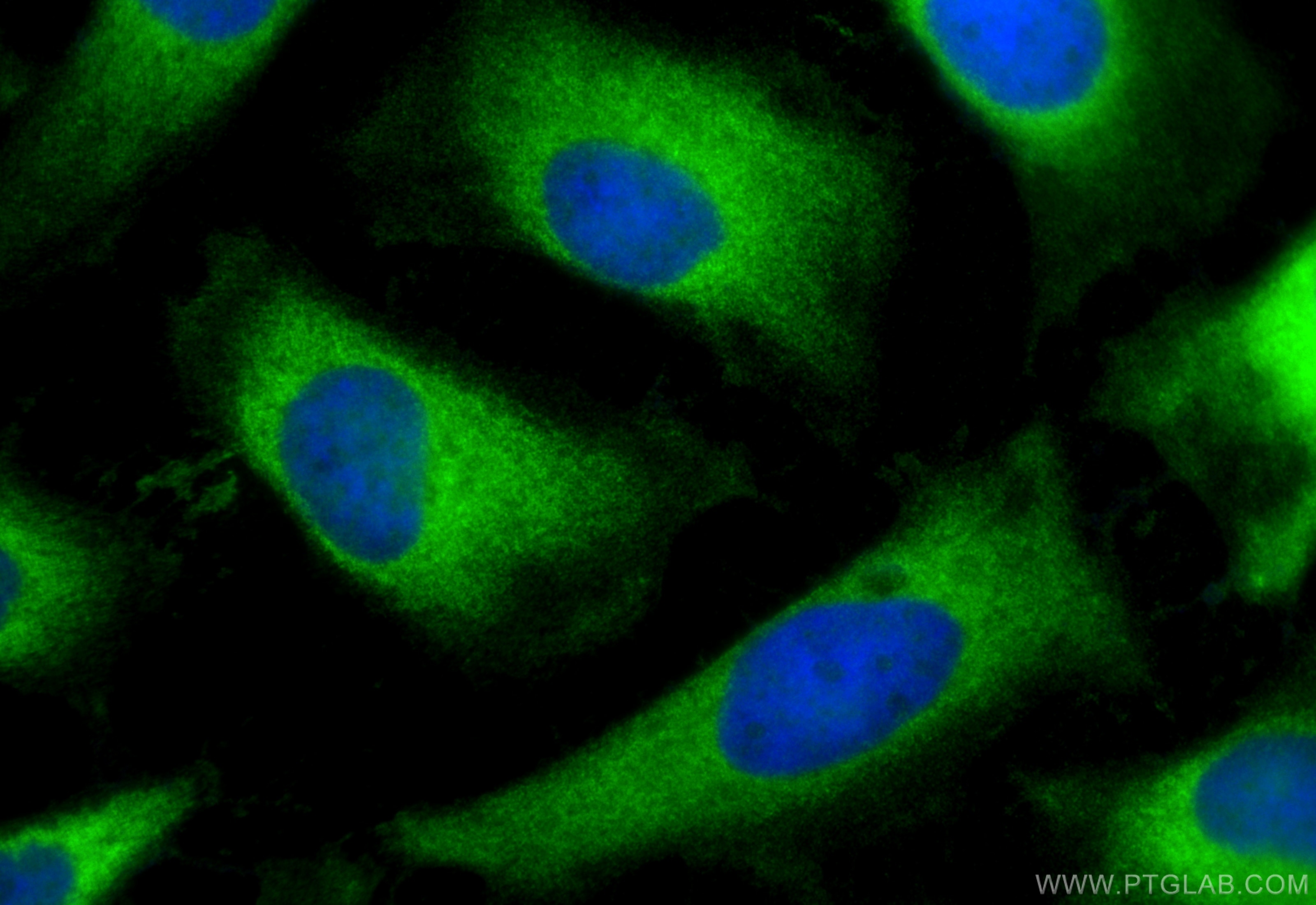
| Positive IF/ICC detected in | HeLa cells |
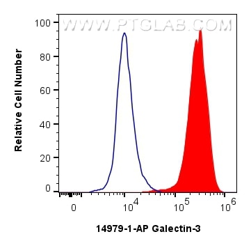
| Positive FC (Intra) detected in | HeLa cells |
Recommended dilution
| Application | Dilution |
|---|---|
| Western Blot (WB) | WB : 1:5000-1:50000 |
| Immunoprecipitation (IP) | IP : 0.5-4.0 ug for 1.0-3.0 mg of total protein lysate |
| Immunohistochemistry (IHC) | IHC : 1:50-1:500 |
| Immunofluorescence (IF)/ICC | IF/ICC : 1:50-1:500 |
| Flow Cytometry (FC) (INTRA) | FC (INTRA) : 0.40 ug per 10^6 cells in a 100 µl suspension |
| It is recommended that this reagent should be titrated in each testing system to obtain optimal results. | |
| Sample-dependent, Check data in validation data gallery. | |
Product Information
14979-1-AP targets Galectin-3 in WB, IHC, IF/ICC, FC (Intra), IP, ELISA applications and shows reactivity with human, mouse, rat samples.
| Tested Reactivity | human, mouse, rat |
| Cited Reactivity | human, mouse, rat, pig |
| Host / Isotype | Rabbit / IgG |
| Class | Polyclonal |
| Type | Antibody |
| Immunogen |
CatNo: Ag6891 Product name: Recombinant human Galectin 3 protein Source: e coli.-derived, PGEX-4T Tag: GST Domain: 1-250 aa of BC001120 Sequence: MADNFSLHDALSGSGNPNPQGWPGAWGNQPAGAGGYPGASYPGAYPGQAPPGAYPGQAPPGAYPGAPGAYPGAPAPGVYPGPPSGPGAYPSSGQPSATGAYPATGPYGAPAGPLIVPYNLPLPGGVVPRMLITILGTVKPNANRIALDFQRGNDVAFHFNPRFNENNRRVIVCNTKLDNNWGREERQSVFPFESGKPFKIQVLVEPDHFKVAVNDAHLLQYNHRVKKLNEISKLGISGDIDLTSASYTMI Predict reactive species |
| Full Name | lectin, galactoside-binding, soluble, 3 |
| Calculated Molecular Weight | 26 kDa |
| Observed Molecular Weight | 31 kDa |
| GenBank Accession Number | BC001120 |
| Gene Symbol | Galectin-3 |
| Gene ID (NCBI) | 3958 |
| RRID | AB_2136768 |
| Conjugate | Unconjugated |
| Form | Liquid |
| Purification Method | Antigen affinity purification |
| UNIPROT ID | P17931 |
| Storage Buffer | PBS with 0.02% sodium azide and 50% glycerol, pH 7.3. |
| Storage Conditions | Store at -20°C. Stable for one year after shipment. Aliquoting is unnecessary for -20oC storage. 20ul sizes contain 0.1% BSA. |
Background Information
Galectins are a family of animal lectins defined by shared characteristic amino-acid sequences and affinity for β-galactose-containing oligosac-charides (PMID: 8063692). Galectin-3, a member of the β-galactoside-binding proteins, contains one carbohydrate recognition domain (CRD) and a proline- and glycine-rich N-terminal domain through which is able to form oligomers (PMID: 14758078). Galectin-3 is widely expressed in many normal tissues and a variety of tumors. It is found intracellularly in nucleus and cytoplasm or secreted outside of cell, being present on the cell surface or in the extracellular space (PMID: 16478649). Galectin-3 is involved in various biological processes including cell growth, adhesion, differentiation, apoptosis, angiogenesis, immune response, neoplastic transformation and metastasis (PMID: 16478649; 14758078).
Protocols
| Product Specific Protocols | |
|---|---|
| FC protocol for Galectin-3 antibody 14979-1-AP | Download protocol |
| IF protocol for Galectin-3 antibody 14979-1-AP | Download protocol |
| IHC protocol for Galectin-3 antibody 14979-1-AP | Download protocol |
| IP protocol for Galectin-3 antibody 14979-1-AP | Download protocol |
| WB protocol for Galectin-3 antibody 14979-1-AP | Download protocol |
| Standard Protocols | |
|---|---|
| Click here to view our Standard Protocols |
Publications
What published studies show
Galectin-3 (LGALS3) polyclonal antibody (Cat# 14979-1-AP) has 79 citations published in journals including Nature, Nature Immunology, Autophagy, Cell Reports, EMBO Journal, Journal of Experimental Medicine, Oncogene, and ACS Nano. Research fields span lysosomal biology and membrane repair, oncology (breast, liver, gastric, pancreatic, glioblastoma), cardiovascular disease, neurodegeneration (Alzheimer's, Parkinson's), immunology/inflammation, lipid metabolism, and fibrosis. Applications include WB, IF, IHC, and IP across human, mouse, rat, and pig models.
| Species | Application | Title |
|---|---|---|
Nat Immunol Heme drives hemolysis-induced susceptibility to infection via disruption of phagocyte functions. | ||
Bioact Mater Biomimetic stress granules replenish lysosomal repair to reinstate macrophage immunometabolic antibacterial programs. | ||
Autophagy E-cigarette aerosols induce the hydrolysis of lysosomal glycerophospholipids through PLA2G4A activation initiated by nicotine binding to CHRNA3/α3 nAchr in airway epithelial cells. | ||
Autophagy The PI4K2A-OSBPL6/ORP6-PS axis mediates lysosomal membrane repair to restore neuronal lipid homeostasis and promote neuronal survival after spinal cord injury. | ||
Exp Hematol Oncol Multi-model analysis of gallbladder cancer reveals the role of OxLDL-absorbing neutrophils in promoting liver invasion |
Reviews
What customers say
Users report positive results across multiple applications. One reviewer rated it 5/5 for immunoprecipitation, calling it "good for IP." For immunofluorescence at 1:200 on reactive astrocytes, a user noted specific staining with minor background, comparable to the product's reference images (4/5). A Western blot user at 1:1000 on HEK293 lysates with overnight membrane incubation and HRP secondary also gave 4/5. Overall, the antibody performs well across IP, IF, and WB with consistent specificity.
The reviews below have been submitted by verified Proteintech customers who received an incentive for providing their feedback.
Ahmad (Verified Customer) (07-02-2026) | Good for IP !
|
Víctor (Verified Customer) (09-18-2024) | The staining appears specific, although there is a bit of background, maybe due to the secondary antibody used. It is quite similar to the staining shown on the product's website
|
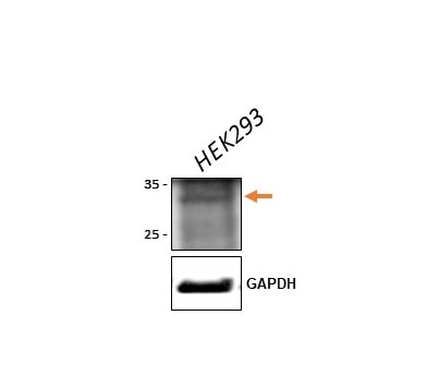
Tom (Verified Customer) (07-07-2021) | HEK293 whole cell lysates on SDS-PAGE. Antibody incubated with membrane overnight in fridge. HRP secondary antibody used for detection.
 |











