
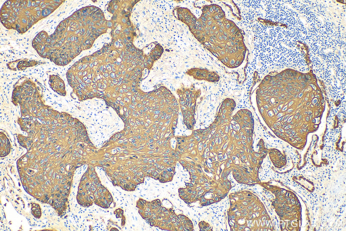
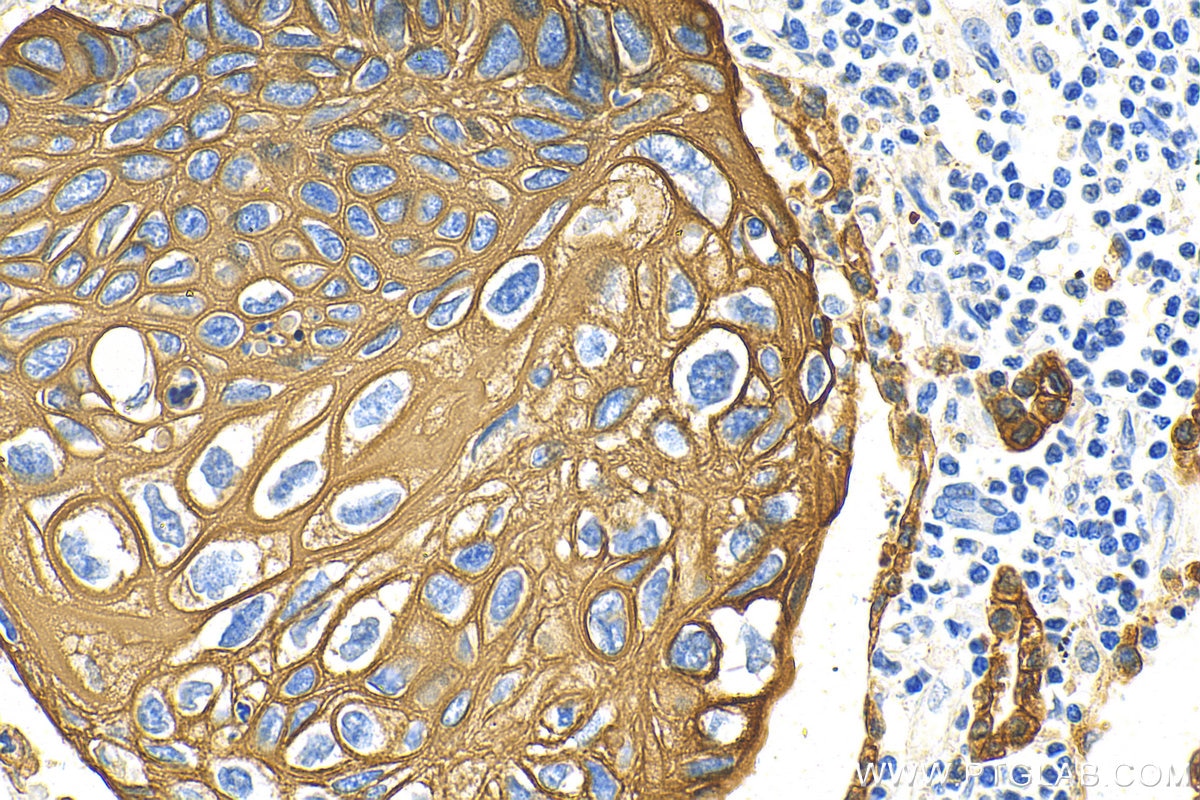
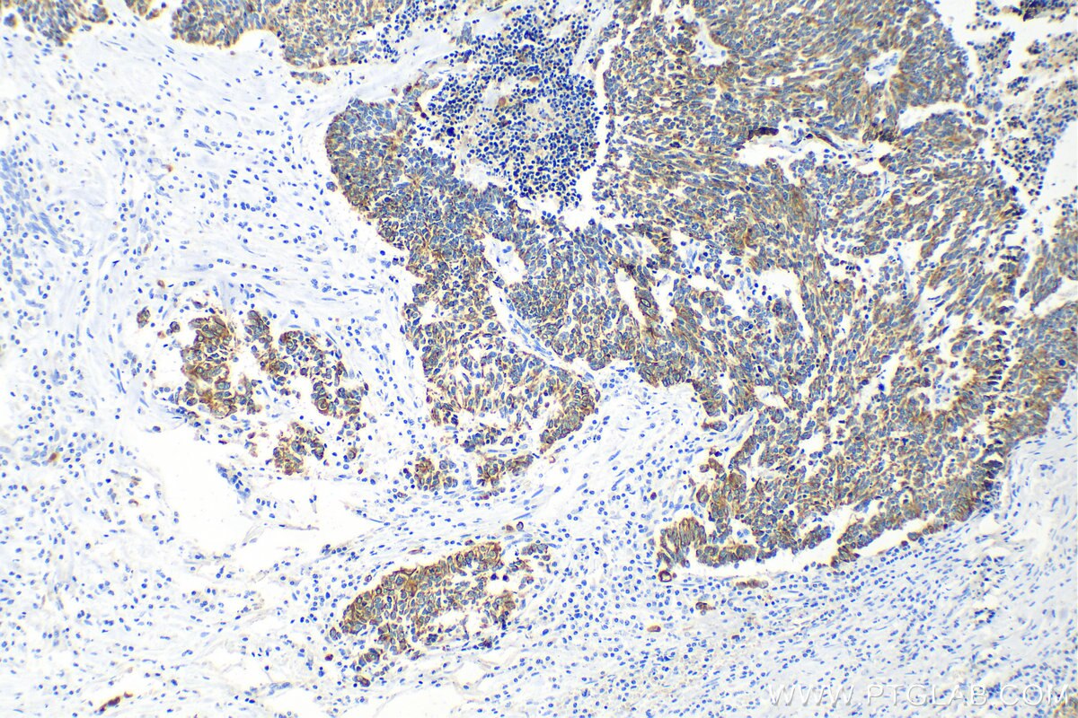
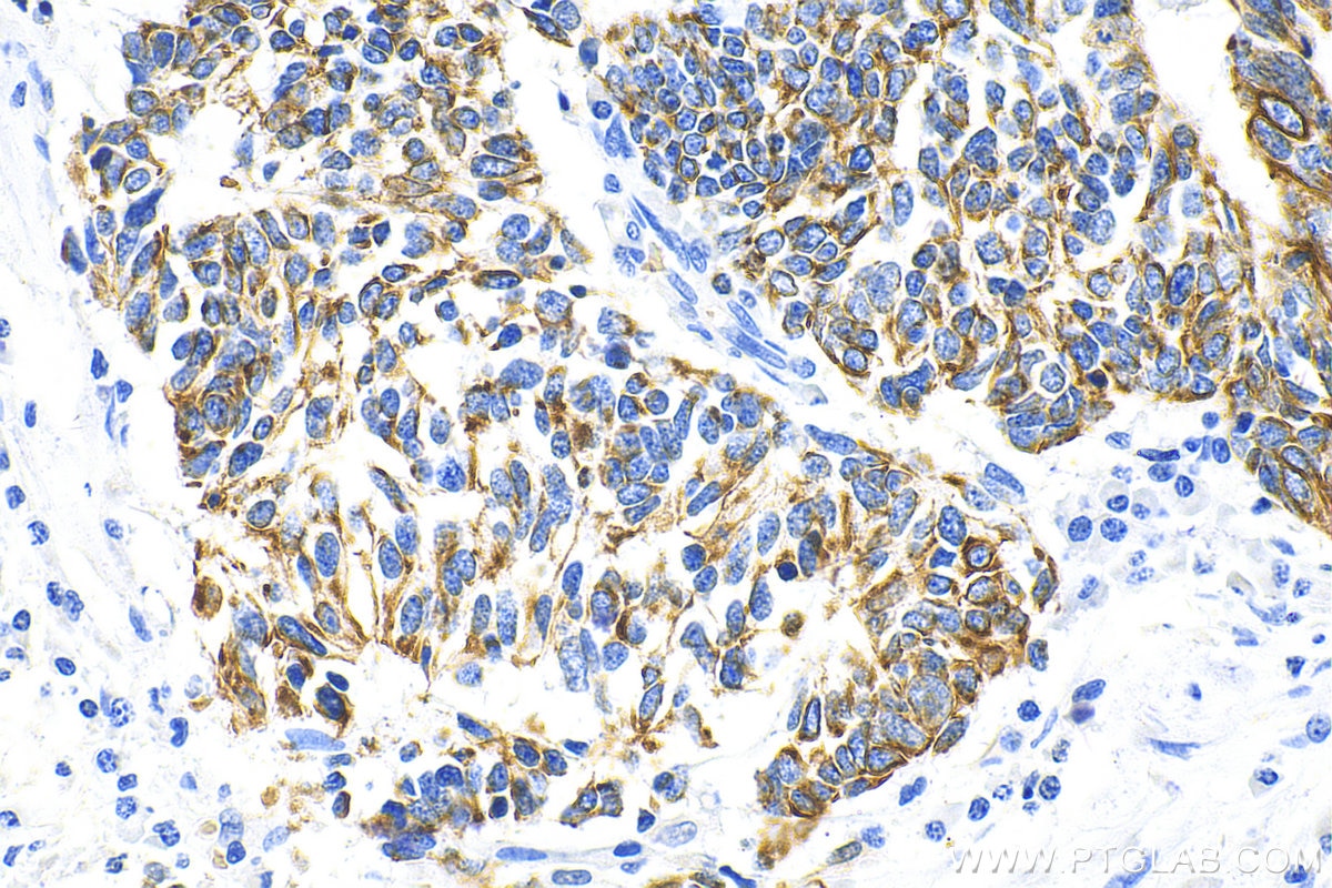
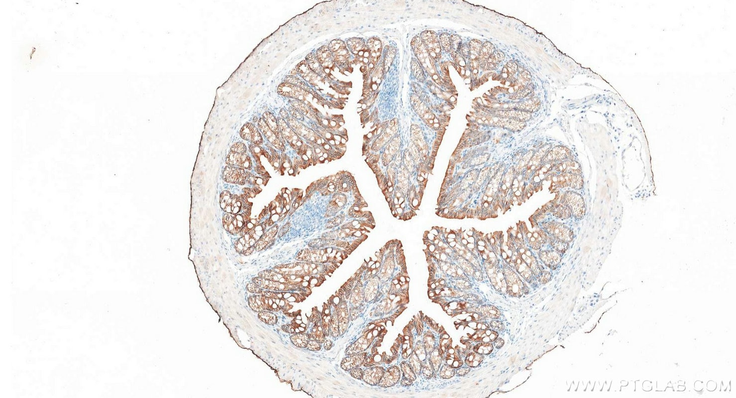
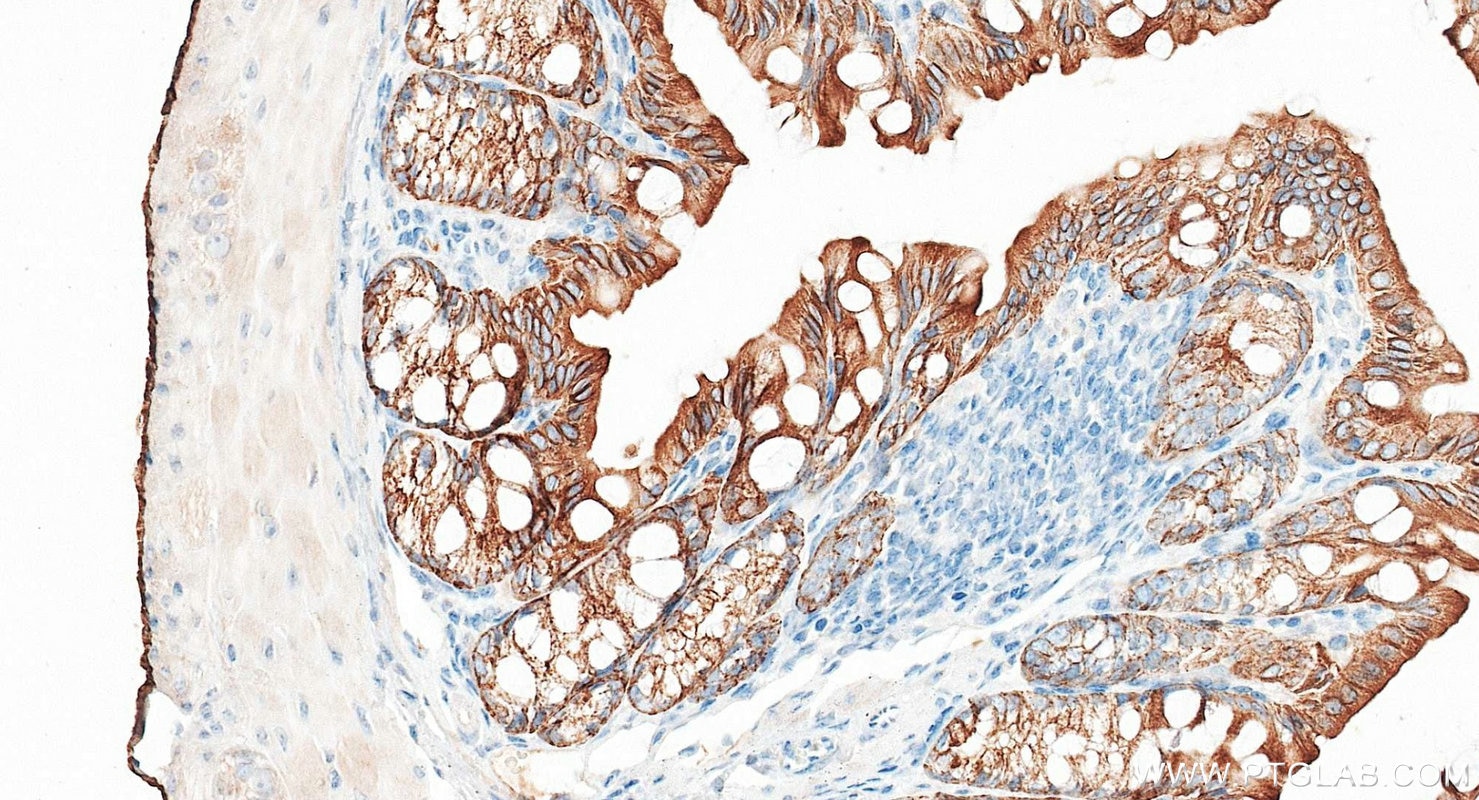
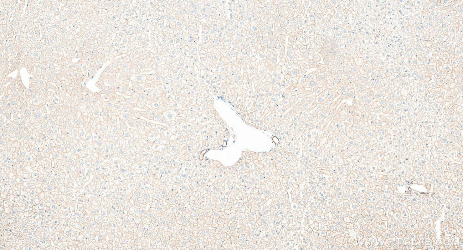
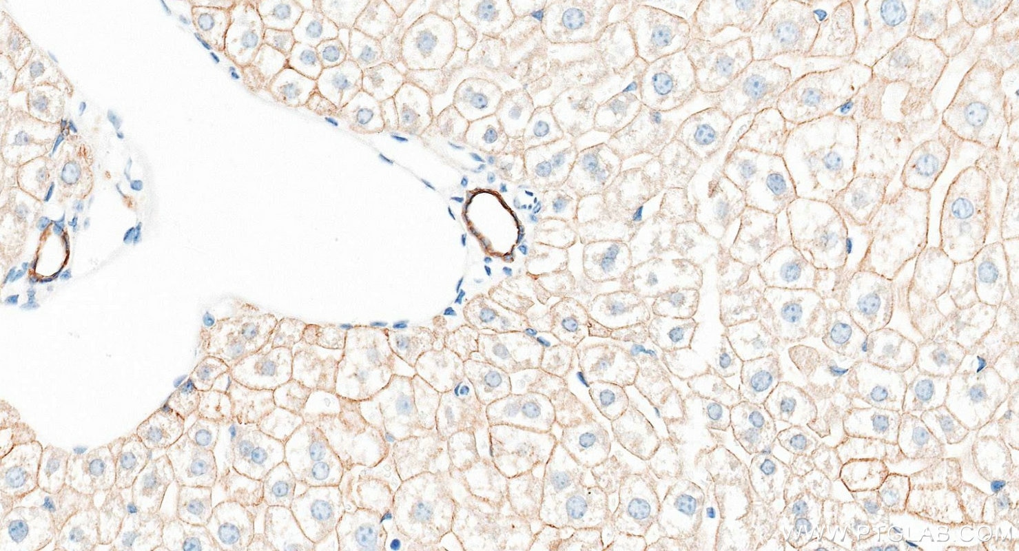
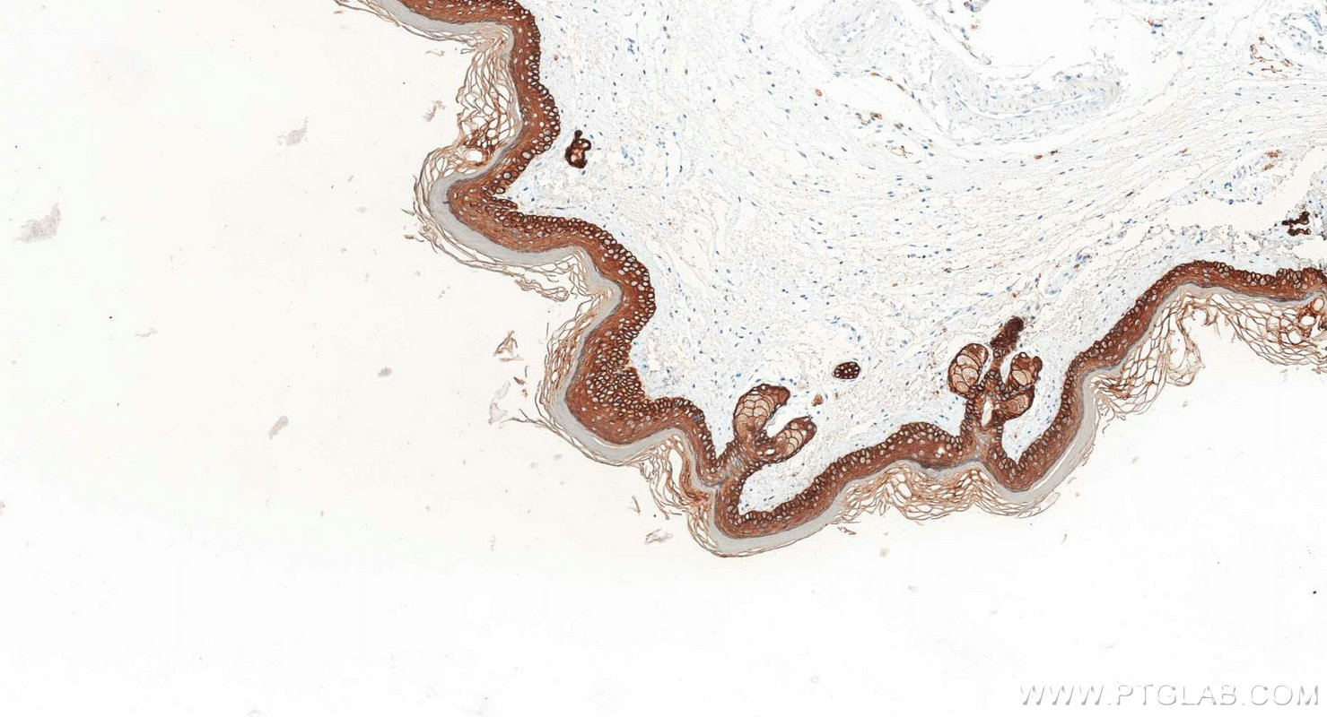
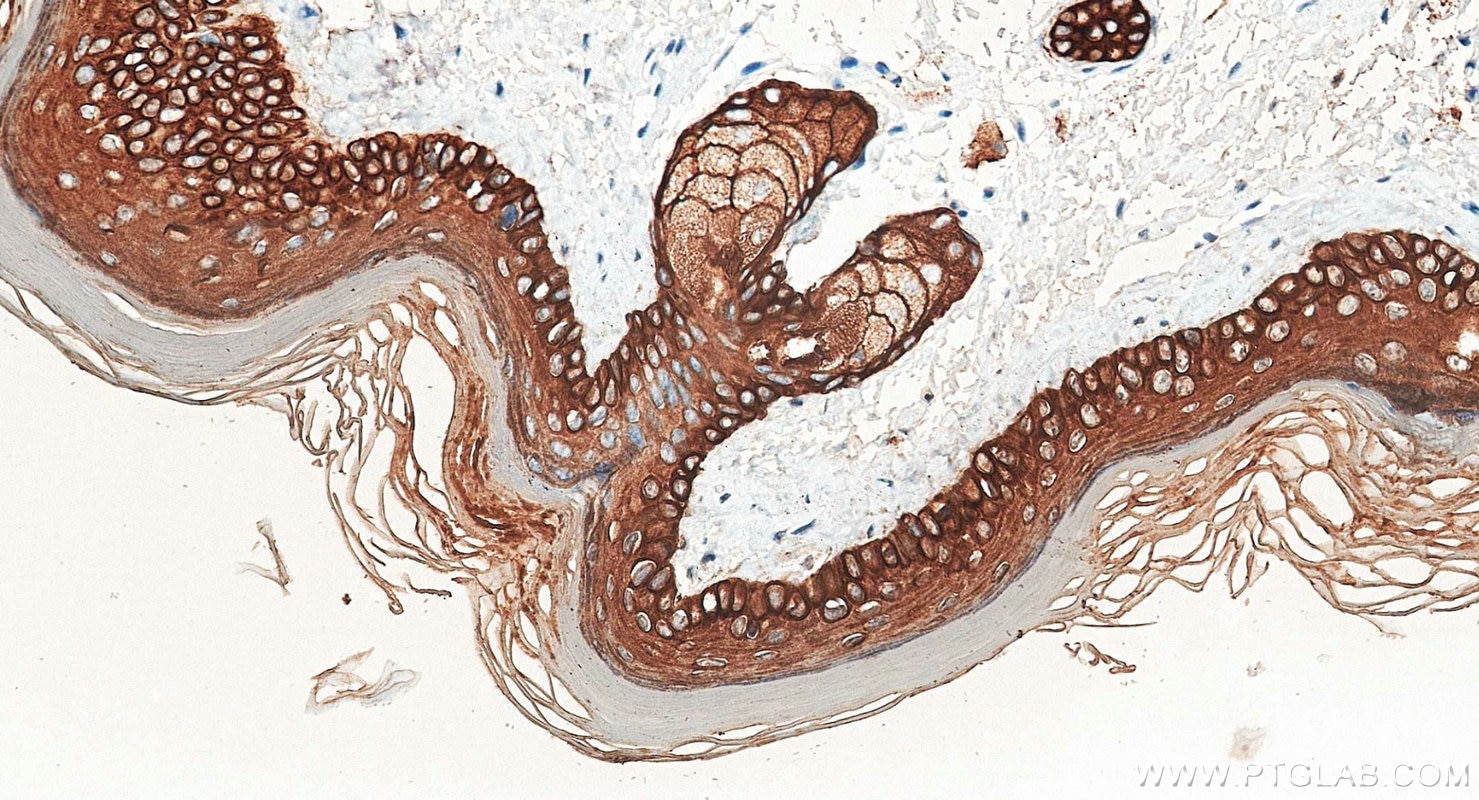
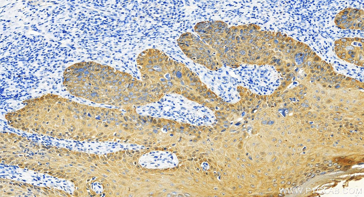
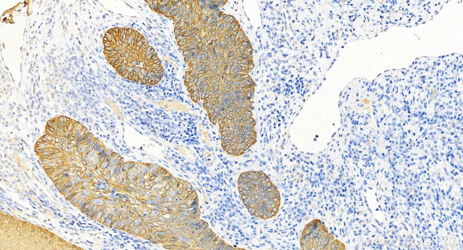

Tested Applications
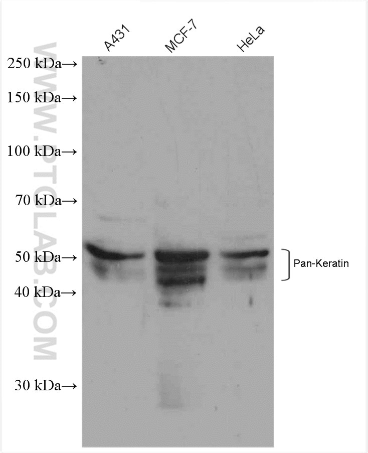
| Positive WB detected in | A431 cells, MCF-7 cells, HeLa cells |
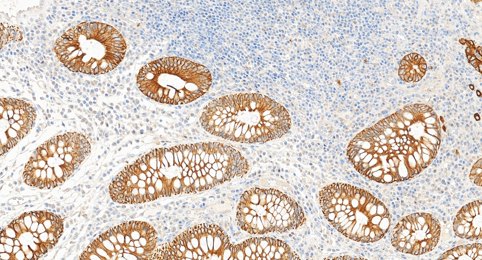
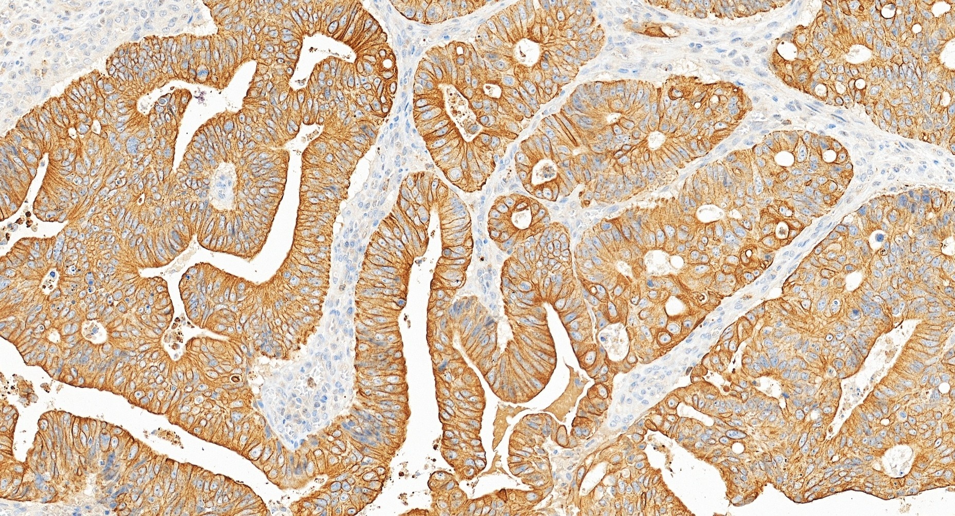
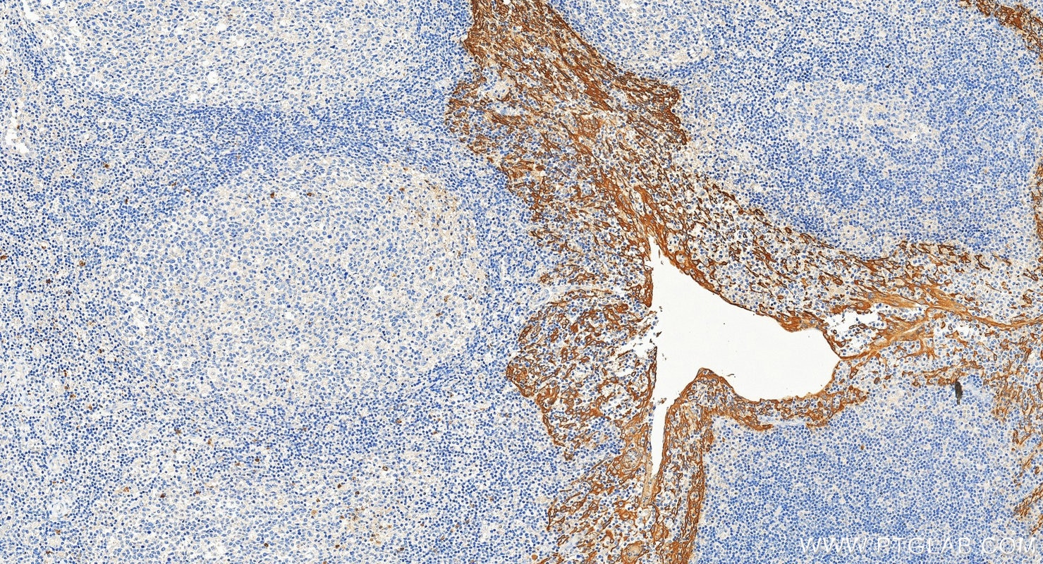
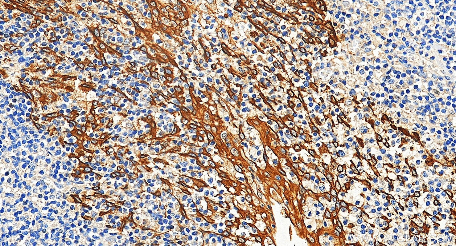
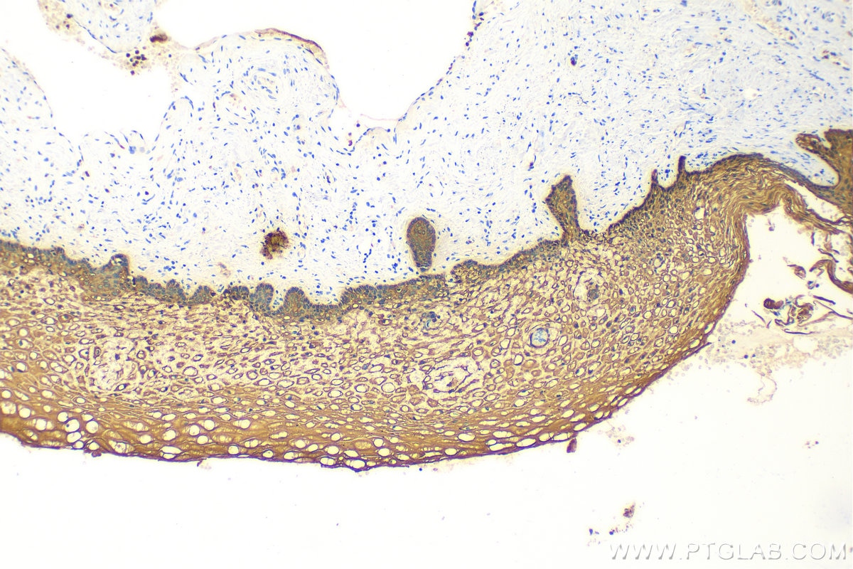
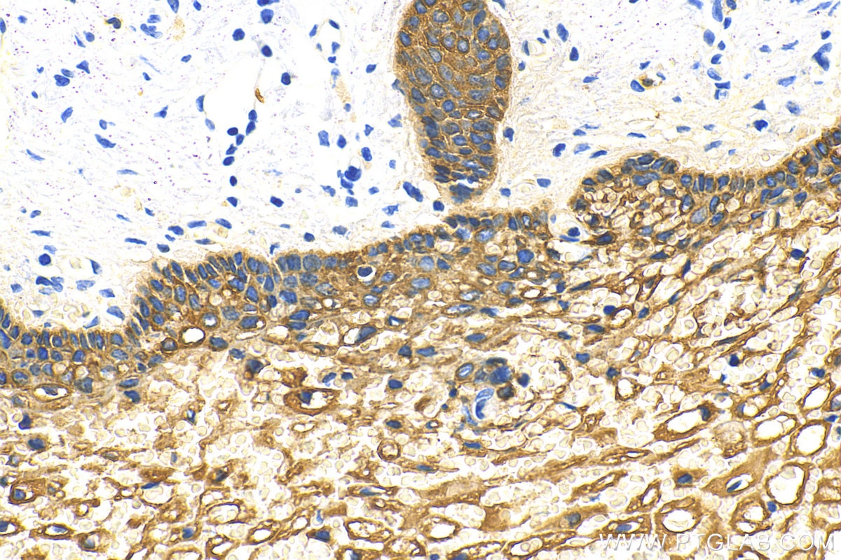
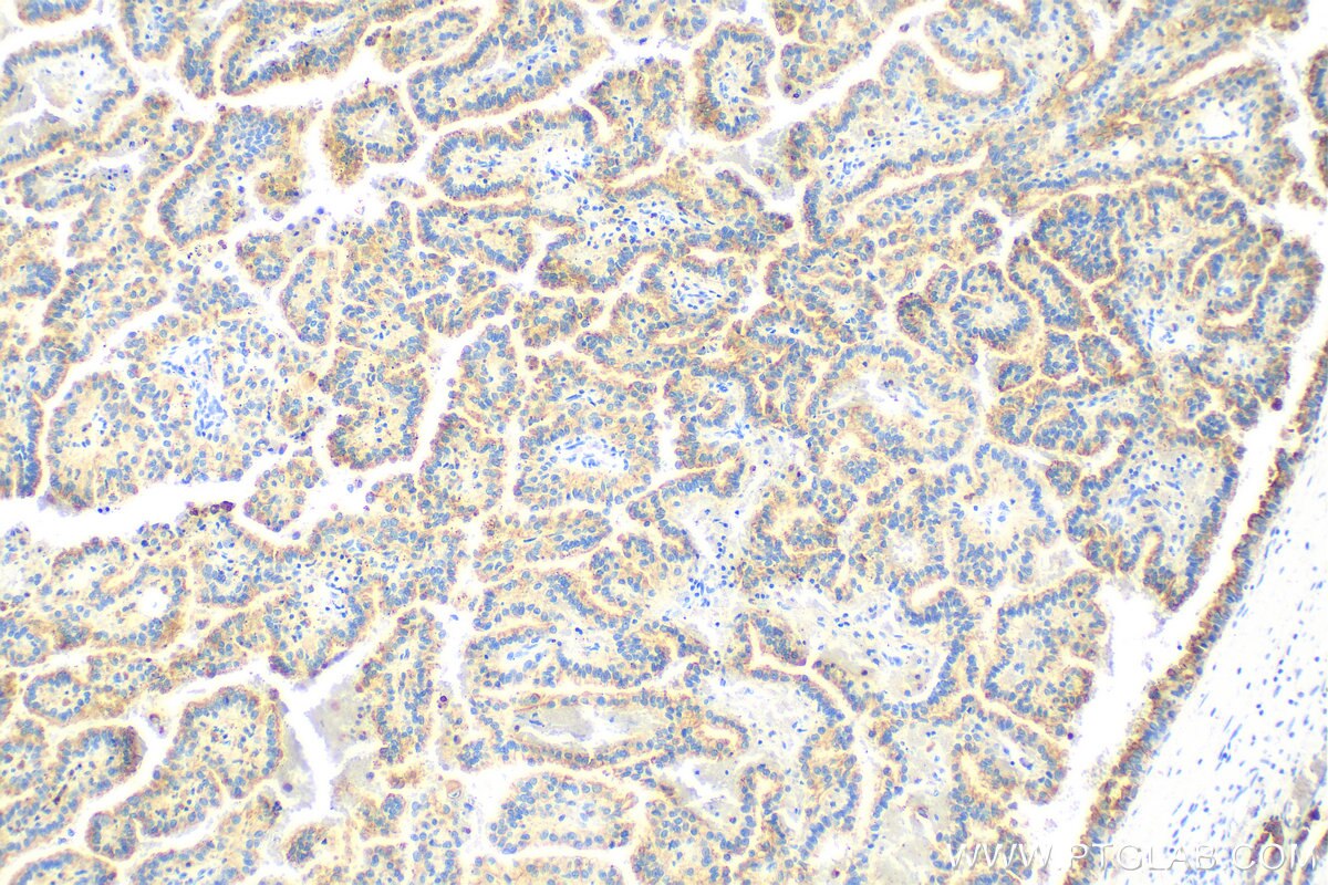
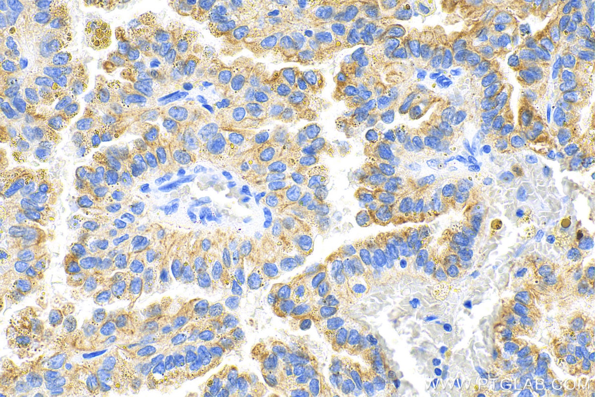
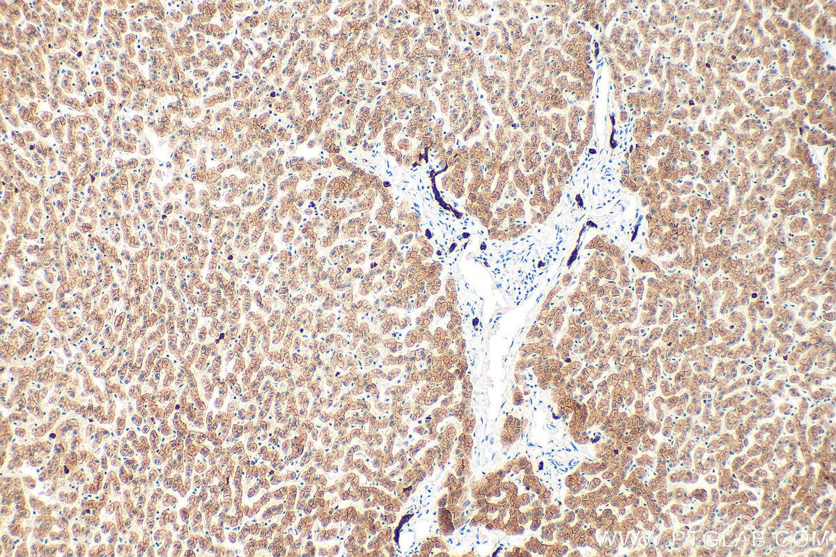
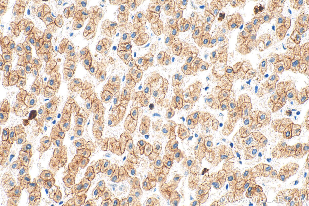
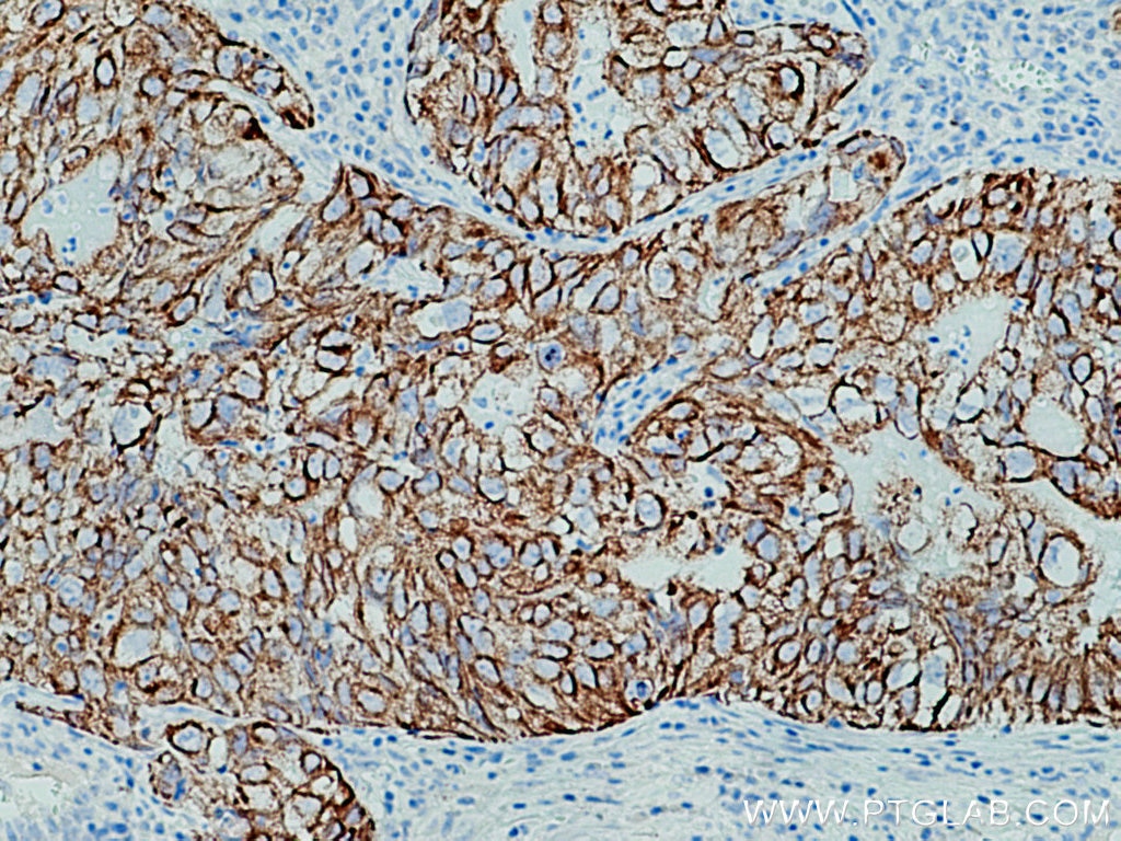
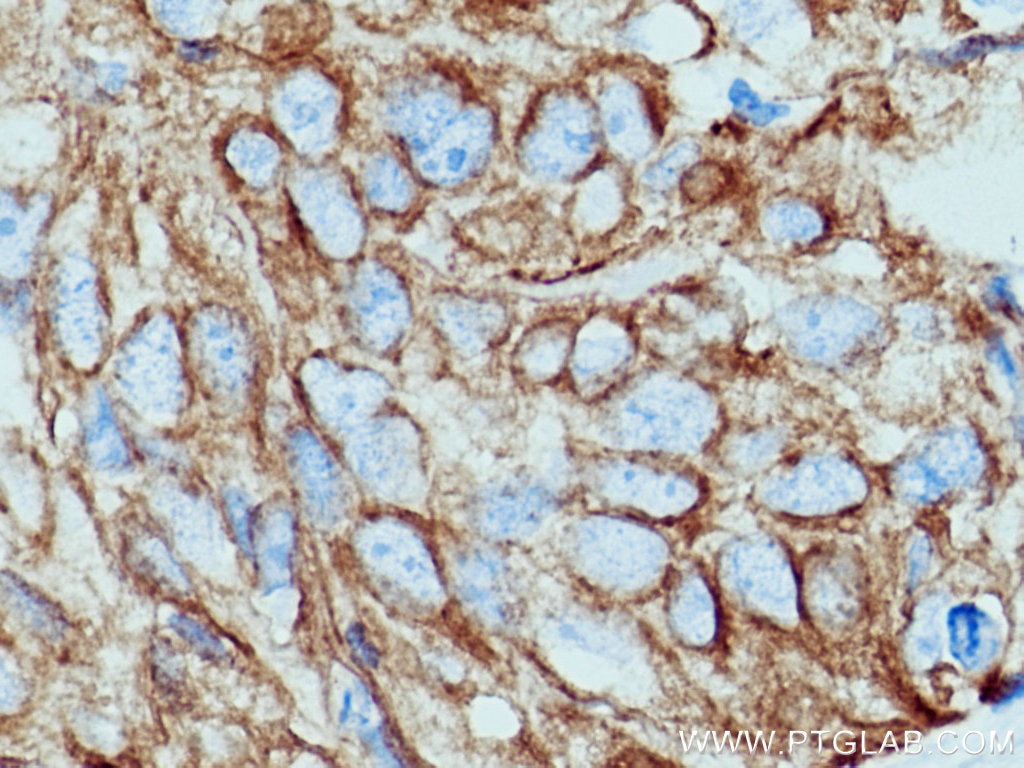
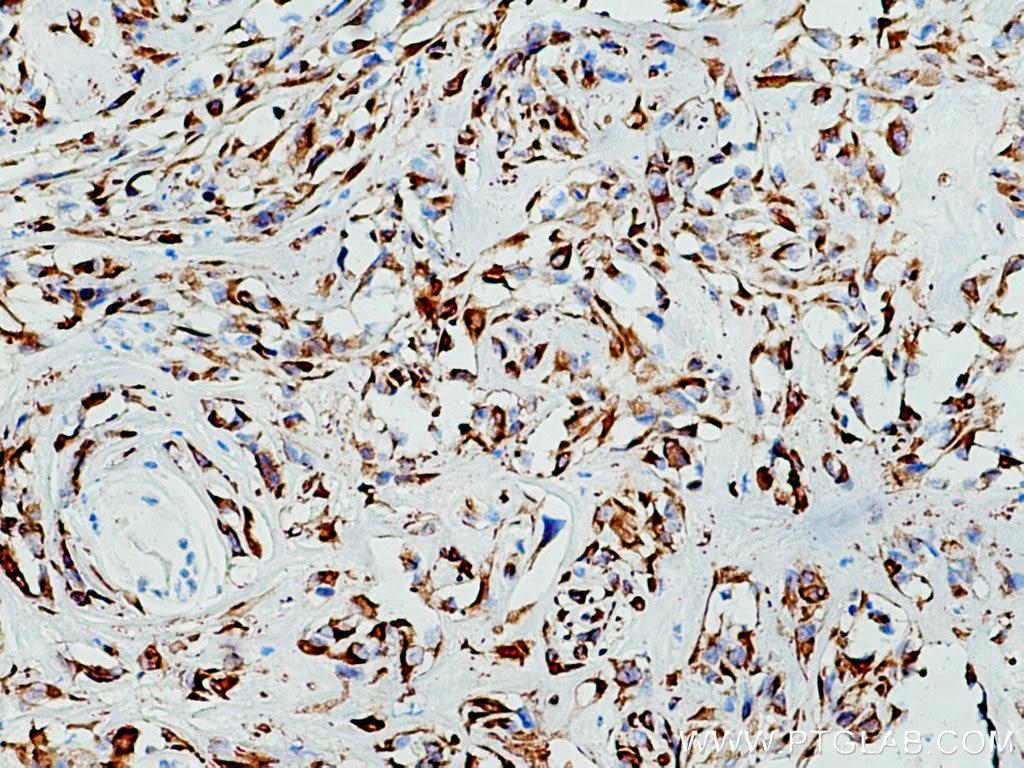
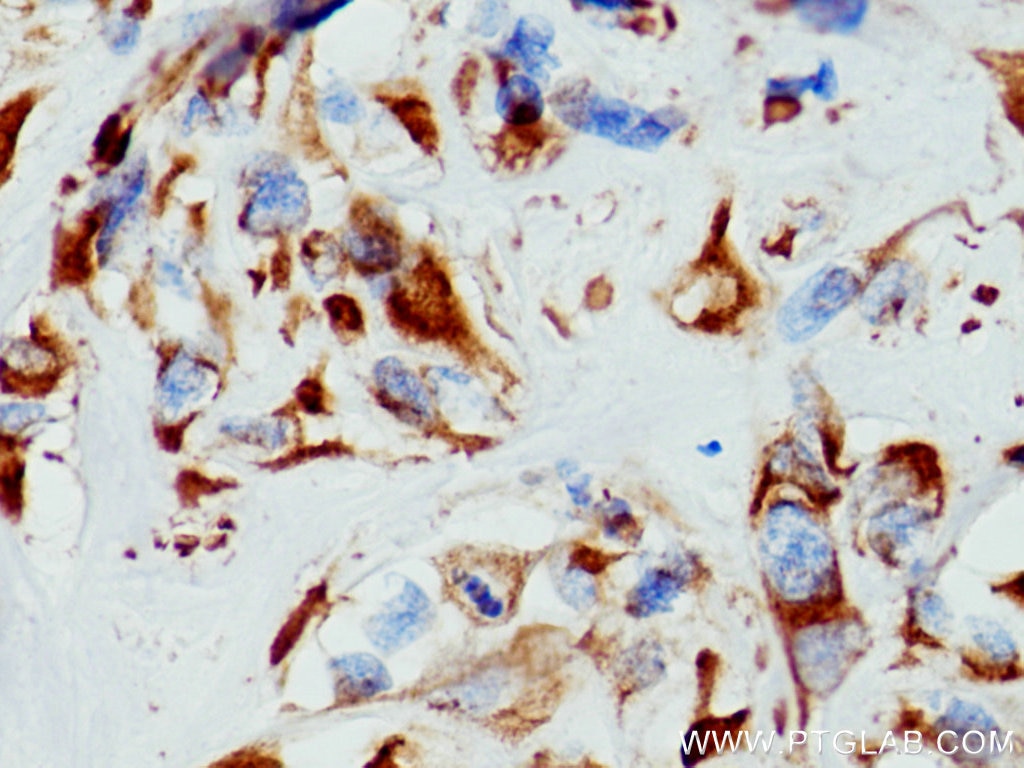
| Positive IHC detected in | human colon cancer tissue, human lung cancer tissue, human renal cell carcinoma tissue, human brown disease, human cervical cancer tissue, human liver tissue, mouse colon tissue, mouse liver tissue, mouse skin tissue, human tonsillitis tissue Note: suggested antigen retrieval with TE buffer pH 9.0; (*) Alternatively, antigen retrieval may be performed with citrate buffer pH 6.0 |
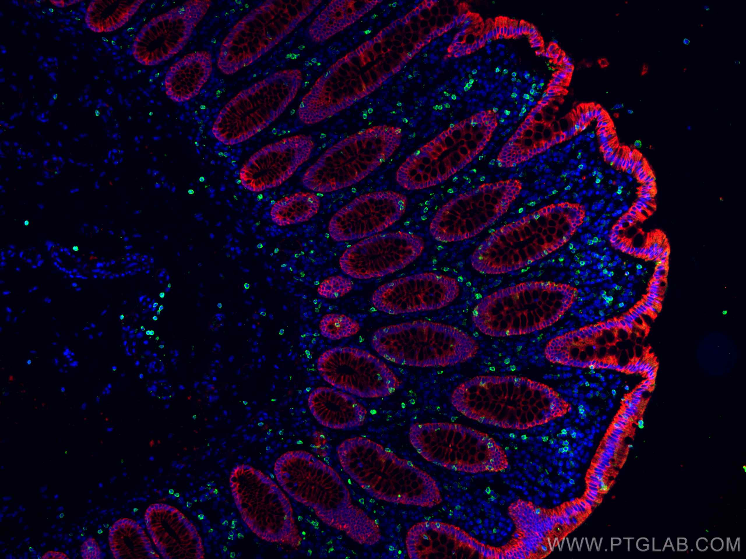
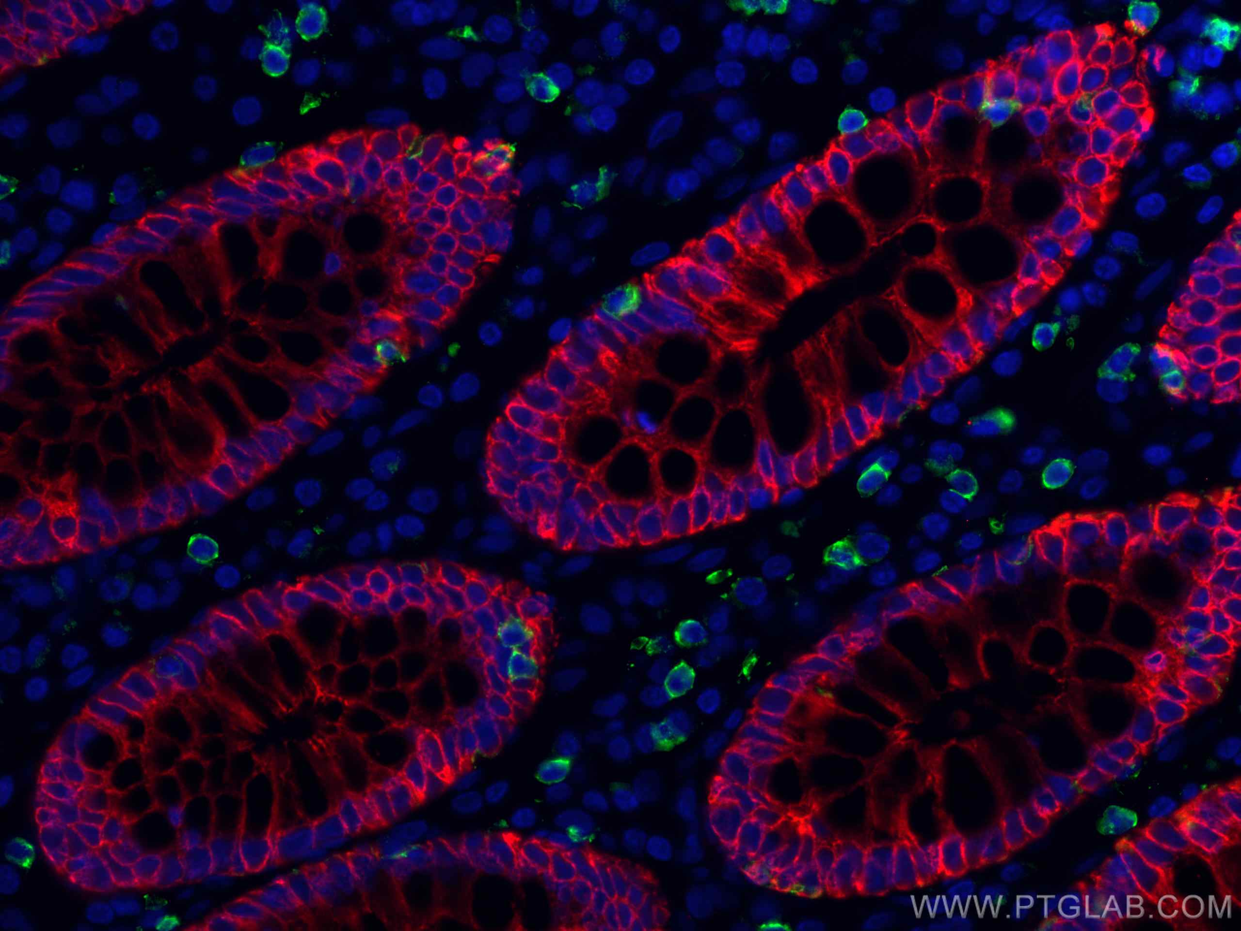
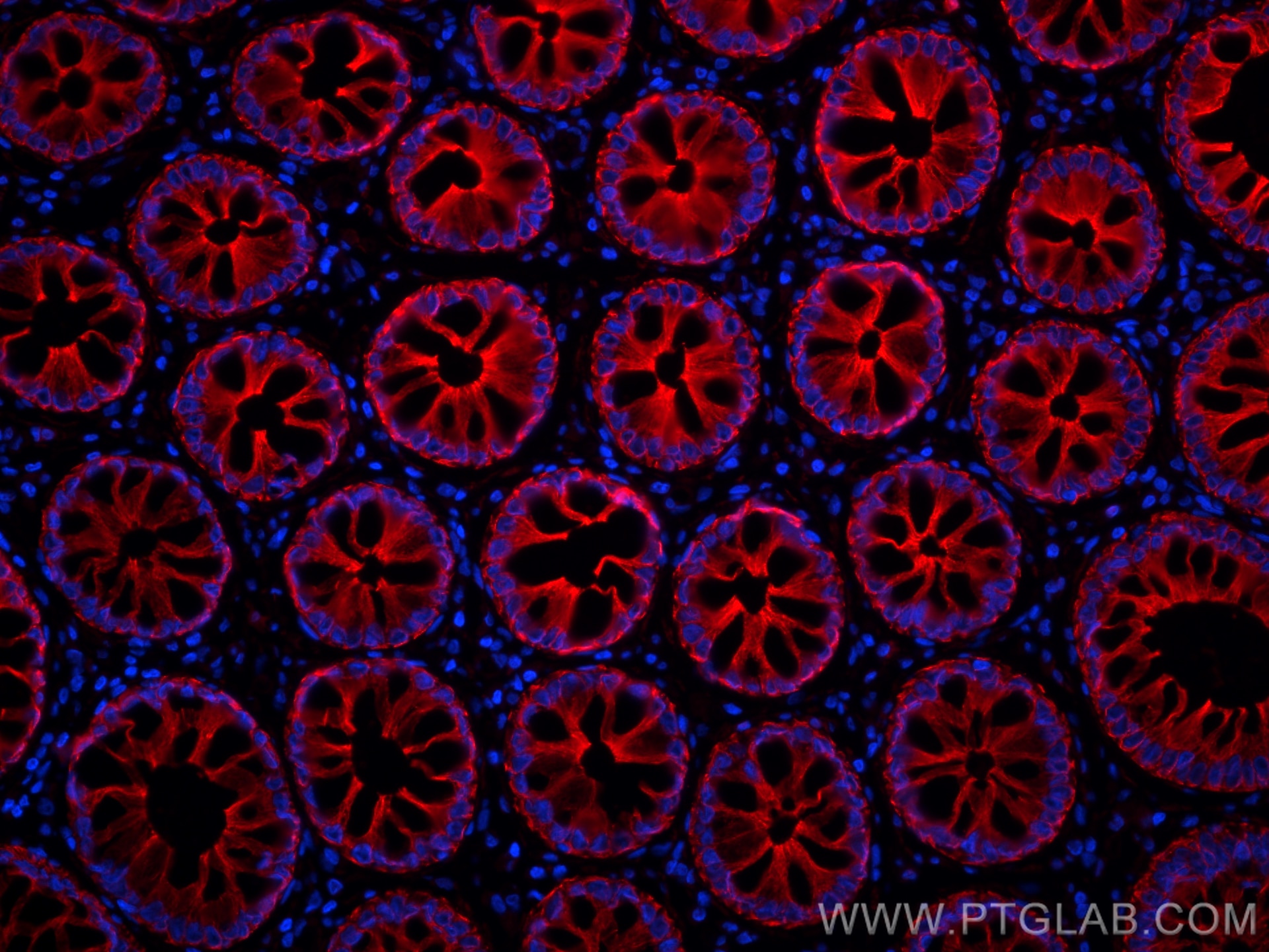
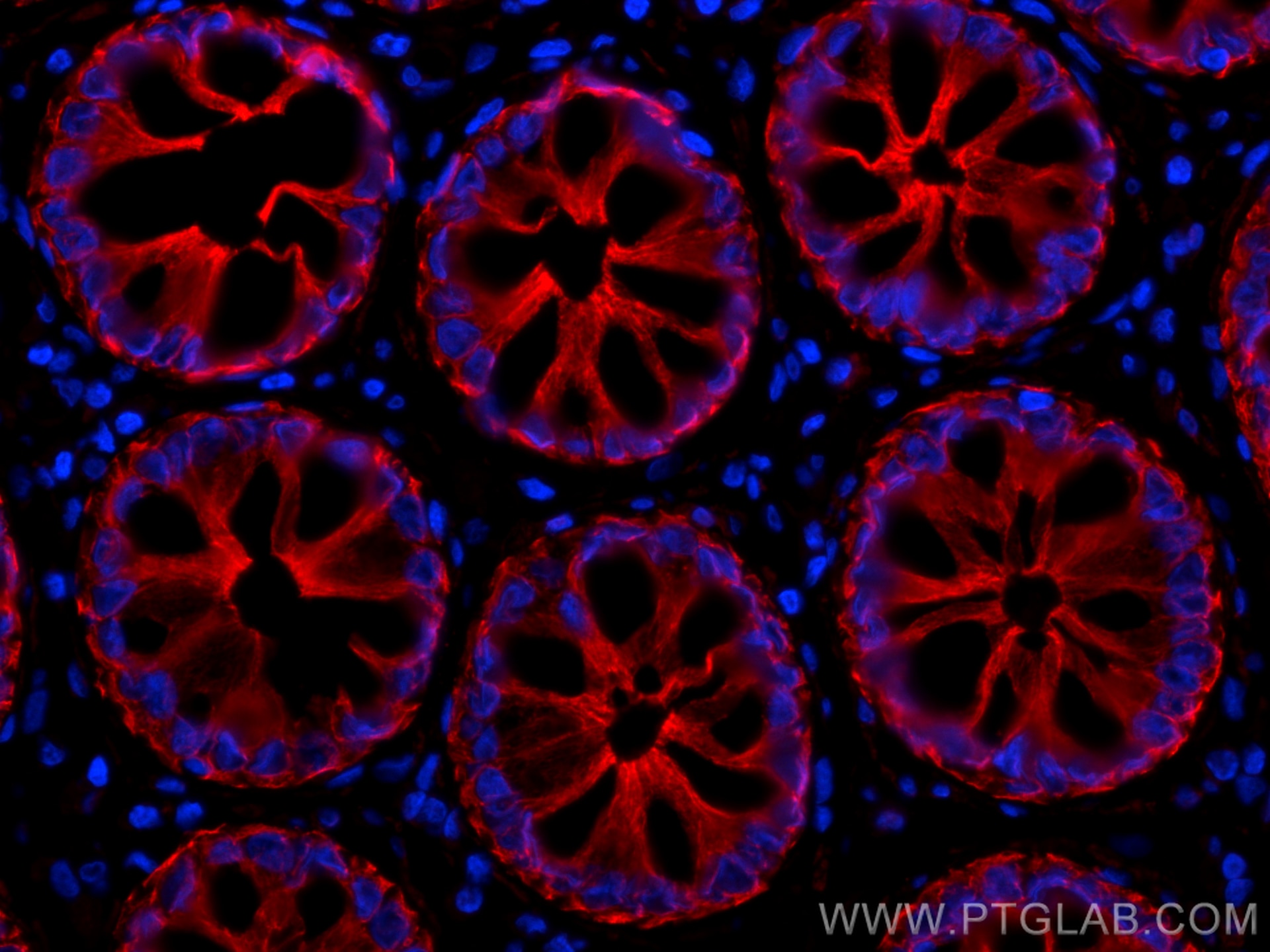
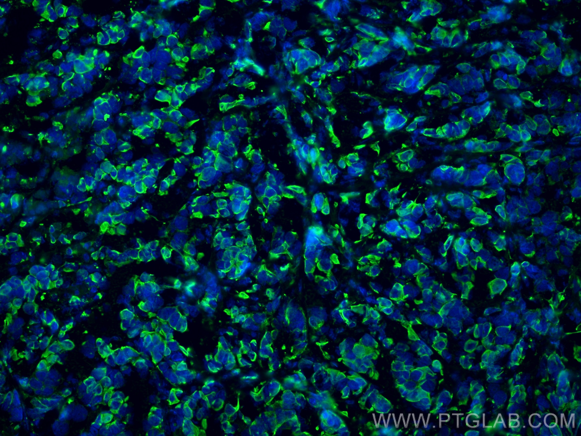
| Positive IF-P detected in | human colon cancer tissue, human rectal cancer tissue |
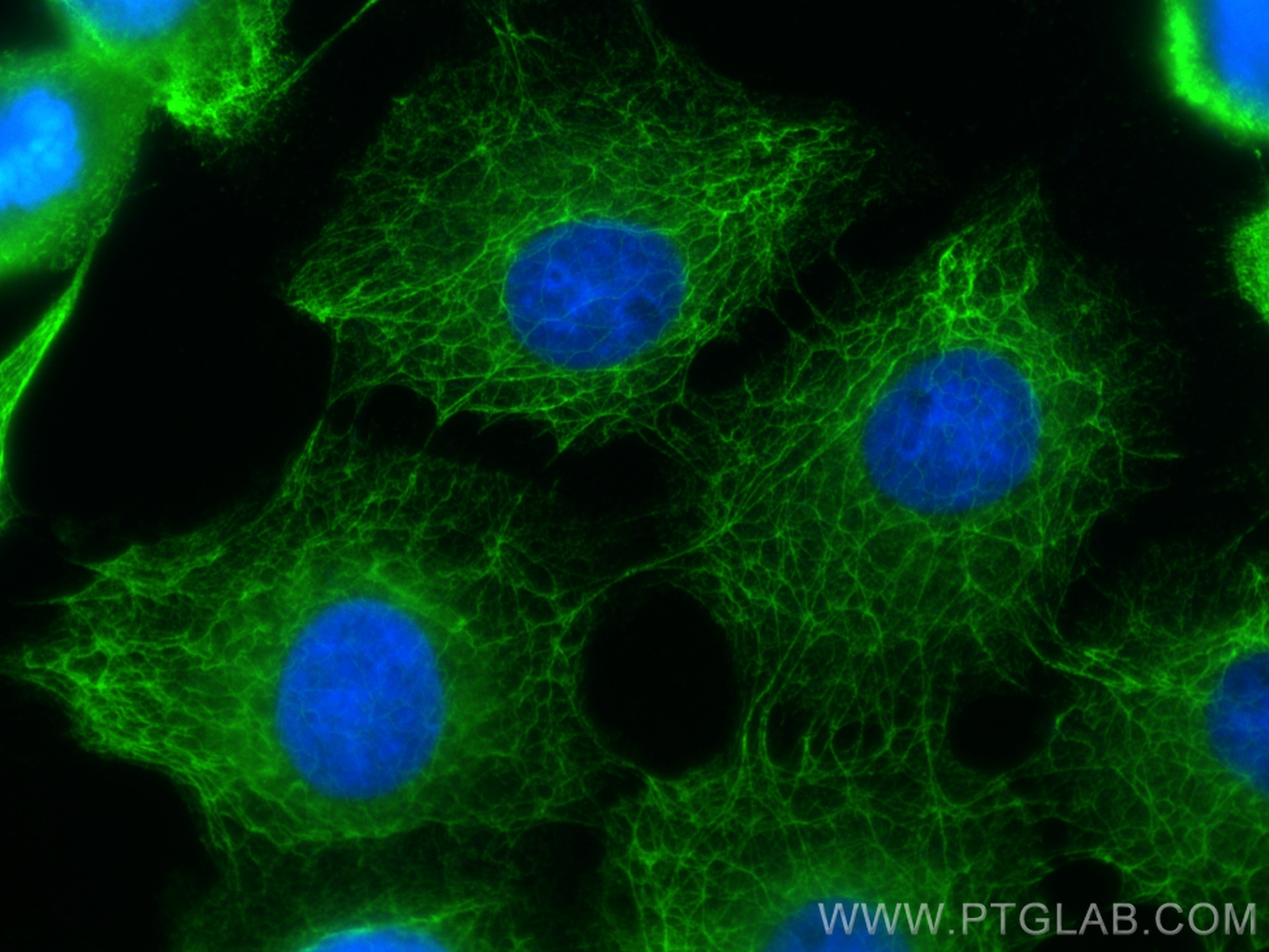
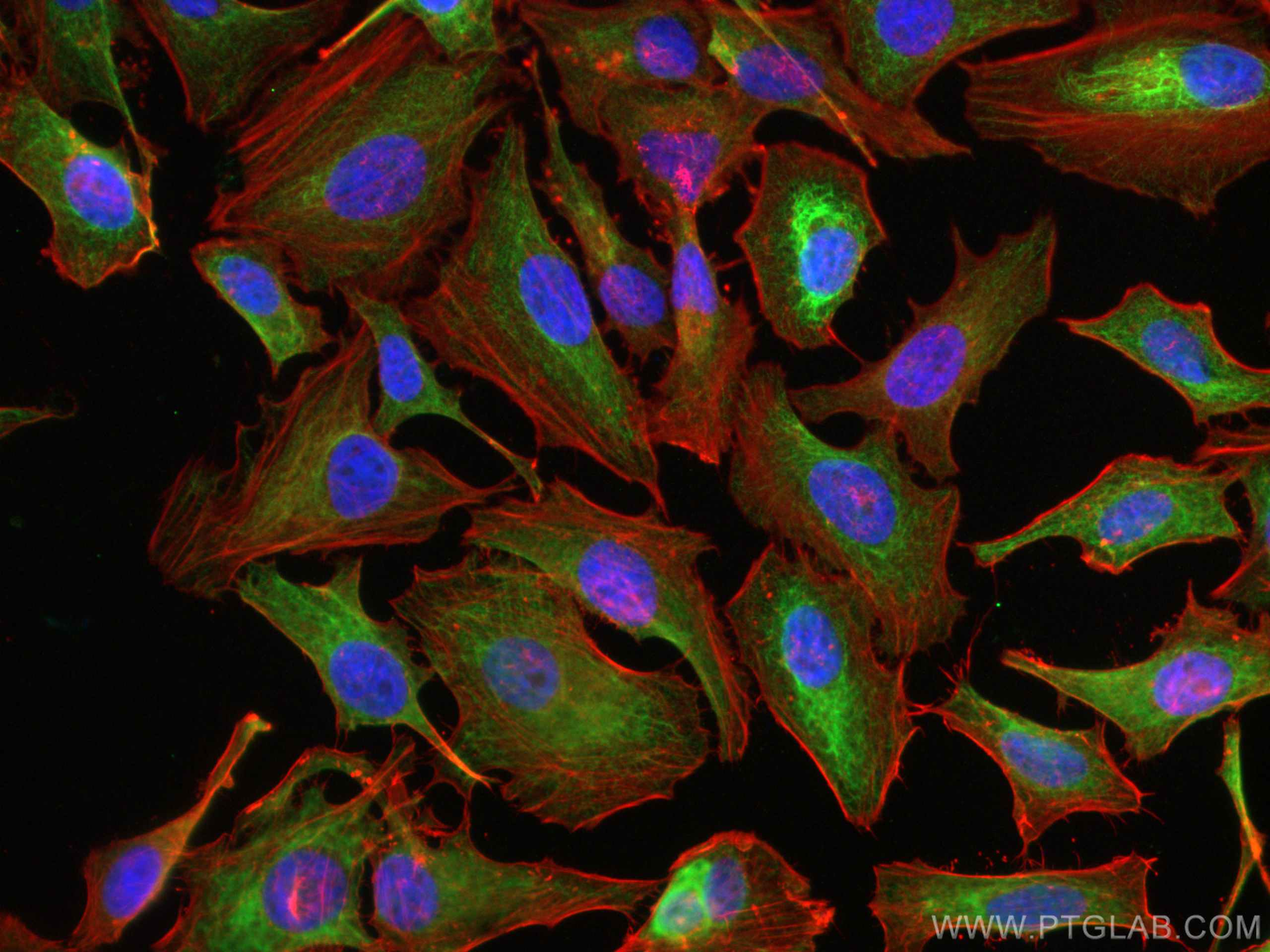
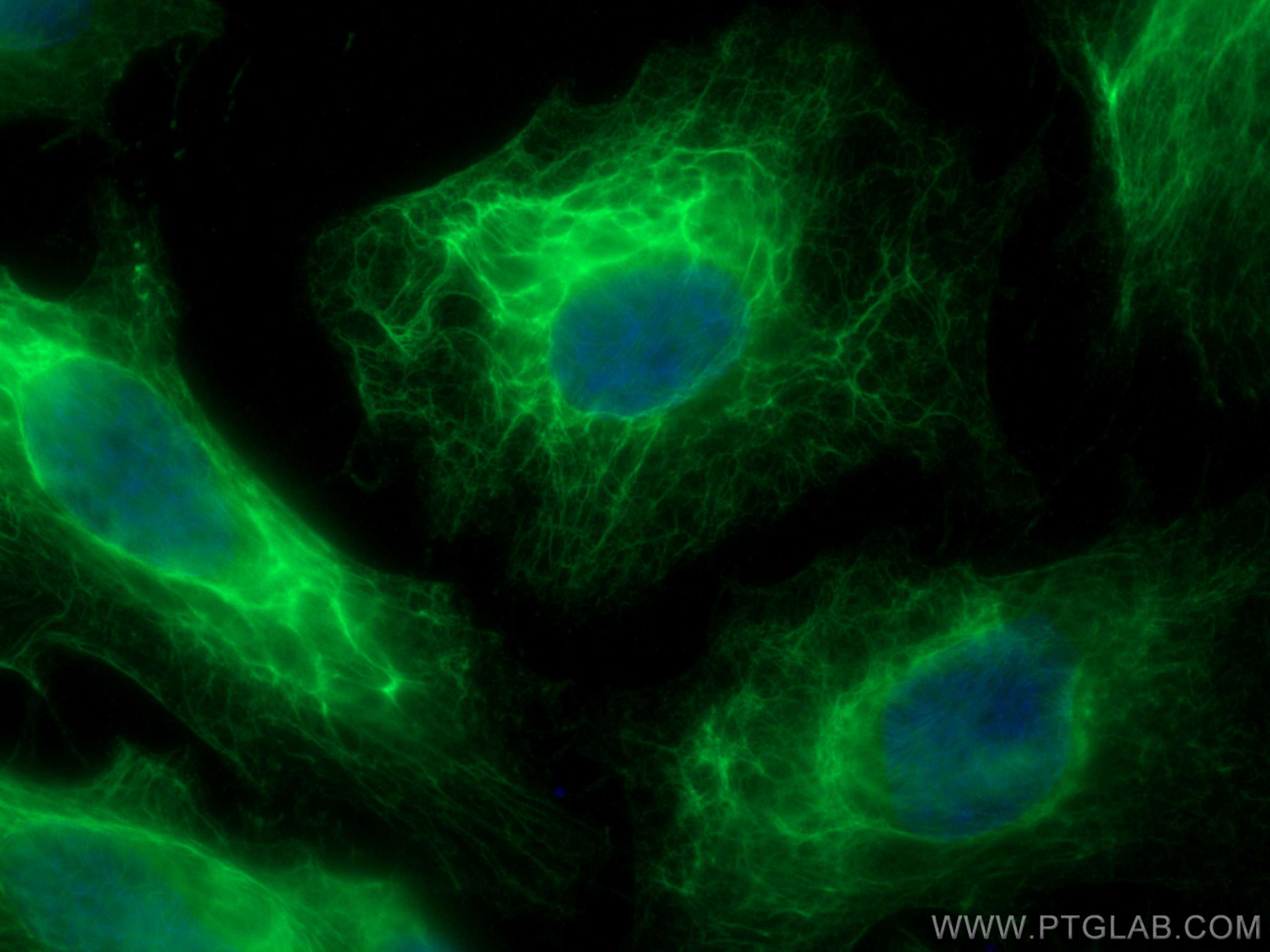
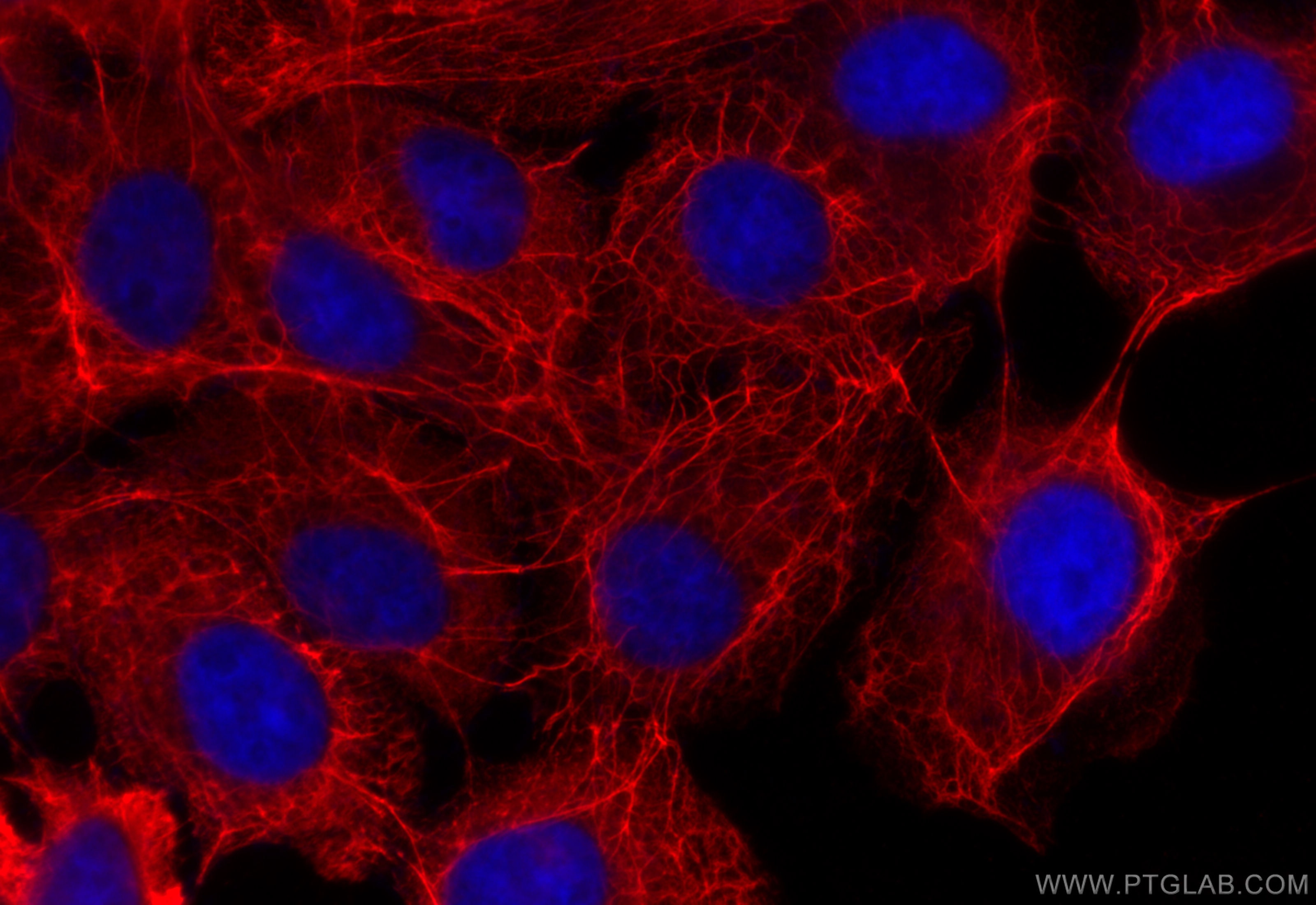
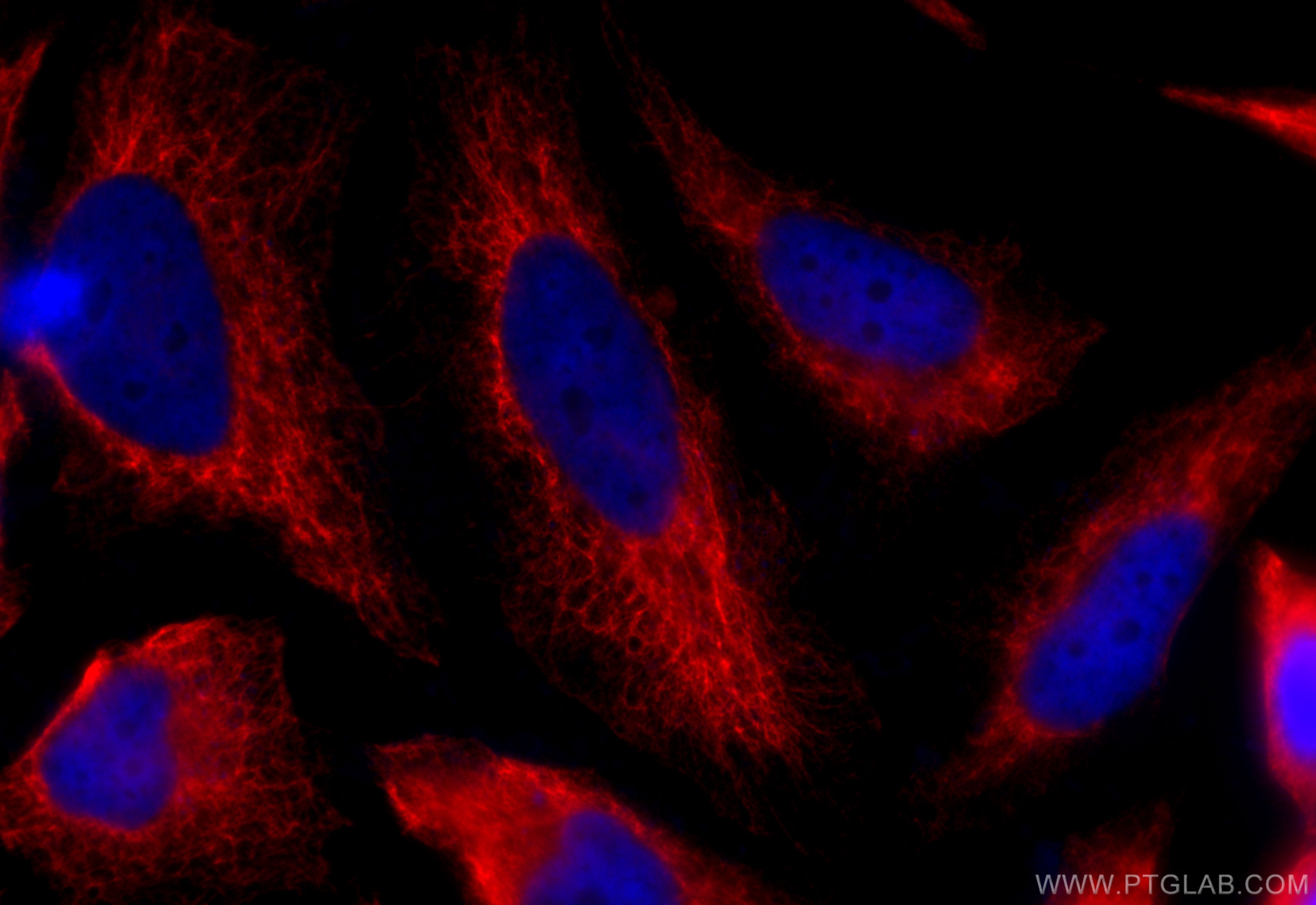
| Positive IF/ICC detected in | A431 cells, HeLa cells, mouse breast cancer |
Recommended dilution
| Application | Dilution |
|---|---|
| Western Blot (WB) | WB : 1:50000-1:200000 |
| Immunohistochemistry (IHC) | IHC : 1:1500-1:6000 |
| Immunofluorescence (IF)-P | IF-P : 1:200-1:800 |
| Immunofluorescence (IF)/ICC | IF/ICC : 1:50-1:500 |
| It is recommended that this reagent should be titrated in each testing system to obtain optimal results. | |
| Sample-dependent, Check data in validation data gallery. | |
Product Information
26411-1-AP targets Pan-Keratin in WB, IHC, IF/ICC, IF-P, ELISA applications and shows reactivity with human, mouse samples.
| Tested Reactivity | human, mouse |
| Cited Reactivity | human, mouse, rat, rabbit, sheep |
| Host / Isotype | Rabbit / IgG |
| Class | Polyclonal |
| Type | Antibody |
| Immunogen |
CatNo: Ag24184 Product name: Recombinant human KRT5 protein Source: e coli.-derived, PGEX-4T Tag: GST Domain: 316-491 aa of BC024292 Sequence: THVSDTSVVLSMDNNRNLDLDSIIAEVKAQYEEIANRSRTEAESWYQTKYEELQQTAGRHGDDLRNTKHEISEMNRMIQRLRAEIDNVKKQCANLQNAIADAEQRGELALKDARNKLAELEEALQKAKQDMARLLREYQELMNTKLALDVEIATYRKLLEGEECRLSGEGVGPVNI Predict reactive species |
| Full Name | keratin 5 |
| Calculated Molecular Weight | 590 aa, 62 kDa |
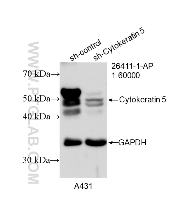
| Observed Molecular Weight | 46-58 kDa |
| GenBank Accession Number | BC024292 |
| Gene Symbol | Cytokeratin 5 |
| Gene ID (NCBI) | 3852 |
| RRID | AB_2880505 |
| Conjugate | Unconjugated |
| Form | Liquid |
| Purification Method | Antigen affinity purification |
| UNIPROT ID | P13647 |
| Storage Buffer | PBS with 0.02% sodium azide and 50% glycerol, pH 7.3. |
| Storage Conditions | Store at -20°C. Stable for one year after shipment. Aliquoting is unnecessary for -20oC storage. 20ul sizes contain 0.1% BSA. |
Background Information
Pan-keratin, also known as pan-cytokeratin, refers to a group of antibodies that recognize a broad array of keratin proteins. Keratins are a large family of intermediate filament (IF) proteins that provide structural integrity to epithelial cells. These proteins are essential for maintaining cell shape, strength, and resilience. The keratin gene family is divided into two major groups: type I keratins (acidic) and type II keratins (basic), with 27 type I and 26 type II keratin genes in humans. Pan-keratin immunostaining is widely used in pathology and research due to its ability to detect a variety of keratin proteins, making it a valuable tool for identifying epithelial cells and tissues. This staining technique is particularly useful in the diagnosis of tumors, as it can help distinguish between epithelial and non-epithelial neoplasms. This antibody is a pan-keratin antibody.
Protocols
| Product Specific Protocols | |
|---|---|
| IF protocol for Pan-Keratin antibody 26411-1-AP | Download protocol |
| IHC protocol for Pan-Keratin antibody 26411-1-AP | Download protocol |
| WB protocol for Pan-Keratin antibody 26411-1-AP | Download protocol |
| Standard Protocols | |
|---|---|
| Click here to view our Standard Protocols |
Publications
What published studies show
The Pan-Keratin Polyclonal antibody (Cat# 26411-1-AP) has 102 citations. Publications span journals including Nature Communications, Nature Cell Biology, Cancer Cell, Molecular Cancer, Science Advances, Advanced Science, Cell Death & Disease, Oncogene, EMBO Molecular Medicine, and Biomaterials. Research fields cover oncology (lung, gastric, ovarian, breast, pancreatic, colorectal cancers), tumor microenvironment, immunotherapy, fibrosis, epithelial biology, biomaterials, and tissue engineering. The antibody is primarily applied in IF, IHC, and WB.
| Species | Application | Title |
|---|---|---|
Cancer Cell Conserved spatial subtypes and cellular neighborhoods of cancer-associated fibroblasts revealed by single-cell spatial multi-omics | ||
Mol Cancer c-Rel drives pancreatic cancer metastasis through fibronectin-integrin signaling-induced isolation stress resistance and EMT. | ||
Nat Cell Biol Aberrant amino acid-sensing promotes immunotherapy resistance via the inflammatory cytokine-ZBTB5-mTORC1 axis | ||
Cancer Res Transfer of Damaged Mitochondria from Cancer Cells to Cancer-Associated Fibroblasts Promotes Tyrosine Kinase Inhibitor Tolerance in EGFR-Mutant Lung Cancer. | ||
J Extracell Vesicles Tissue-Derived Extracellular Vesicles Define Diagnostic Biomarkers for Renal Cell Carcinoma. | ||
Nat Commun Spatial heterogeneity of MDSCs mediated by ANXA1-FPRs signaling drives immune suppression in OSCC progression. |







































