

Tested Applications
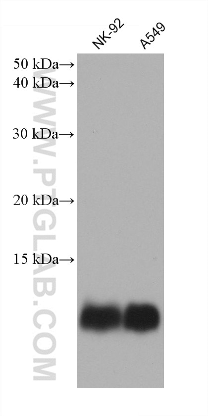
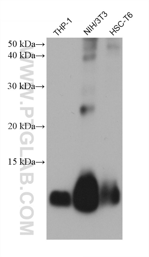
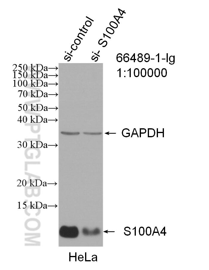
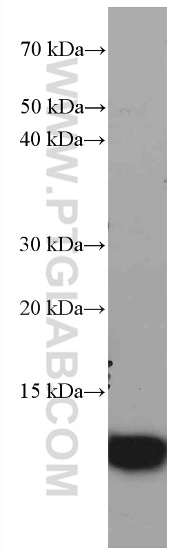
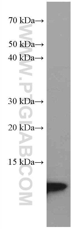
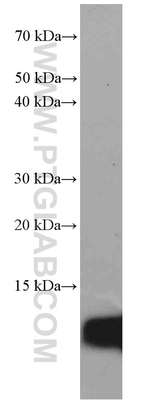
| Positive WB detected in | NK-92 cells, A375 cells, HeLa cells, PC-3 cells, THP-1 cells, A549 cells, NIH/3T3 cells, HSC-T6 cells |
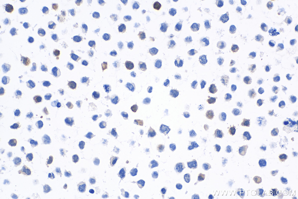
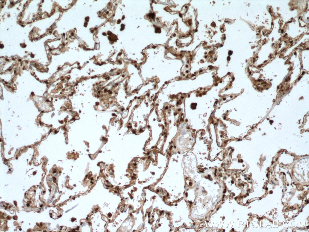
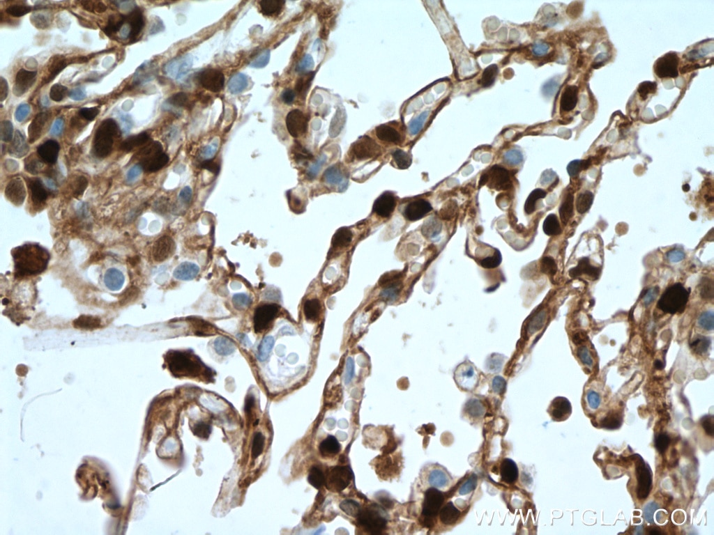
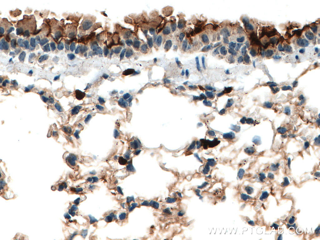
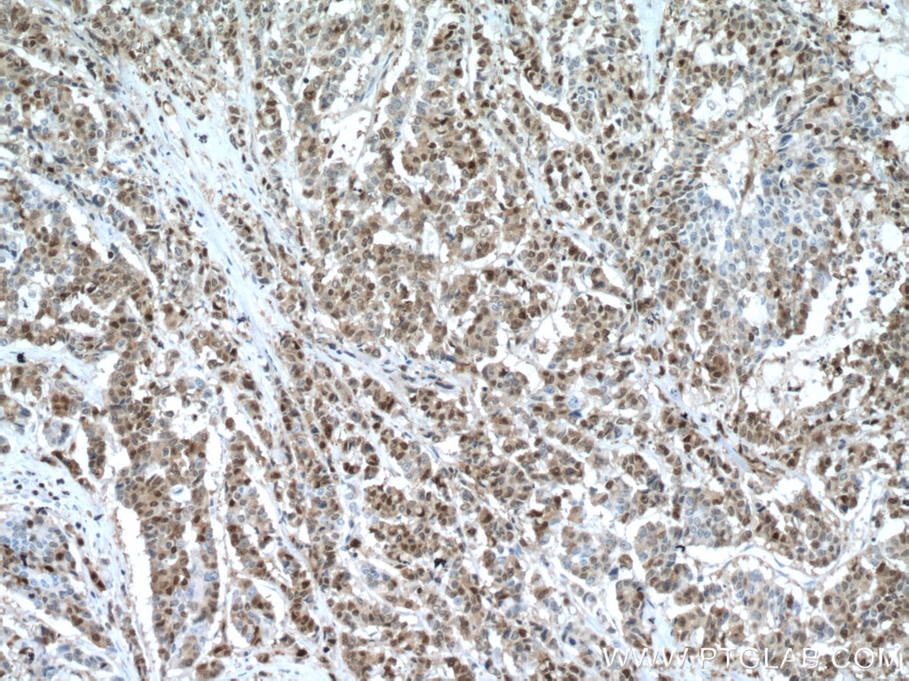
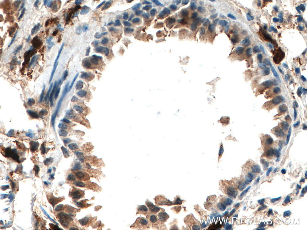
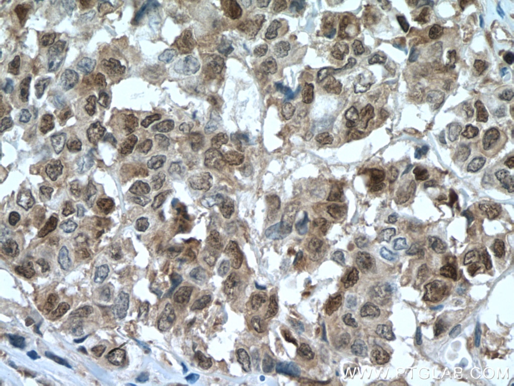
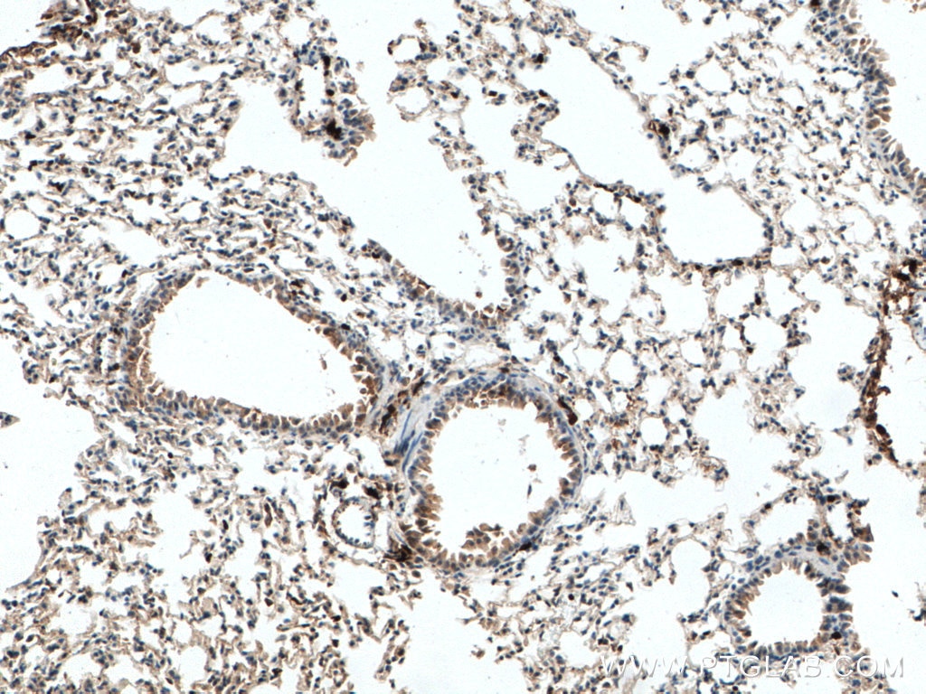
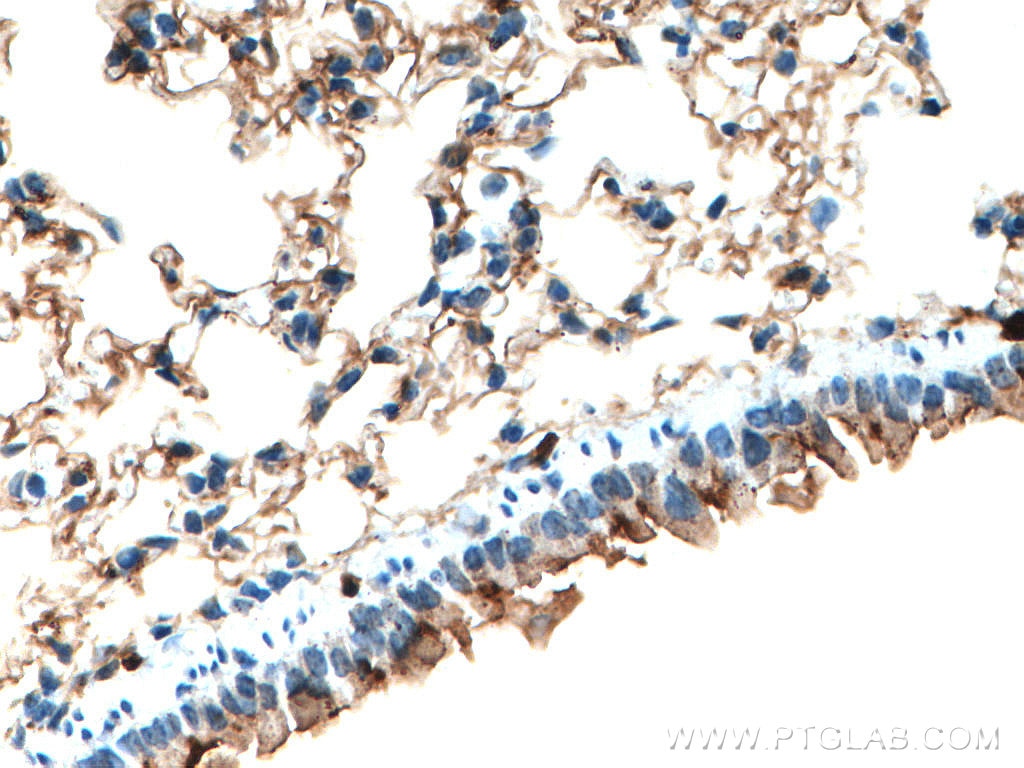
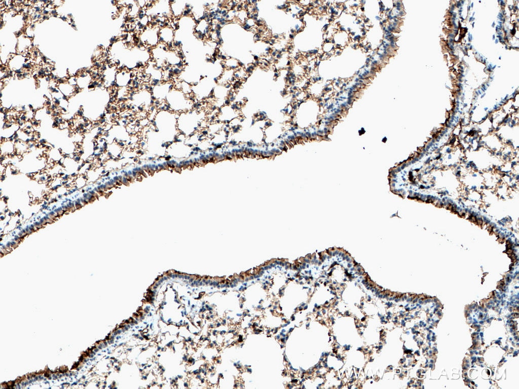
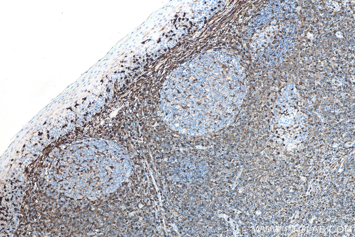
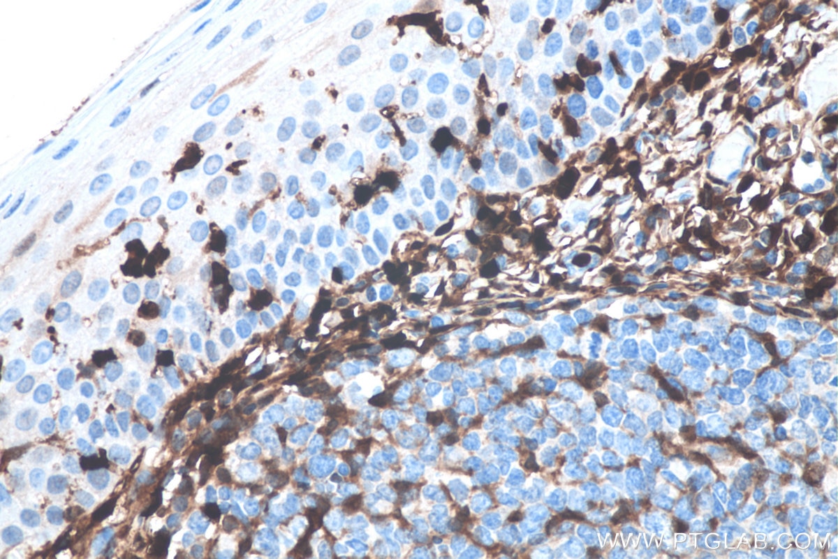
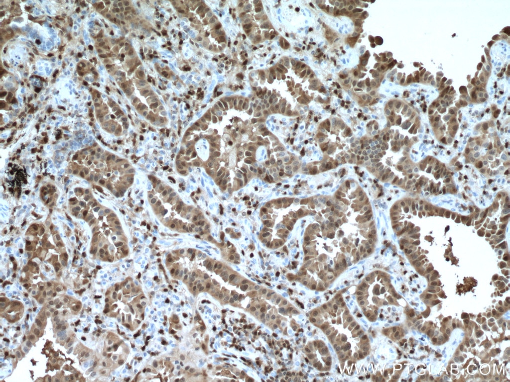
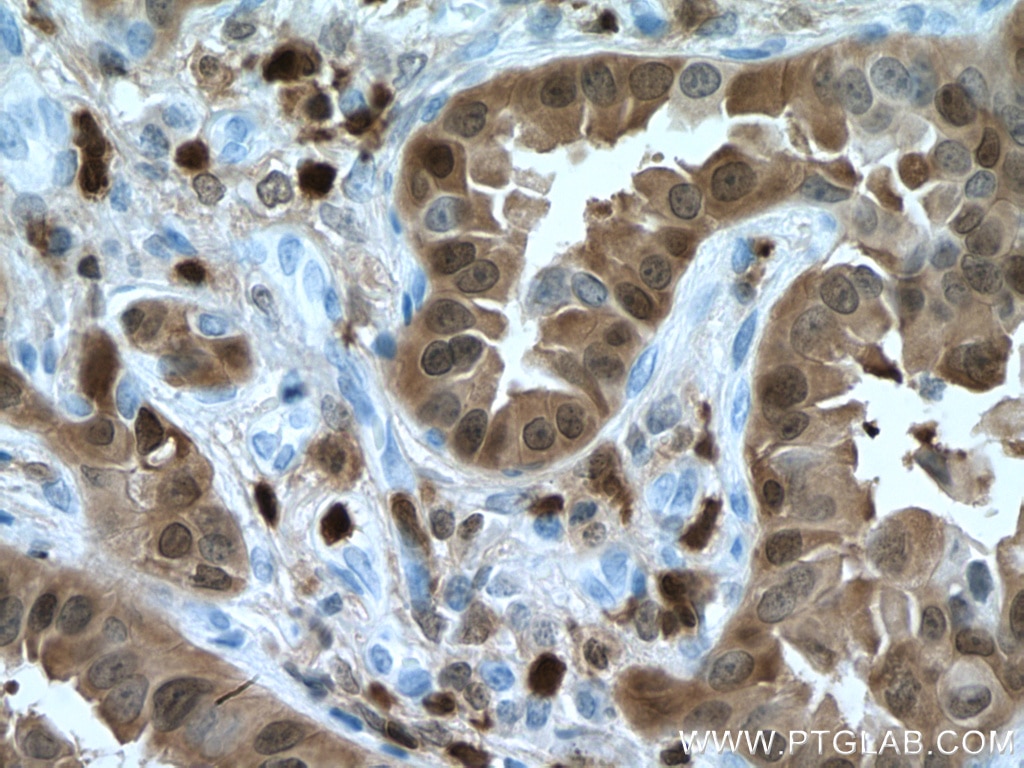
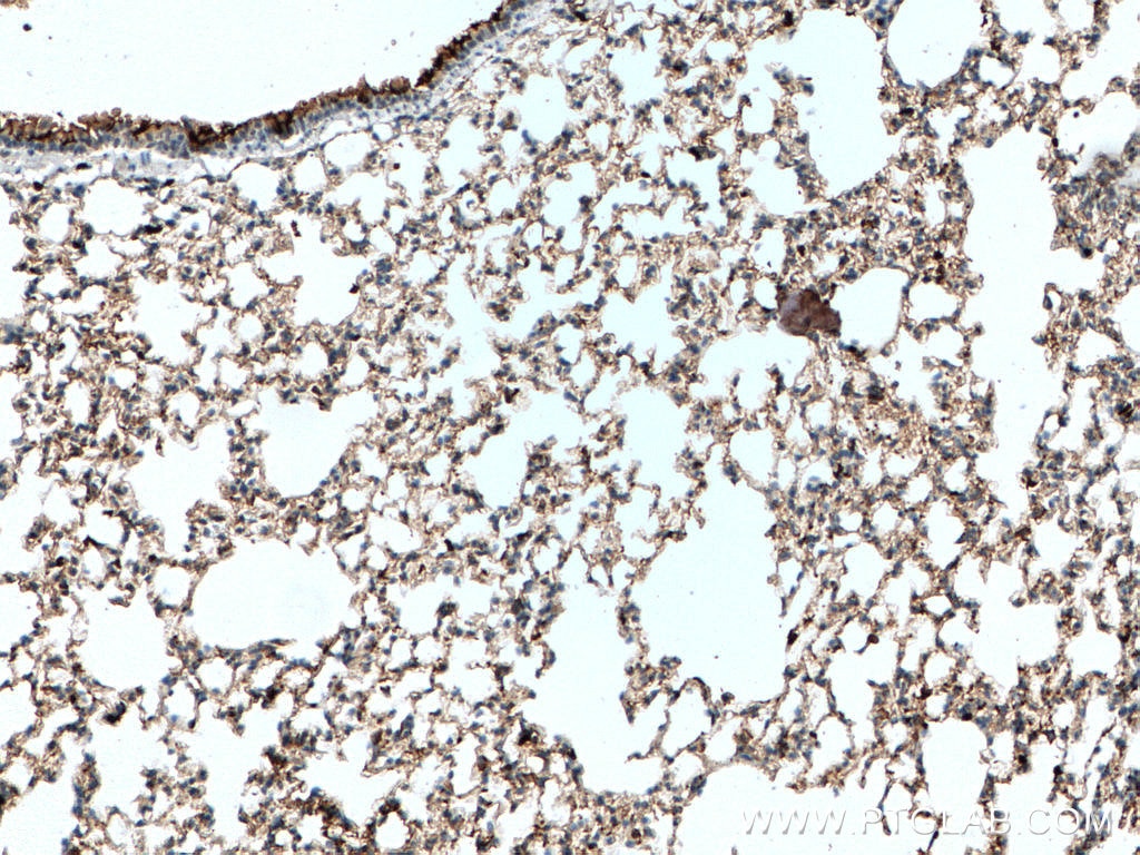
| Positive IHC detected in | human lung cancer tissue, human colon cancer tissue, human tonsillitis tissue, K-562 cells, mouse lung tissue Note: suggested antigen retrieval with TE buffer pH 9.0; (*) Alternatively, antigen retrieval may be performed with citrate buffer pH 6.0 |
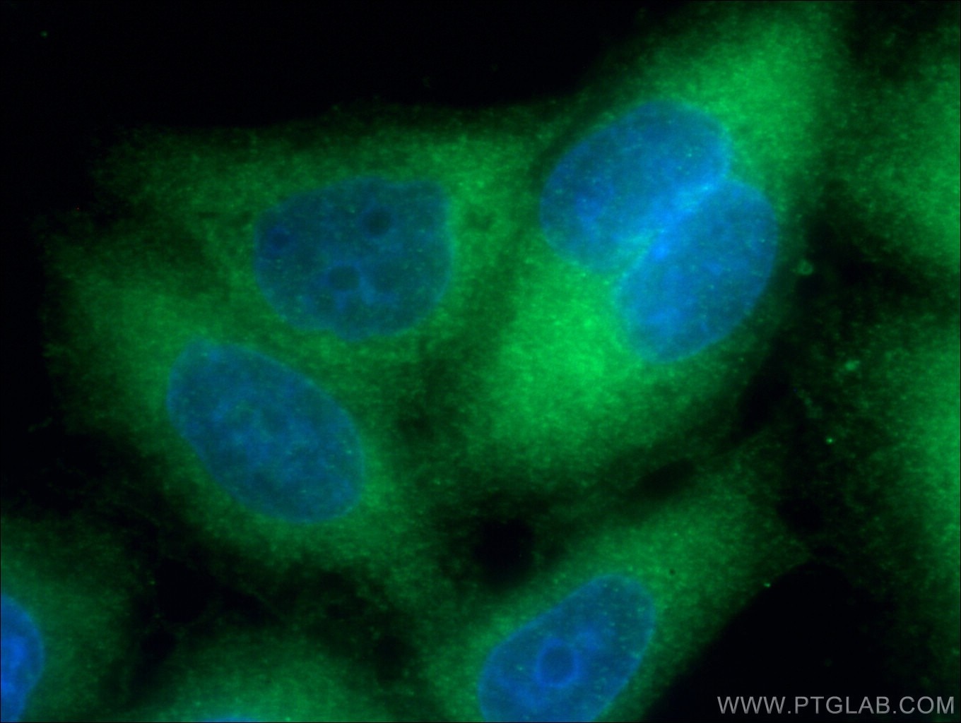
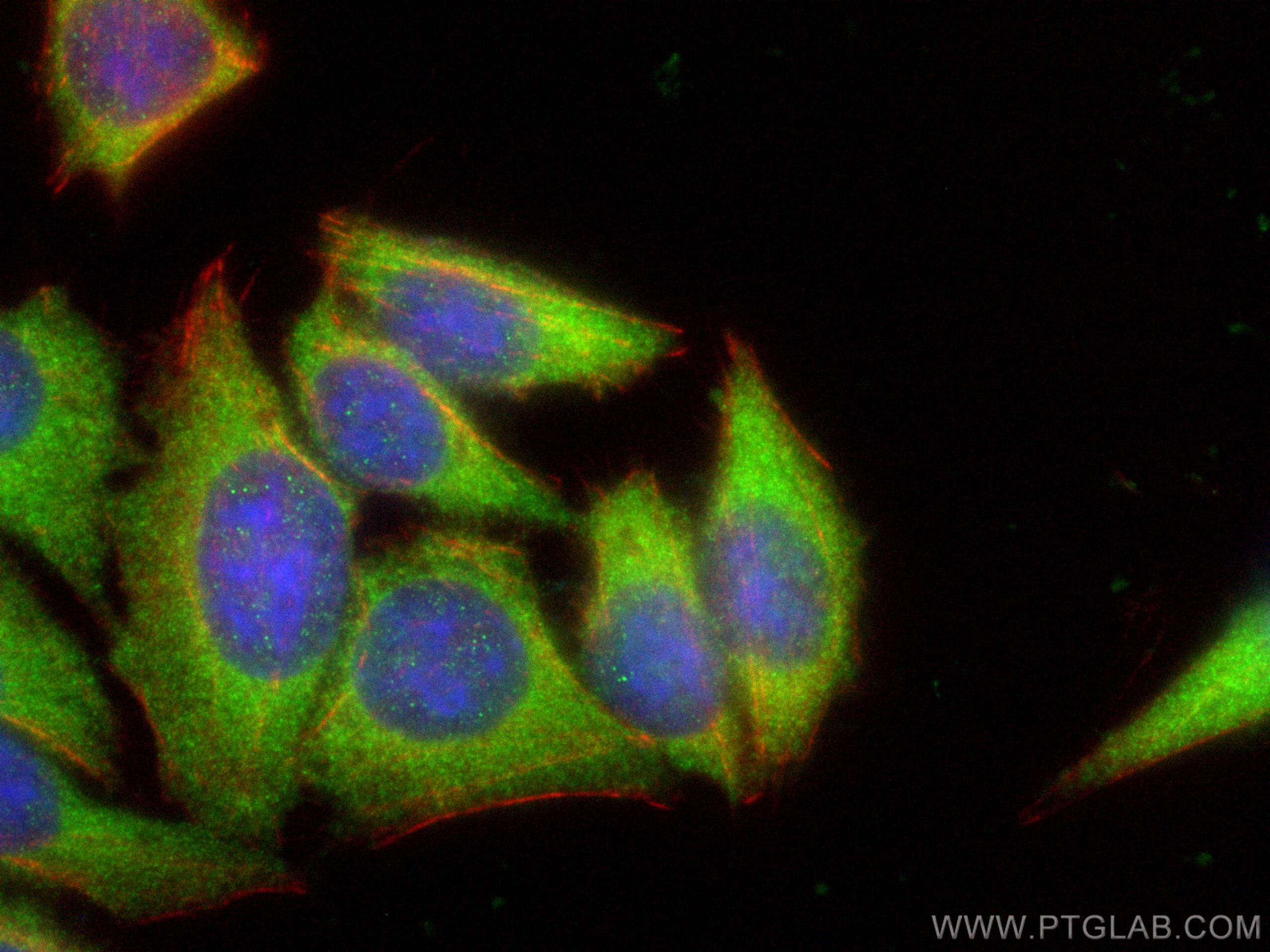
| Positive IF/ICC detected in | HepG2 cells, HeLa cells |
Recommended dilution
| Application | Dilution |
|---|---|
| Western Blot (WB) | WB : 1:5000-1:50000 |
| Immunohistochemistry (IHC) | IHC : 1:1000-1:4000 |
| Immunofluorescence (IF)/ICC | IF/ICC : 1:50-1:500 |
| It is recommended that this reagent should be titrated in each testing system to obtain optimal results. | |
| Sample-dependent, Check data in validation data gallery. | |
Product Information
66489-1-Ig targets S100A4 in WB, IHC, IF/ICC, ELISA applications and shows reactivity with human, mouse samples.
| Tested Reactivity | human, mouse |
| Cited Reactivity | human, mouse, rat |
| Host / Isotype | Mouse / IgG2a |
| Class | Monoclonal |
| Type | Antibody |
| Immunogen |
CatNo: Ag9019 Product name: Recombinant human S100A4 protein Source: e coli.-derived, PGEX-4T Tag: GST Domain: 1-101 aa of BC016300 Sequence: MACPLEKALDVMVSTFHKYSGKEGDKFKLNKSELKELLTRELPSFLGKRTDEAAFQKLMSNLDSNRDNEVDFQEYCVFLSCIAMMCNEFFEGFPDKQPRKK Predict reactive species |
| Full Name | S100 calcium binding protein A4 |
| Calculated Molecular Weight | 101 aa, 12 kDa |
| Observed Molecular Weight | 12 kDa |
| GenBank Accession Number | BC016300 |
| Gene Symbol | S100A4 |
| Gene ID (NCBI) | 6275 |
| ENSEMBL Gene ID | ENSG00000196154 |
| RRID | AB_2881854 |
| Conjugate | Unconjugated |
| Form | Liquid |
| Purification Method | Protein A purification |
| UNIPROT ID | P26447 |
| Storage Buffer | PBS with 0.02% sodium azide and 50% glycerol, pH 7.3. |
| Storage Conditions | Store at -20°C. Stable for one year after shipment. Aliquoting is unnecessary for -20oC storage. 20ul sizes contain 0.1% BSA. |
Background Information
S100A4 is a member of the S100 family of calcium-binding proteins. The S100 family members have been involved in the regulation of a number of cellular processes such as cell cycle progression and differentiation. S100A4 is known to localize to and function in the nucleus, cytoplasm of cells, and the extracellular space. S100A4 has also been shown to be associated with tumor growth, motility, invasion, metastasis, angiogenesis, apoptosis, and chemoresistance. It is a fibroblast-specific protein associated with mesenchymal cell morphology and motility, is expressed during epithelial-mesenchymal transformations (EMT) in vivo (PMID: 9362334). It is a specific prognostic marker for renal survival in patients with IgAN (PMID: 16105038). It is also an improved marker for lung fibroblasts that could be useful for investigating the pathogenesis of pulmonary fibrosis(PMID: 15618458). Overexpression of S100A4 is correlated with a worse prognosis inpatients with various types of cancer.
Protocols
| Product Specific Protocols | |
|---|---|
| IF protocol for S100A4 antibody 66489-1-Ig | Download protocol |
| IHC protocol for S100A4 antibody 66489-1-Ig | Download protocol |
| WB protocol for S100A4 antibody 66489-1-Ig | Download protocol |
| Standard Protocols | |
|---|---|
| Click here to view our Standard Protocols |
Publications
What published studies show
The anti-S100A4 antibody (Cat# 66489-1-Ig) has 37 citations across journals including Blood, Cancer Research, Advanced Science, Cell Death & Differentiation, Journal of Hepatology, American Journal of Pathology, PLoS Pathogens, Investigative Ophthalmology & Visual Science, and Frontiers in Pharmacology. Research fields span fibrosis (cardiac, renal, pulmonary, hepatic), cancer (ovarian, colorectal, gastric, glioblastoma, leukemia), EMT/EndMT, macrophage biology, Zika virus, diabetic retinopathy, and preeclampsia.
| Species | Application | Title |
|---|---|---|
J Hepatol Disruption of carbon metabolism by the hepatic mononuclear phagocytic system drives liver injury in a mouse model of severe dengue. | ||
Blood C1Q labels a highly aggressive macrophage-like leukemia population indicating extramedullary infiltration and relapse | ||
Cancer Res Cancer-Associated Fibroblasts Expressing Sulfatase 1 Facilitate VEGFA-Dependent Microenvironmental Remodeling to Support Colorectal Cancer | ||
Adv Sci (Weinh) TP53RK Drives the Progression of Chronic Kidney Disease by Phosphorylating Birc5 | ||
Adv Sci (Weinh) Meis1 Targets Protein Tyrosine Phosphatase Receptor J in Fibroblast to Retard Chronic Kidney Disease Progression | ||
Cell Death Differ Upregulation of Wilms' Tumor 1 in epicardial cells increases cardiac fibrosis in dystrophic mice. |
Reviews
What customers say
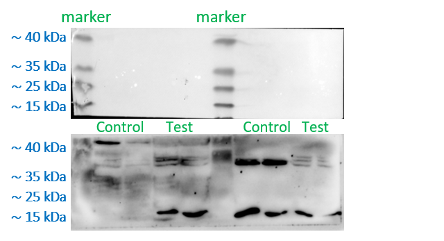
Users report that this antibody performs well in Western Blot with high specificity, even at dilutions far exceeding the recommended range (up to 1:50,000). One reviewer noted excellent band specificity on A2780 cells at both 1:1,000 and 1:50,000. Another reviewer raised concerns about the low molecular weight of the target protein, which caused bands to migrate lower in the gel and produce less ideal band shapes despite using optimized 10% and 12% gels. Overall, users find it reliable and cost-effective due to its ability to work at high dilutions.
The reviews below have been submitted by verified Proteintech customers who received an incentive for providing their feedback.
CAITLIN (Verified Customer) (03-04-2025) | Worked great, highly specific even when used at higher dilution than recommended
|
Caitlin (Verified Customer) (10-15-2024) | Works well, very convenient that the antibody can be used at such high dilutions and still achieve band specificity!
|
Shinford (Verified Customer) (10-26-2023) | I have some reservations about the antibody because of its relatively low molecular weight. We attempted to create separate gels with 10% and 12% concentrations and made an effort to run the gels for a limited time to ensure that the entire protein remained within the original gels and membranes. While we were able to detect several bands, their positions lower in the gel led to less than ideal band shapes.
 |
























