



Tested Applications
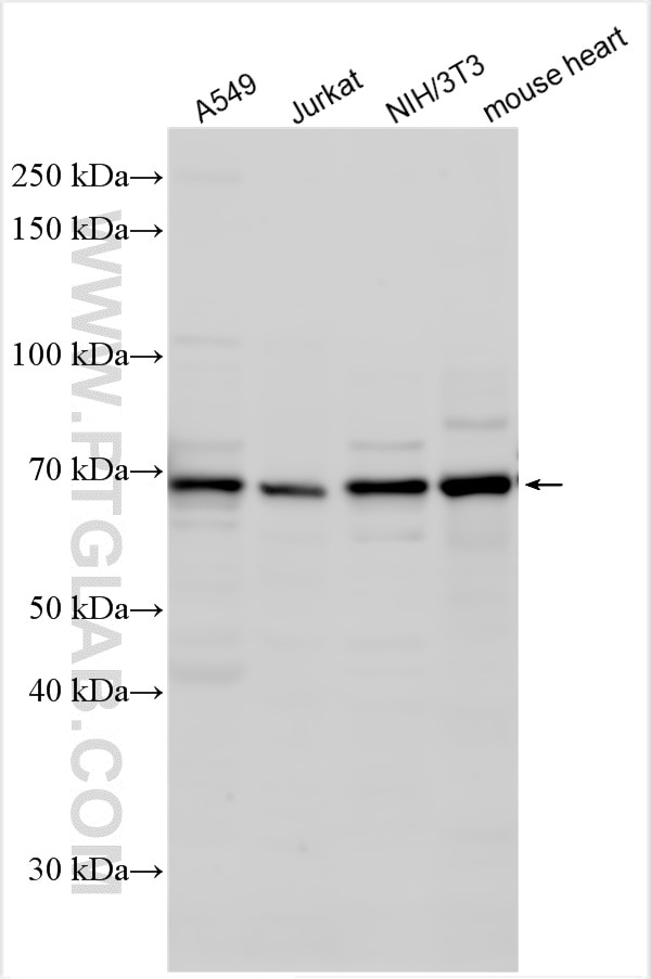
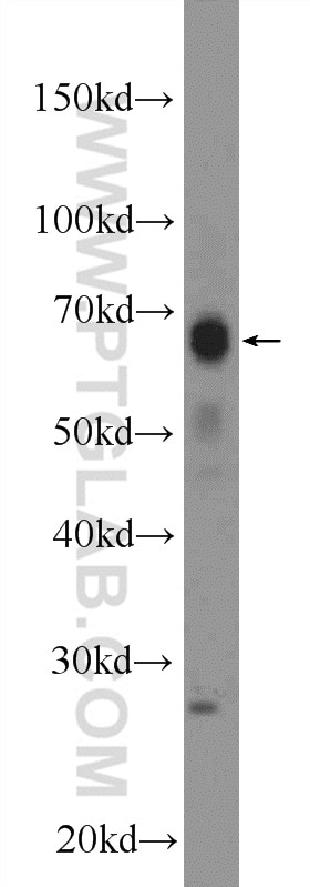
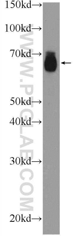
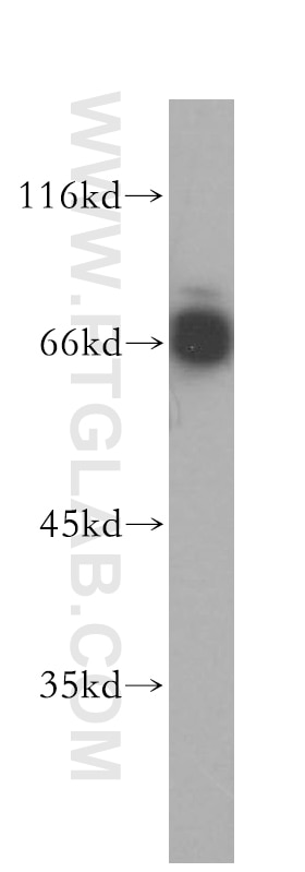
| Positive WB detected in | A549 cells, Jurkat cells, mouse heart tissue, rat skin tissue, NIH/3T3 cells |
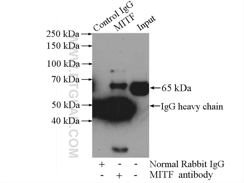
| Positive IP detected in | mouse heart tissue |
| Positive IHC detected in | mouse skin tissue Note: suggested antigen retrieval with TE buffer pH 9.0; (*) Alternatively, antigen retrieval may be performed with citrate buffer pH 6.0 |
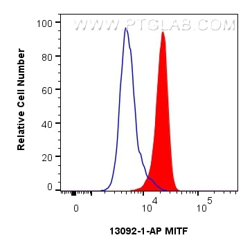
| Positive FC (Intra) detected in | HeLa cells |
Recommended dilution
| Application | Dilution |
|---|---|
| Western Blot (WB) | WB : 1:500-1:3000 |
| Immunoprecipitation (IP) | IP : 0.5-4.0 ug for 1.0-3.0 mg of total protein lysate |
| Immunohistochemistry (IHC) | IHC : 1:500-1:2000 |
| Flow Cytometry (FC) (INTRA) | FC (INTRA) : 0.25 ug per 10^6 cells in a 100 µl suspension |
| It is recommended that this reagent should be titrated in each testing system to obtain optimal results. | |
| Sample-dependent, Check data in validation data gallery. | |
Product Information
13092-1-AP targets MITF in WB, IHC, IF, FC (Intra), IP, ELISA applications and shows reactivity with human, mouse, rat samples.
| Tested Reactivity | human, mouse, rat |
| Cited Reactivity | human, mouse, rat, duck |
| Host / Isotype | Rabbit / IgG |
| Class | Polyclonal |
| Type | Antibody |
| Immunogen |
CatNo: Ag3679 Product name: Recombinant human MITF protein Source: e coli.-derived, PGEX-4T Tag: GST Domain: 1-91 aa of BC012503 Sequence: MLEMLEYNHYQVQTHLENPTKYHIQQAQRQQVKQYLSTTLANKHANQVLSLPCPNQPGDHVMPPVPGSSAPNSPMAMLTLNSNCEKEFMKQ Predict reactive species |
| Full Name | microphthalmia-associated transcription factor |
| Calculated Molecular Weight | 91 aa, 10 kDa, 59 kDa |
| Observed Molecular Weight | 59-65 kDa |
| GenBank Accession Number | BC012503 |
| Gene Symbol | MITF |
| Gene ID (NCBI) | 4286 |
| RRID | AB_10597698 |
| Conjugate | Unconjugated |
| Form | Liquid |
| Purification Method | Antigen affinity purification |
| UNIPROT ID | O75030 |
| Storage Buffer | PBS with 0.02% sodium azide and 50% glycerol, pH 7.3. |
| Storage Conditions | Store at -20°C. Stable for one year after shipment. Aliquoting is unnecessary for -20oC storage. 20ul sizes contain 0.1% BSA. |
Background Information
The retinal pigment epithelium (RPE) has a essential role in maintaining visual function and dedifferentiation of RPE contributes to the pathophysiology of several ocular diseases[PMID: 22523078]. Microphthalmia-associated transcription factor (MITF) is a key regulator of RPE differentiation that is also down-regulated in dedifferentiated hfRPE cells. MITF is a basic helix-loop-helix (hHLH)-leucine zipper protein that involves in the development of various cell types, including neural crest-derived melanocytes and optic cup-derived retinal pigment epithelial cells [PMID: 10578055].
Protocols
| Product Specific Protocols | |
|---|---|
| FC protocol for MITF antibody 13092-1-AP | Download protocol |
| IHC protocol for MITF antibody 13092-1-AP | Download protocol |
| IP protocol for MITF antibody 13092-1-AP | Download protocol |
| WB protocol for MITF antibody 13092-1-AP | Download protocol |
| Standard Protocols | |
|---|---|
| Click here to view our Standard Protocols |
Publications
What published studies show
The anti-MITF antibody (Cat# 13092-1-AP) has accumulated 57 citations across journals including Cancer Communications, Autophagy, Theranostics, Journal of Clinical Investigation, Cell Death & Differentiation, Acta Pharmaceutica Sinica B, Phytomedicine, International Journal of Molecular Sciences, Scientific Reports, and Pigment Cell & Melanoma Research. Key research fields include melanogenesis and skin pigmentation disorders, melanoma oncology, autophagy and lysosomal biology, natural product-based drug discovery, and biomaterials/tissue engineering applications.
| Species | Application | Title |
|---|---|---|
Cancer Commun (Lond) HMMR alleviates endoplasmic reticulum stress by promoting autophagolysosomal activity during endoplasmic reticulum stress-driven hepatocellular carcinoma progression | ||
Autophagy Feedback loops between DNMT1 and autophagy as well as senescence promotes organ aging and canities. | ||
Autophagy The human vault RNA enhances tumorigenesis and chemoresistance through the lysosome in hepatocellular carcinoma | ||
Theranostics TFE3, a potential therapeutic target for Spinal Cord Injury via augmenting autophagy flux and alleviating ER stress. | ||
Acta Pharm Sin B EHMT2 promotes tumorigenesis in GNAQ/11-mutant uveal melanoma via ARHGAP29-mediated RhoA pathway | ||
J Clin Invest mTORC1 feedback to AKT modulates lysosomal biogenesis through MiT/TFE regulation. |
Reviews
What customers say
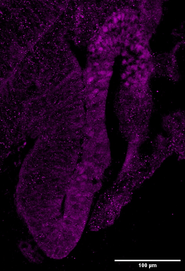
Both reviewers rated this antibody 5 stars for immunofluorescence. One user tested it on retinal organoids at 1:500 dilution, reporting specific nuclear staining in pigmented cells with no nonspecific signal in other tissue types. They noted that antigen retrieval reduces signal specificity and is not recommended. The second user simply reported no unspecific staining and great outcomes. Overall, customers find the antibody highly specific and effective for IF applications, with the key tip being to avoid antigen retrieval for best results.
The reviews below have been submitted by verified Proteintech customers who received an incentive for providing their feedback.
Eva (Verified Customer) (07-01-2025) | The antibody is functional both with and without antigen retrieval (AR: 110ºC for 1 min, a second cycle of 90ºC for 10s, in a citrate buffer 10mM pH=6); however, signal specificity appears reduced when antigen retrieval is applied, and its use is therefore not recommended. After x3 PBST (PBS + 0,2% Tween) washes, cryostat sample slides were incubated 2 hours with blocking buffer (PBST 0,2% + 5% FBS + 1% BSA) at Room Temperature (RT). The primary antibody was diluted 1:500 in Blocking Buffer and incubated with the samples overnight at 4 °C. Next day, wash x3 with PBST 0,2%, and incubate second antibody with fluorocrome at 1:500 2-3 hours at RT. Some background staining is observed, likely due to a high concentration of the secondary antibody, as it does not correspond to a specific signal. The staining is specifically localized in nuclei of pigmented cells, as expected, and no nonspecific signal is detected in other tissue types within the sample.
 |
Alessandro (Verified Customer) (11-06-2022) | no unspecific staining, great outcome
|









